. 2004 Jun;164(6):2027-38.
doi: 10.1016/S0002-9440(10)63762-5.

Keratinocytes from patients lacking collagen XVII display a migratory phenotype

Affiliations

Keratinocytes from patients lacking collagen XVII display a migratory phenotype

Kaisa Tasanen et al. Am J Pathol. 2004 Jun.

Abstract

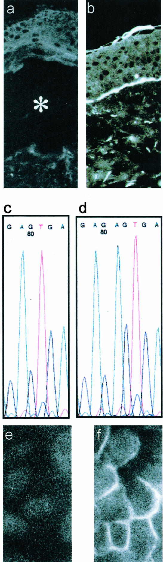
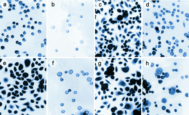
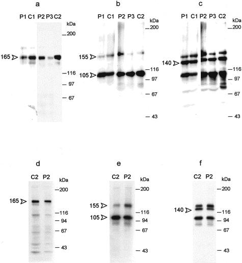
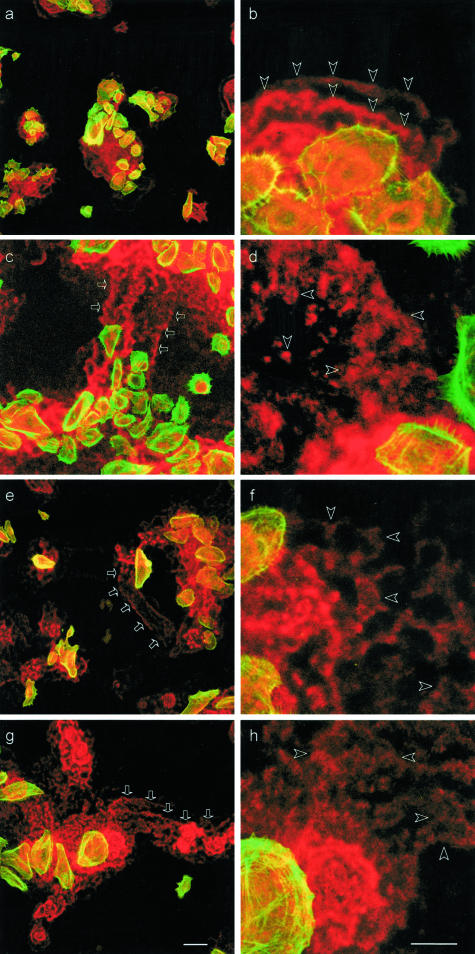
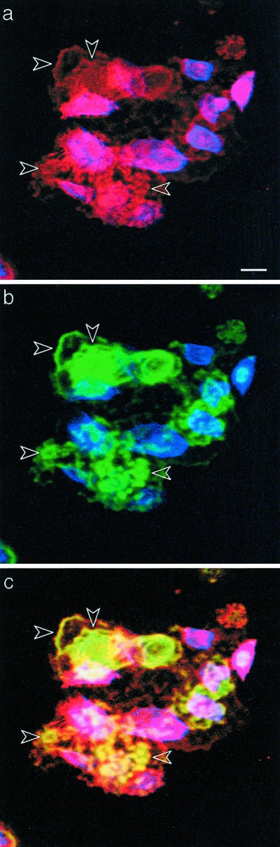
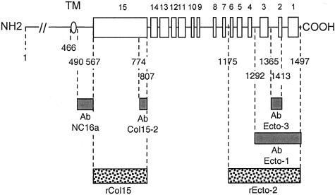
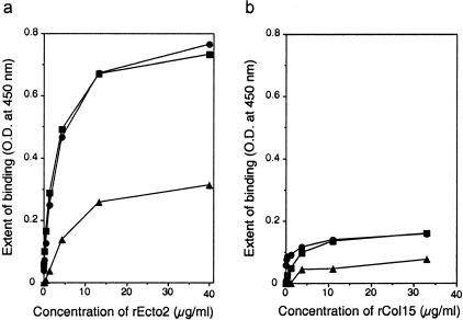
Acquired or inherited junctional epidermolysis bullosa are skin diseases characterized by a separation between the epidermis and the dermis. In inherited nonlethal junctional epidermolysis bullosa, genetic analysis has identified mutations in the COL17A1 gene coding for the transmembrane collagen XVII whereas patients with acquired diseases have autoantibodies against this protein. This suggests that collagen XVII participates in the adhesion of basal keratinocytes to the extracellular matrix. To test this hypothesis, we studied the behavior of keratinocytes with null mutations in the COL17A1 gene. Initial adhesion of mutant cells to laminin 5 was comparable to controls and similarly dependent on alpha3beta1 integrins. The spreading of mutant cells was, however, enhanced, suggesting a propensity to migrate, which was confirmed by migration assays. In addition, laminin 5 deposited by collagen XVII-deficient keratinocytes was scattered and poorly organized, suggesting that correct integration of laminin 5 within the matrix requires collagen XVII. This assumption was supported by the co-distribution of the two proteins in the matrix of normal human keratinocytes and by protein-protein-binding assays showing that the C-terminus of collagen XVII binds to laminin 5. Together, the results unravel an unexpected role of collagen XVII in the regulation of keratinocyte migration.


Figures

Figure 1
Figure 1
Characterization of C17−/− keratinocytes. Immunostaining of collagen XVII with Ab NC16a is negative in the skin of P1 (a) whereas it is positive in normal human skin (b). In a the asterisk denotes the blister cavity. Analysis of genomic DNA of P1 showed a homozygous 2-bp deletion at nucleotide position 520, 520delAG, of the COL17A1 gene XVII (c). The normal sequence is shown in d. Immunofluorescence staining of keratinocytes from P1 shows lack of collagen XVII expression (e), whereas the NC16a Ab decorates the plasma membrane of NHKs (f).
Figure 2
Figure 2
Adhesion and spreading of C17−/− keratinocytes and NHKs. NHKs (a–d) and keratinocytes from P1 (e–h) were seeded on laminin 1 (a, b, e, f) or laminin 5 (c, d, g, h) in the presence (b, f, d, h) or absence (a, e, c, g) of mAb P4C10 against the integrin β1 subunit (1:1000 dilution). After 30 minutes of adhesion, adherent cells were fixed, stained, and photographed as described in Materials and Methods. Note that in all cases mAb P4C10 inhibits cell adhesion.
Figure 3
Figure 3
Adhesion of C17−/− keratinocytes to laminin 5 is mediated by α3β1 integrins. C17−/− keratinocytes from P2 (a, b) and NHKs (c, d) were seeded on laminin 5 (a, c) or collagen IV (b, d) in the absence (white columns) or presence of function-blocking Abs against β1 (black columns) or α3 (hatched columns) integrins. After 30 minutes, adherent cells were fixed, stained, and the extent of adhesion was measured by color reading as described in Materials and Methods. Adhesion to laminin 5 of NHKs and C17−/− keratinocytes is inhibited by mAbs against β1 (clone P4C10,1:500) or α3 (clone P1B5, 1:400) integrins. Adhesion to collagen IV is inhibited by P4C10 only. The values represent the average of triplicate wells ± SEM.
Figure 4
Figure 4
C17−/− keratinocytes display more lamellipodias than NHKs. NHKs (a–c) and C17−/− keratinocytes from P2 (d–f) were seeded on laminin 1 (a, d), laminin 5 (b, e), or collagen IV (c, f). After 30 minutes of adhesion, adherent cells were fixed, stained, and photographed under phase contrast microscopy. Compared to NHKs (top), most C17−/− keratinocytes (bottom) form lamellipodia.
Figure 5
Figure 5
C17−/− keratinocytes are more motile than NHKs. a–c: In vitro wound closure assays. NHKs (a) and C17−/− keratinocytes from P2 (b) were seeded at the same density on duplicate wells coated with laminin 5. After 2.5 hours a scratch was made in the monolayers and photographs of the wound edge were taken immediately (T0) and after 2 (T2) and 5 hours (T5). The dotted line indicates the position of the wound margin at T0. c: The migration of NHKs (white columns) and C17−/− keratinocytes (black columns) was measured on photographs and expressed in arbitrary units (distance covered by the cells between photographs T0 and T5; mean of 10 measurements ± SEM). The results are shown for two experiments (experiment 1 and experiment 2) performed on different days. d–f: Scattering assays. Equal numbers of NHKs (d) and C17−/− keratinocytes from P2 (e) were seeded as small colonies in the center of tissue culture wells. After 60 minutes the wells were filled with keratinocyte growth medium and photographs of the colony margins were taken immediately (T0) and after 2, 4, 6, and 8 hours (T8). The dotted line marks the margin of the colonies at T0. f: The number of cells that had emigrated from three individual colonies of NHKs (open circles) and C17−/− keratinocytes (filled circles) was counted on photographs taken at different times after onset of the experiment as indicated. Both assays were repeated on 2 consecutive days.
Figure 6
Figure 6
The α6β4 integrin is targeted to cell-matrix contacts in both C17−/− keratinocytes and NHKs. Immunostainings for β4 (a, c, e, g) or α6 (b, d, f, h) integrins (in red) are positive in NHKs (a, b) and keratinocytes from nJEB patients (P1, c and d; P2, e and f; P3, g and h). Labeling of fibrillar actin (superimposed green) was used to delineate cell bodies. The stainings were observed with a laser confocal microscope at the plane of cell-substrate contacts. In NHK, the β4 (a) and α6 (b) integrins are restricted to patches underneath or in the vicinity of cell bodies. For keratinocytes of nJEB patients (c–h), both subunits are targeted to cell-matrix adhesions underneath the cells and, in addition, integrin remnants are scattered on the culture support. Scale bar, 50 μm.
Figure 7
Figure 7
NHKs and C17−/− keratinocytes produce and process laminin 5 similarly. The media (a–c) or cell extracts (d–f) of NHKs (C1, C2) and cells from nJEB patients (P1, P2, P3) were fractionated by SDS-PAGE, electrophoretically transferred to nitrocellulose membranes, and immunoblotted with BM165 (a, d) or D4B5 (b, e) against the laminin α3 or γ2 chains, respectively. The blots shown in b and e were reblotted with a goat Ab against a C-terminal peptide of the laminin β3 chain (c, f). Molecular weight markers are indicated at the right of the blots. Note that similar amounts of the processed α3 chain (165 kd, arrowhead in a and d), the unprocessed and processed γ2 chain (155 and 105 kd, arrowheads in b and e) and the β3 chain (140 kd, arrowhead in c and f) are present in NHKs and C17−/− keratinocytes.
Figure 8
Figure 8
Scattered deposition of laminin 5 by C17−/− keratinocytes. NHKs (a, b) and keratinocytes of nJEB patients (P1, c and d; P2, e and f; P3 g and h) were double stained for laminin 5 (mAb BM165) and fibrillar actin. Laser-scanning microscopy images at low and high magnification recorded at the interface between cells and the culture support show the superimposed stainings of laminin 5 (red) and fibrillar actin (green) used to visualize the cell bodies. Laminin 5 is deposited by NHKs in well-limited areas (a), in the typical belt-like manner, and exclusively in the close vicinity of cells (b, arrowheads). Keratinocytes from nJEB patients deposit laminin 5 in a lacy manner underneath and away from the cells (d, f, h, arrowheads) and leave laminin 5-rich paths on the culture support (c, e, and g, arrows). Scale bars: 100 μm (a, c, e, g); 50 μm (b, d, f, h).
Figure 9
Figure 9
Collagen XVII and laminin 5 co-distribute in the matrix of NHKs. NHKs were sequentially stained with NC16a against collagen XVII (a, red), BM165 against laminin 5 (b, green) followed by appropriate fluorochrome-conjugated second Abs and phalloidin coumarin phenyl isothiocyanate to delineate the cell bodies (a, b; blue). Two photon-scanning microscopy images were recorded at the cell/substrate interface. Collagen XVII (a, red) and laminin 5 (b, green) are present underneath the cells (blue) and in the ECM left by cells on the culture support. Superimposition of the three fluorochromes (c) shows that collagen XVII and laminin 5 co-distribute in cell-free deposits. Scale bar, 100 μm.
Figure 10
Figure 10
Deoxycholate-insoluble forms of collagen XVII are present in the ECM of NHKs. The medium of NHKs was collected and the cell layer was extracted by successive incubations with 0.5% sodium deoxycholate. The detergent-insoluble material was resuspended into Laemmli buffer containing 1 mmol/L dithiothreitol and fractionated by SDS-PAGE. After electrophoretic transfer to nitrocellulose, immunoblotting with Abs against collagen XVII was used to detect full-length (180 kd) and shed (120 kd) forms of collagen XVII (arrows). Lane 1, cell layer, deoxycholate extract; lanes 2 and 3, deoxycholate-insoluble matrix; lane 4, medium.
Figure 11
Figure 11
Representation of collagen XVII and mapping of the reagents used in the study. The ectodomain of collagen XVII contains 15 collagenous (white boxes) and 16 noncollagenous (black lines) segments. The oval marks the transmembrane domain (TM). Dotted boxes correspond to the recombinant fragments rEcto2 and rCol15 and the gray boxes indicates previously described domain-specific Abs used in this study.
Figure 12
Figure 12
The C-terminal domain of collagen XVII binds to laminin 5. Multiwell plates coated with laminin 1 (triangles) or two different batches of laminin 5 (squares and circles) were incubated with increasing concentrations of rEcto2 (a) or rCol15 (b) recombinant fragments. Binding of the fragments was detected by ELISA using domain-specific Ab Ecto3 and Ab Col15-2, respectively, followed by horseradish peroxidase-conjugated second Abs and color reaction. A distinct binding is detected for rEcto2 on laminin 5 coats only.

Similar articles

Cited by

References

    1. Fuchs E, Yang Y. Crossroads on cytoskeletal highways. Cell. 1999;98:547–550. - PubMed
    1. Schäcke H, Schumann H, Hammami-Hauasli N, Raghunath R, Bruckner-Tuderman L. Two forms of collagen XVII in keratinocytes. A full-length transmembrane protein and a soluble ectodomain. J Biol Chem. 1998;273:25937–25943. - PubMed
    1. Hirako Y, Usukura J, Uematsu U, Hashimoto T, Kitajima Y, Owaribe K. Cleavage of BP180, a 180-kDa bullous pemphigoid antigen, yields a 120-kDa collagenous extracellular polypeptide. J Biol Chem. 1998;273:9711–9717. - PubMed
    1. Franzke CW, Tasanen K, Schäcke H, Zhou Z, Tryggvason K, Mauch C, Zigrino P, Sunnarborg S, Lee DC, Fahrenholz F, Bruckner-Tuderman L. Transmembrane collagen XVII, an epithelial adhesion protein, is shed from the cell surface by ADAMs. EMBO J. 2002;21:5026–5035. - PMC - PubMed
    1. Koster J, Geerts D, Favre B, Borradori L, Sonnenberg A. Analysis of the interactions between BP180, BP230, plectin and the integrin α6β4 important for hemidesmosome assembly. J Cell Sci. 2003;116:387–399. - PubMed

Publication types

MeSH terms

LinkOut - more resources