. 2004 Jun;78(11):5619-32.
doi: 10.1128/JVI.78.11.5619-5632.2004.

Multiple enzymatic activities associated with severe acute respiratory syndrome coronavirus helicase

Affiliations

Multiple enzymatic activities associated with severe acute respiratory syndrome coronavirus helicase

Konstantin A Ivanov et al. J Virol. 2004 Jun.

Abstract

Severe acute respiratory syndrome coronavirus (SARS-CoV), a newly identified group 2 coronavirus, is the causative agent of severe acute respiratory syndrome, a life-threatening form of pneumonia in humans. Coronavirus replication and transcription are highly specialized processes of cytoplasmic RNA synthesis that localize to virus-induced membrane structures and were recently proposed to involve a complex enzymatic machinery that, besides RNA-dependent RNA polymerase, helicase, and protease activities, also involves a series of RNA-processing enzymes that are not found in most other RNA virus families. Here, we characterized the enzymatic activities of a recombinant form of the SARS-CoV helicase (nonstructural protein [nsp] 13), a superfamily 1 helicase with an N-terminal zinc-binding domain. We report that nsp13 has both RNA and DNA duplex-unwinding activities. SARS-CoV nsp13 unwinds its substrates in a 5'-to-3' direction and features a remarkable processivity, allowing efficient strand separation of extended regions of double-stranded RNA and DNA. Characterization of the nsp13-associated (deoxy)nucleoside triphosphatase ([dNTPase) activities revealed that all natural nucleotides and deoxynucleotides are substrates of nsp13, with ATP, dATP, and GTP being hydrolyzed slightly more efficiently than other nucleotides. Furthermore, we established an RNA 5'-triphosphatase activity for the SARS-CoV nsp13 helicase which may be involved in the formation of the 5' cap structure of viral RNAs. The data suggest that the (d)NTPase and RNA 5'-triphosphatase activities of nsp13 have a common active site. Finally, we established that, in SARS-CoV-infected Vero E6 cells, nsp13 localizes to membranes that appear to be derived from the endoplasmic reticulum and are the likely site of SARS-CoV RNA synthesis.


Figures
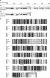
FIG.1.
FIG.1.
Expression and primary structure of the SARS-CoV nsp13 helicase. (A) Overview of the domain organization and (predicted) proteolytic processing of the SARS-CoV replicase polyproteins pp1a and pp1ab. Nsp13 is encoded by ORF 1b and is processed from pp1ab by the 3C-like proteinase. The processing end products of pp1a are designated nsp1 to nsp11, and those of pp1ab are designated nsp1 to nsp10 and nsp12 to nsp16. Note that nsp1 to nsp10 may be released by proteolytic processing of either pp1a or pp1ab, whereas nsp11 is processed from pp1a and nsp12 to nsp16 are processed from pp1ab. Nsp11 and nsp12 have a number of common residues at their N termini. Cleavage sites that are (predicted to be) processed by the viral main proteinase are indicated by grey arrowheads, and sites that are processed by the papain-like proteinase 2 are indicated by black arrowheads. Ac, acidic domain (92); X, X domain (21), which is predicted to have ADP-ribose 1"-phosphatase activity (65); SUD, SARS-CoV unique domain (65); PL2, papain-like cysteine proteinase 2 (72); Y, Y domain containing a transmembrane domain and a putative metal-binding domain (65, 72, 92); TM1, TM2, and TM3, putative transmembrane domains 1 to 3, respectively; 3CL, 3C-like main proteinase (3, 72); RdRp, putative RNA-dependent RNA polymerase domain (19, 29, 43, 52); HEL, superfamily 1 helicase domain (72); ExoN, putative 3′-to-5′ exonuclease (65); XendoU, putative poly(U)-specific endoribonuclease (65); MT, putative S-adenosylmethionine-dependent ribose 2′-O-methyltransferase (65, 81); C/H, domains containing conserved Cys and His residues and predicted to bind metal ions. (B) Sequence comparison of coronavirus helicases. The alignment was generated with the ClustalW program (version 1.82) (http://www.ebi.ac.uk/clustalw/) and used as the input for the ESPript program, version 2.1 (http://prodes.toulouse.inra.fr/ESPript/cgi-bin/ESPript.cgi). The nsp13 sequences of SARS-CoV (isolate Frankfurt 1; accession no. AY291315), mouse hepatitis virus (MHV, strain A59; NC_001846), bovine coronavirus (BCoV, isolate LUN; AF391542), human coronavirus 229E (HCoV-229E; X69721), porcine epidemic diarrhea virus (PEDV, strain CV777; AF353511); transmissible gastroenteritis virus (TGEV, strain Purdue 46; AJ271965), and avian infectious peritonitis virus (IBV, strain Beaudette; M95169) were derived from the replicative polyproteins of these viruses, whose sequences were obtained from the DDBJ, EMBL, and GenBank databases. Conserved helicase motifs I to VI (18) are indicated. Near the N terminus, the 12 conserved Cys and His residues predicted to form a binuclear zinc-binding cluster (77) are indicated by @. Also indicated is the conserved Lys288 residue (corresponding to Lys5589 in pp1ab), which, in the MBP-nsp13_KA control protein, was replaced with Ala. Lys288 is part of conserved helicase motif I (18), which is also called the Walker A box (82). Highlighted in grey is the C-terminal nsp13 sequence against which the rabbit antiserum, α-nsp13, used in this study was raised.
FIG. 2.
FIG. 2.
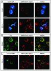
Immunofluorescence microscopy analysis showing the intracellular distribution of the SARS-CoV nsp13 helicase in infected Vero E6 cells. Cells were fixed at 6 h postinfection (A, top row) or 9 h postinfection (A [bottom row], B, and C) and analyzed with a conventional fluorescence microscope (A) or laser scanning confocal microscope (B and C). The SARS-CoV nsp13 staining developed from a punctatedispersed pattern at 6 h postinfection to a large, mainly perinuclear staining at 9 h postinfection. Part of the nsp13 signal overlapped the labeling for the ECFP-ER and PDI marker proteins used in this study, whereas no colocalization with a Golgi marker protein (EGFP-Golgi) was observed. Bar, 20 μm. (A) Prior to infection, cell cultures were transfected with an expression plasmid encoding endoplasmic reticulum-targeted ECFP. Part of the cells remained untransfected or uninfected, explaining the cells positive for nsp13 or the marker protein only. (B) Nontransfected cells were infected with SARS-CoV at a multiplicity of infection of 1 and used for double labeling with antisera recognizing nsp13 and the cellular protein PDI, a resident protein of the endoplasmic reticulum and intermediate compartment. (C) Prior to infection, cell cultures were transfected with an expression plasmid encoding Golgi complex-targeted EGFP.
FIG. 3.
FIG. 3.
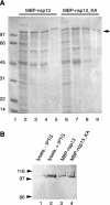
Purification of recombinant MBP-nsp13 and MBP-nsp13_KA fusion proteins from E. coli cells. (A) Aliquots taken at each step of the purification protocol were analyzed on a sodium dodecyl sulfate-12% polyacrylamide gel, and the proteins were stained with Coomassie brilliant blue dye. Lanes: 1, protein molecular mass markers, with masses indicated on the left (in kilodaltons); 2, cleared lysate of IPTG-induced E. coli TB1 bacteria transformed with the expression plasmid pMal-SARS-CoV-nsp13; 3 and 4, peak fractions 1 and 2, respectively, from the amylose-agarose chromatography column; 5, pooled peak fractions from the Superdex 200 column; 6, cleared lysate of IPTG-induced E. coli TB1 bacteria transformed with the expression plasmid pMal-SARS-CoV-nsp13_KA; 7 and 8, peak fractions 1 and 2, respectively, from the amylose-agarose chromatography column; 9, pooled peak fractions from the Superdex 200 column. The fusion proteins are indicated by an arrowhead. (B) Western immunoblot analysis with SARS-CoV nsp13-specific rabbit antiserum. Lanes: 1, cleared lysate of E. coli TB1 bacteria transformed with the expression plasmid pMal-SARS-CoV-nsp13; 2, cleared lysate of IPTG-induced E. coli TB1 bacteria transformed with the expression plasmid pMal-SARS-CoV-nsp13; 3, purified MBP-nsp13; 4, purified MBP-nsp13_KA. The positions of protein molecular mass markers are indicated on the left (in kilodaltons).
FIG. 4.
FIG. 4.
RNA and DNA duplex-unwinding activities of SARS-CoV nsp13 have 5′-to-3′ polarity. The reaction conditions were as described in Materials and Methods. The structures of the substrates are shown schematically, with the radiolabeled strands marked by asterisks. (A) Helicase assay with RNA substrates 5′-RNA4 (lanes 1 to 4) and 3′-RNA2 (lanes 5 to 9), containing 5′ and 3′ single-stranded regions, respectively. Both RNA substrates contained a 22-bp duplex region. The reaction products were separated on a nondenaturing 10% polyacrylamide gel and visualized by autoradiography. Lanes 1 and 5, reactions without protein; lanes 2 and 6, heat-denatured RNA substrate; lanes 3 and 7, reactions containing MBP-nsp13; lanes 4 and 8, reactions containing MBP-nsp13_KA. (B) Helicase assay with DNA substrates. With the exception of DNA-0, which was entirely double stranded, the substrates consisted of identical 22-bp duplexes to which 30-nucleotide-long, single-stranded oligo(dT) tails were attached at different positions. Lanes 1, 5, 9, and 13, reactions without protein; lanes 2, 6, 10, and 14, heat-denatured DNA substrates; lanes 3, 7, 11, and 14, reactions containing MBP-nsp13_KA; lanes 4, 8, 12, and 16, reactions containing MBP-nsp13.
FIG. 5.
FIG. 5.
Effective unwinding of DNA and RNA substrates containing extended duplex regions by SARS-CoV nsp13. Reaction products were separated on 4.5% (left panel) and 5% (right panel) polyacrylamide gels. Lanes 1 and 5, reactions without protein; lanes 2 and 6, heat-denatured substrates; lanes 3 and 7, reactions containing MBP-nsp13; lanes 4 and 8, reactions containing MBP-nsp13_KA.
FIG. 6.
FIG. 6.
SARS-CoV nsp13 has RNA 5′-triphosphatase activity. 5′-γ-32P-labeled RNA was prepared as described in Materials and Methods and incubated with MBP-nsp13 or MBP-nsp13_KA. The reaction products were separated by thin-layer chromatography (A) and polyacrylamide gel electrophoresis (B) and visualized by autoradiography. (A) Lane 1, [γ-32P]GTP without protein; lane 2, [γ-32P]GTP and MBP-nsp13 (GTPase activity); lane 3, 5′-γ-32P-labeled RNA without protein; lane 4, 5′-γ-32P-labeled RNA and MBP-nsp13; lane 5, 5′-γ-32P-labeled RNA and MBP-nsp13_KA. (B) Lane 1, [γ-32P]GTP without protein; lane 2, [γ-32P]GTP and MBP-nsp13 (GTPase activity); lane 3, 5′-γ-32P-labeled RNA without protein; lanes 4, 5, and 6, 5′-γ-32P-labeled RNA and MBP-nsp13. Reactions were terminated by the addition of EDTA after 10 min (lane 4), 30 min (lane 5), and 60 min (lanes 2 and 6).
FIG. 7.
FIG. 7.
Substrate specificity of the SARS-CoV nsp13-associated RNA 5′-triphosphatase activity. The substrate RNA, 5′-GGGAAAAA-3′, was synthesized by in vitro transcription in the presence of [α-32P]GTP (lanes 1 to 4) or [γ-32P]GTP (lanes 5 to 8). Reactions were performed as described in Materials and Methods. The RNA substrates were incubated without protein (lanes 1 and 5), with MBP-nsp13 (lanes 2 and 6), with MBP-nsp13_KA (lanes 3 and 7), or with alkaline phosphatase from calf intestine (CIP) (lanes 4 and 8). Reaction products were separated by thin-layer chromatography on polyethyleneimine cellulose-F plates and visualized by autoradiography. Reaction mixtures were incubated for 60 min (lanes 2, 3, 6, and 7) or 30 min (lanes 4 and 8).
FIG. 8.
FIG. 8.
Inhibition of the SARS-CoV nsp13-associated RNA 5′-triphosphatase activity by ATP. The plot illustrates the effect of including 2 mM ATP, ADP, AMP, or the ATP analog AMP-PNP on the RNA 5′-triphosphatase activity of MBP-nsp13. The average values of two experiments are plotted.

Similar articles

Cited by

References

    1. Ahlquist, P., A. O. Noueiry, W. M. Lee, D. B. Kushner, and B. T. Dye. 2003. Host factors in positive-strand RNA virus genome replication. J. Virol. 77:8181-8186. - PMC - PubMed
    1. Almazán, F., J. M. González, Z. Pénzes, A. Izeta, E. Calvo, J. Plana-Durán, and L. Enjuanes. 2000. Engineering the largest RNA virus genome as an infectious bacterial artificial chromosome. Proc. Natl. Acad. Sci. USA 97:5516-5521. - PMC - PubMed
    1. Anand, K., J. Ziebuhr, P. Wadhwani, J. R. Mesters, and R. Hilgenfeld. 2003. Coronavirus main proteinase (3CLpro) structure: basis for design of anti-SARS drugs. Science 300:1763-1767. - PubMed
    1. Bautista, E. M., K. S. Faaberg, D. Mickelson, and E. D. McGruder. 2002. Functional properties of the predicted helicase of porcine reproductive and respiratory syndrome virus. Virology 298:258-270. - PMC - PubMed
    1. Bird, L. E., H. S. Subramanya, and D. B. Wigley. 1998. Helicases: a unifying structural theme? Curr. Opin. Struct. Biol. 8:14-18. - PubMed

Publication types

MeSH terms

LinkOut - more resources