

A prospective functional MR imaging study of mild traumatic brain injury in college football players
- PMID: 15140712
- PMCID: PMC7974462
A prospective functional MR imaging study of mild traumatic brain injury in college football players
Abstract
Background and purpose: Although concussion is common among athletes, evidence-based methods for clinical evaluation, treatment, and recovery are lacking. We used a prospective, functional neuroimaging approach to assess sports-related concussion in which imaging was performed before injury so that brain changes resulting from concussion could be better understood.
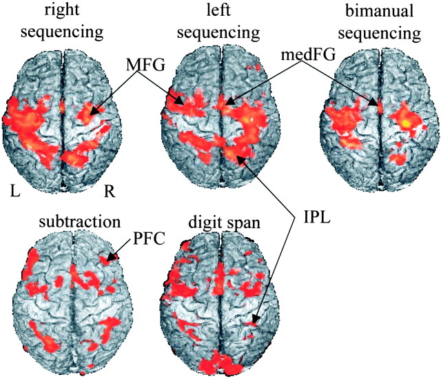
Methods: Neurophysiologic correlates of sports-related concussion were investigated in eight college football players by using functional MR imaging. Preseason baseline levels of blood oxygen level-dependent (BOLD) activity were acquired during the performance of a test battery that included mathematical, memory, and sensorimotor coordination tasks. Four players who had a concussion repeated these baseline procedures within 1 week of injury. The remaining control players were retested at the end of the season.
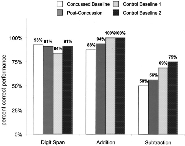
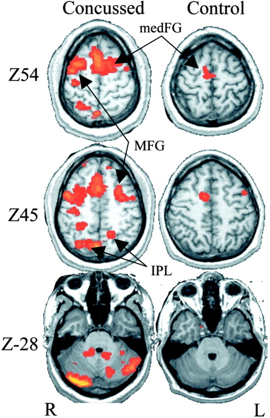
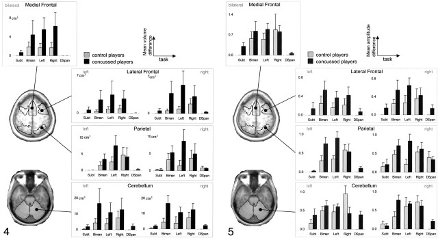
Results: Specific neural signatures of concussion were detected in individual players by comparing postconcussion results to preconcussion baseline values. The validity of these indicators was confirmed by comparing them with the same measures in noninjured control subjects. When compared with control subjects, concussed players had marked within-subject increases in the amplitude and extent of BOLD activity during a finger-sequencing task. Effects were observed primarily in the parietal and lateral frontal and cerebellar regions.
Conclusion: Differences in neural functioning were observed in the absence of observed deficits in behavioral performance, suggesting that this approach may increase sensitivity to concussion compared with neuropsychological evaluation alone. Though preliminary, the proposed prospective neuroimaging approach may have great potential for understanding mild traumatic brain injury and identifying mechanisms underlying recovery.
Figures
Similar articles
-
Acute effects and recovery time following concussion in collegiate football players: the NCAA Concussion Study.JAMA. 2003 Nov 19;290(19):2556-63. doi: 10.1001/jama.290.19.2556. JAMA. 2003. PMID: 14625332
-
Relationship between concussion and neuropsychological performance in college football players.JAMA. 1999 Sep 8;282(10):964-70. doi: 10.1001/jama.282.10.964. JAMA. 1999. PMID: 10485682
-
Cumulative effects associated with recurrent concussion in collegiate football players: the NCAA Concussion Study.JAMA. 2003 Nov 19;290(19):2549-55. doi: 10.1001/jama.290.19.2549. JAMA. 2003. PMID: 14625331
-
Effects of Career Duration, Concussion History, and Playing Position on White Matter Microstructure and Functional Neural Recruitment in Former College and Professional Football Athletes.Radiology. 2018 Mar;286(3):967-977. doi: 10.1148/radiol.2017170539. Epub 2017 Oct 31. Radiology. 2018. PMID: 29087238 Free PMC article.
-
Preliminary evidence from a prospective DTI study suggests a posterior-to-anterior pattern of recovery in college athletes with sports-related concussion.Brain Behav. 2018 Dec;8(12):e01165. doi: 10.1002/brb3.1165. Epub 2018 Nov 22. Brain Behav. 2018. PMID: 30566282 Free PMC article.
Cited by
-
Reduced Functional Connectivity in Adults with Persistent Post-Concussion Symptoms: A Functional Near-Infrared Spectroscopy Study.J Neurotrauma. 2018 Jun 1;35(11):1224-1232. doi: 10.1089/neu.2017.5365. Epub 2018 Mar 23. J Neurotrauma. 2018. PMID: 29373947 Free PMC article.
-
Neuroimaging coordination dynamics in the sport sciences.Methods. 2008 Aug;45(4):325-35. doi: 10.1016/j.ymeth.2008.06.001. Epub 2008 Jul 9. Methods. 2008. PMID: 18602998 Free PMC article. Review.
-
Brain activation during neurocognitive testing using functional near-infrared spectroscopy in patients following concussion compared to healthy controls.Brain Imaging Behav. 2014 Dec;8(4):621-34. doi: 10.1007/s11682-014-9289-9. Brain Imaging Behav. 2014. PMID: 24477579 Free PMC article.
-
Functional MRI of mild traumatic brain injury (mTBI): progress and perspectives from the first decade of studies.Brain Imaging Behav. 2012 Jun;6(2):193-207. doi: 10.1007/s11682-012-9173-4. Brain Imaging Behav. 2012. PMID: 22618832 Free PMC article. Review.
-
Neuroimaging in Pediatric Patients with Mild Traumatic Brain Injury: Relating the Current 2018 Centers for Disease Control Guideline and the Potential of Advanced Neuroimaging Modalities for Research and Clinical Biomarker Development.J Neurotrauma. 2021 Jan 1;38(1):44-52. doi: 10.1089/neu.2020.7100. Epub 2020 Oct 21. J Neurotrauma. 2021. PMID: 32640874 Free PMC article. Review.
References
-
- Thurman DJ, Branche CM, Sneizek JE. The epidemiology of sports-related traumatic brain injuries in the United States: recent developments. J Head Trauma Rehab 1998;13:1–18 - PubMed
-
- Powell JW, Barber-Foss KD. Traumatic brain injury in high school athletes. JAMA 1999;282:958–963 - PubMed
-
- Maroon JC, Podell K, Powell JW. Cerebral concussion in athletes: evaluation and neuropsychological testing. Neurosurgery 2000;47:659–669 - PubMed
-
- Echemendia RJ, Julian LJ. Mild traumatic brain injury in sports: neuropsychology’s contribution to a developing field. Neuropsychology Review 2001;11:69–88 - PubMed
-
- Denny-Brown D, Russel W. Experimental cerebral concussion. Brain 1941;64:93–164
Publication types
MeSH terms
Grants and funding
LinkOut - more resources
Full Text Sources
Other Literature Sources
Medical