In vivo tracking of T cell development, ablation, and engraftment in transgenic zebrafish
- PMID: 15123839
- PMCID: PMC409925
- DOI: 10.1073/pnas.0402248101
In vivo tracking of T cell development, ablation, and engraftment in transgenic zebrafish
Abstract
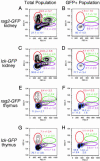
Transgenic zebrafish that express GFP under control of the T cell-specific tyrosine kinase (lck) promoter were used to analyze critical aspects of the immune system, including patterns of T cell development and T cell homing after transplant. GFP-labeled T cells could be ablated in larvae by either irradiation or dexamethasone added to the water, illustrating that T cells have evolutionarily conserved responses to chemical and radiation ablation. In transplant experiments, thymocytes from lck-GFP fish repopulated the thymus of irradiated wild-type fish only transiently, suggesting that the thymus contains only short-term thymic repopulating cells. By contrast, whole kidney marrow permanently reconstituted the T lymphoid compartment of irradiated wild-type fish, suggesting that long-term thymic repopulating cells reside in the kidney.
Figures


Similar articles
-
Suppression of apoptosis by bcl-2 overexpression in lymphoid cells of transgenic zebrafish.Blood. 2005 Apr 15;105(8):3278-85. doi: 10.1182/blood-2004-08-3073. Epub 2004 Dec 23. Blood. 2005. PMID: 15618471
-
Isolating Malignant and Non-Malignant B Cells from lck:eGFP Zebrafish.J Vis Exp. 2019 Feb 22;(144). doi: 10.3791/59191. J Vis Exp. 2019. PMID: 30855581
-
Dynamic Changes in Lymphocyte Populations Establish Zebrafish as a Thymic Involution Model.J Immunol. 2024 Jun 1;212(11):1733-1743. doi: 10.4049/jimmunol.2300495. J Immunol. 2024. PMID: 38656392
-
The zebrafish: a new model of T-cell and thymic development.Nat Rev Immunol. 2005 Apr;5(4):307-17. doi: 10.1038/nri1590. Nat Rev Immunol. 2005. PMID: 15803150 Review.
-
Ontogeny of the immune system of fish.Fish Shellfish Immunol. 2006 Feb;20(2):126-36. doi: 10.1016/j.fsi.2004.09.005. Fish Shellfish Immunol. 2006. PMID: 15939627 Review.
Cited by
-
Real-time imaging of polymersome nanoparticles in zebrafish embryos engrafted with melanoma cancer cells: Localization, toxicity and treatment analysis.EBioMedicine. 2020 Aug;58:102902. doi: 10.1016/j.ebiom.2020.102902. Epub 2020 Jul 21. EBioMedicine. 2020. PMID: 32707448 Free PMC article.
-
Definitive hematopoiesis is dispensable to sustain erythrocytes and macrophages during zebrafish ontogeny.iScience. 2024 Jan 17;27(2):108922. doi: 10.1016/j.isci.2024.108922. eCollection 2024 Feb 16. iScience. 2024. PMID: 38327794 Free PMC article.
-
The axillary lymphoid organ - an external, experimentally accessible immune organ in the zebrafish.bioRxiv [Preprint]. 2024 Jul 25:2024.07.25.605139. doi: 10.1101/2024.07.25.605139. bioRxiv. 2024. PMID: 39091802 Free PMC article. Preprint.
-
One Host-Multiple Applications: Zebrafish (Danio rerio) as Promising Model for Studying Human Cancers and Pathogenic Diseases.Int J Mol Sci. 2022 Sep 6;23(18):10255. doi: 10.3390/ijms231810255. Int J Mol Sci. 2022. PMID: 36142160 Free PMC article. Review.
-
Identification of dendritic antigen-presenting cells in the zebrafish.Proc Natl Acad Sci U S A. 2010 Sep 7;107(36):15850-5. doi: 10.1073/pnas.1000494107. Epub 2010 Aug 23. Proc Natl Acad Sci U S A. 2010. PMID: 20733076 Free PMC article.
References
-
- Driever, W., Solnica-Krezel, L., Schier, A. F., Neuhauss, S. C., Malicki, J., Stemple, D. L., Stainier, D. Y., Zwartkruis, F., Abdelilah, S., Rangini, Z., et. al. (1996) Development (Cambridge, U.K.) 123, 37-46. - PubMed
-
- Haffter, P., Granato, M., Brand, M., Mullins, M. C., Hammerschmidt, M., Kane, D. A., Odenthal, J., van Eeden, F. J., Jiang, Y. J., Heisenberg, C. P., et. al. (1996) Development (Cambridge, U.K.) 123, 1-36. - PubMed
-
- Golling, G., Amsterdam, A., Sun, Z., Antonelli, M., Maldonado, E., Chen, W., Burgess, S., Haldi, M., Artzt, K., Farrington, S., et. al. (2002) Nat. Genet. 31, 135-140. - PubMed
-
- Poss, K. D., Nechiporuk, A., Hillam, A. M., Johnson, S. L. & Keating, M. T. (2002) Development (Cambridge, U.K.) 129, 5141-5149. - PubMed
-
- Thisse, C. & Zon, L. I. (2002) Science 295, 457-462. - PubMed
Publication types
MeSH terms
Substances
Associated data
- Actions
Grants and funding
LinkOut - more resources
Full Text Sources
Other Literature Sources
Molecular Biology Databases
Miscellaneous

