. 2004 Jul;15(7):3132-45.
doi: 10.1091/mbc.e04-02-0103. Epub 2004 Apr 30.

Disturbed cholesterol traffic but normal proteolytic function in LAMP-1/LAMP-2 double-deficient fibroblasts

Affiliations

Disturbed cholesterol traffic but normal proteolytic function in LAMP-1/LAMP-2 double-deficient fibroblasts

Eeva-Liisa Eskelinen et al. Mol Biol Cell. 2004 Jul.

Abstract

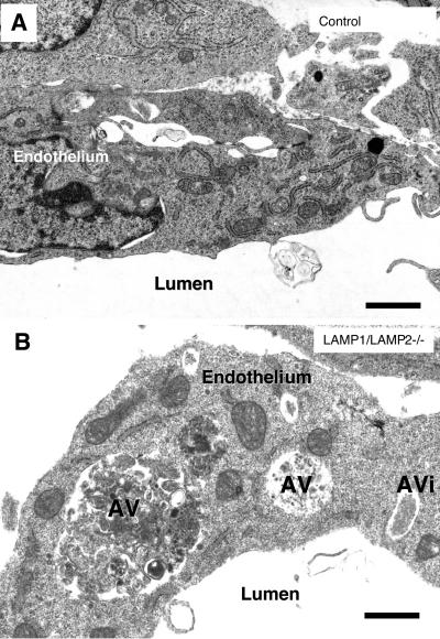
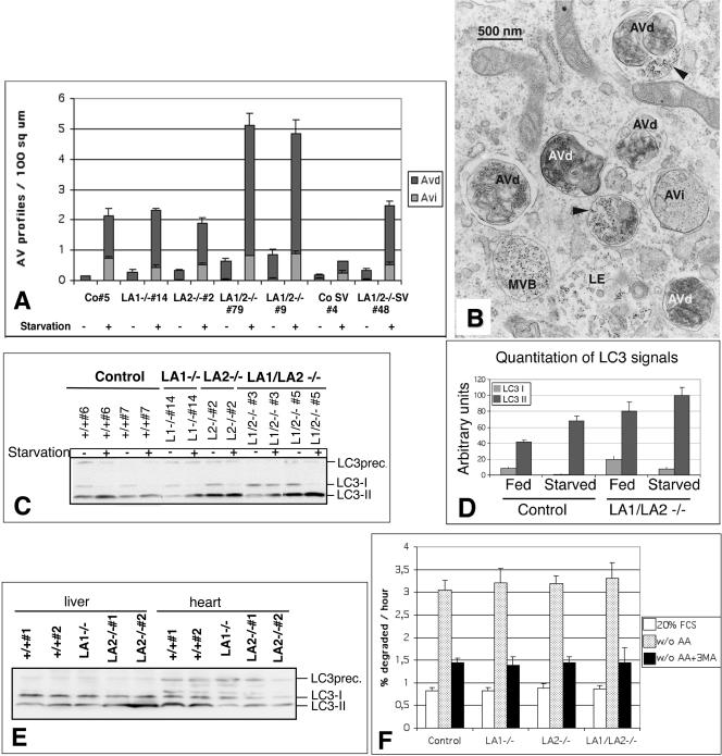
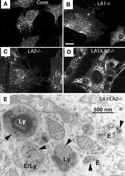
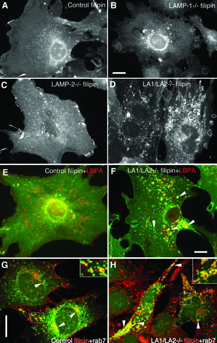
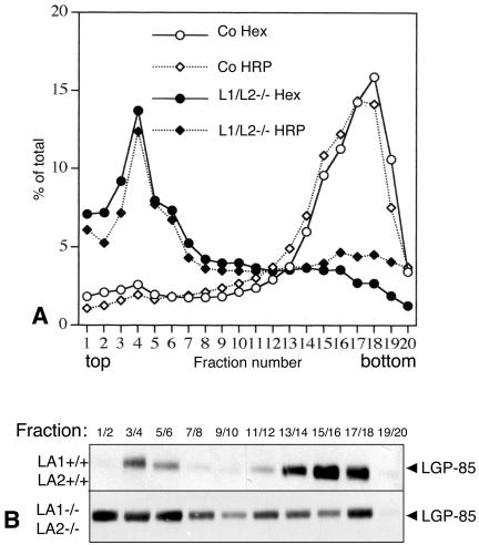
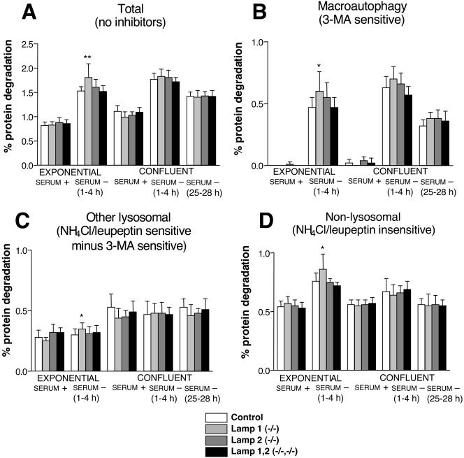
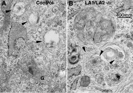
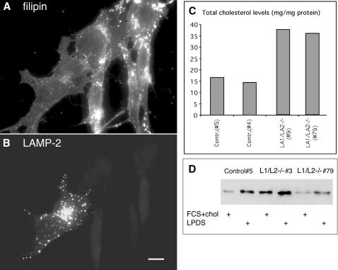
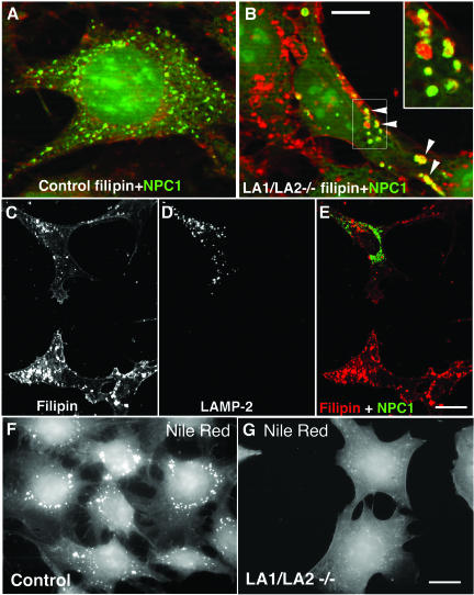
Mice double deficient in LAMP-1 and -2 were generated. The embryos died between embryonic days 14.5 and 16.5. An accumulation of autophagic vacuoles was detected in many tissues including endothelial cells and Schwann cells. Fibroblast cell lines derived from the double-deficient embryos accumulated autophagic vacuoles and the autophagy protein LC3II after amino acid starvation. Lysosomal vesicles were larger and more peripherally distributed and showed a lower specific density in Percoll gradients in double deficient when compared with control cells. Lysosomal enzyme activities, cathepsin D processing and mannose-6-phosphate receptor expression levels were not affected by the deficiency of both LAMPs. Surprisingly, LAMP-1 and -2 deficiencies did not affect long-lived protein degradation rates, including proteolysis due to chaperone-mediated autophagy. The LAMP-1/2 double-deficient cells and, to a lesser extent, LAMP-2 single-deficient cells showed an accumulation of unesterified cholesterol in endo/lysosomal, rab7, and NPC1 positive compartments as well as reduced amounts of lipid droplets. The cholesterol accumulation in LAMP-1/2 double-deficient cells could be rescued by overexpression of murine LAMP-2a, but not by LAMP-1, highlighting the more prominent role of LAMP-2. Taken together these findings indicate partially overlapping functions for LAMP-1 and -2 in lysosome biogenesis, autophagy, and cholesterol homeostasis.


Figures

Figure 1.
Figure 1.
(A) Immunoblot analysis of extracts from mouse embryonic fibroblasts derived from E12.5 embryos. The expression of lysosomal membrane proteins LAMP-1, LAMP-2, and LIMP-2/LGP-85 was analyzed. (B-G) Macroscopic phenotype of LAMP-1/LAMP-2-deficient embryos (C, D, E, and G) in comparison with control (LAMP-1+/+/LAMP-2+/+ littermates; B and F). (H) Microscopic organization of the organs of a double-deficient embryo (E12) as seen in a transversal section. NT, neural tube. DRG, dorsal root ganglion. Br, bronchus. H, heart. Semithin section, toluidine blue. Bars in B-E, 5 mm; F and G, 2 mm; H, 0.5 mm.
Figure 2.
Figure 2.
Autophagic vacuoles accumulated in cells of LAMP-1/LAMP-2 double-deficient embryos. (A) Endothelium of a wild-type embryo. (B) Endothelium of a blood vessel in the neural tube in a LAMP-1/LAMP-2 double-deficient embryo. The endothelial cells displayed several cytoplasmic vacuoles with polymorphous contents. Some of the vacuoles could be identified as autophagic vacuoles (AV) or autophagosomes (AVi). Bars, 0.5 μm.
Figure 3.
Figure 3.
Accumulation of autophagic vacuoles in LAMP-1/LAMP-2 double-deficient MEFs. (A) Primary and SV-40 large T-antigen immortalized (CoSV, LA1/2-/-SV) MEFs were cultured in the presence or absence of serum and amino acids for 2 h. The amounts of early (Avi) and late autophagic vacuoles (Avd) were determined by quantitative electron microscopy. Co, control cells; LA1-/-, LAMP-1-deficient cells; LA2-/-, LAMP-2-deficient cells, LA1/2-/- double-deficient cells. The numbers (5, etc.) indicate individual cell lines. (B) Electron micrograph of LAMP-1/LAMP-2 double-deficient cells (79), which were fed with BSA gold in serum-free medium for 2 h to label endosomes and lysosomes. Early (AVi) and late autophagic vacuoles (AVd) were abundant. Multivesicular bodies (MVB) and late endosomes (LE) containing BSA gold are also indicated. Arrowheads indicate late autophagic vacuoles which have fused with BSA-gold positive endosomes. (C) Western blotting of the autophagic marker protein LC3 in nonstarved cells and in cells incubated in serum and amino acid free medium for 2 h. The locations of LC3 precursor (LC3prec.) as well as LC3I and LC3II are indicated on the right. (D) Quantitation of the absolute levels of LC3I and LC3II in the Western blot shown in C for control and double-deficient cells. The values represent the average and SD of cell lines with identical genotype. (E) LC3 Western blot of LAMP-1 and LAMP-2 single-deficient liver and heart extracts. (F) Degradation of long lived proteins in the absence or presence of 3-methyladenine (3MA). MEFs were metabolically labeled with [3H]valine or [3H]leucine and then chased for 24 h in full medium. Cells were then switched to culture medium with serum (20% FCS) or to serum and amino acid free medium (w/o AA) for 4 h. TCA-soluble and -precipitable radioactivities were measured from the culture medium. Protein degradation was measured as the net release of TCA-soluble radioactivity, expressed as percentage of the total TCA-precipitable radioactivity present in cells at the beginning of the incubation. The results are mean and SD from 9-14 (20% FCS) or 6 experiments (w/o AA) with 2-3 parallel samples. Three (control, LA1-/-, LA2-/-) or one (LA1/LA2-/-, 79) independent cell lines were used.
Figure 4.
Figure 4.
The lysosomal compartment in LAMP double-deficient MEFs. Immunofluorescence staining of LIMP-2/LGP-85 in (A) control, (B) LAMP-1-deficient, (C) LAMP-2-deficient, and (D) LAMP-1/LAMP-2 double-deficient MEFs. Note the larger size and the more peripheral location of lysosomes in LAMP double knockout cells. Bar, 20 μm. The images show representative cells, based on experiments with at least two independent cell lines for each genotype. (E) Electron micrograph of LAMP-1/LAMP-2 double-deficient cells fed with the fluid-phase endocytic marker BSA-gold for 2 h. Structures with a typical morphology of dense lysosomes (Ly) or endosomes (E) contain the endocytic marker (arrowheads). Note also the multilamellar membranes inside the lysosome on the left.
Figure 7.
Figure 7.
Cholesterol accumulation in LAMP-1/LAMP-2 double-deficient MEFs. Filipin staining revealed distribution of unesterified cholesterol in (A) control, (B) LAMP-1-/-, (C) LAMP-2-/-, and (D) LAMP-1/LAMP-2-/- cells. Note increased vesicular staining in LAMP-2 single-deficient cells and prominent vesicular accumulation in LAMP double-deficient cells. (E and F) Double labeling with filipin (green) and the late endosomal marker LBPA (red) in control (E) and LAMP double-deficient cells (F). Yellow color (arrowheads) indicates colocalization in a subset of the endosomes of LAMP double knockout cells (F). (G and H) Double labeling of filipin (red) and GFP-rab7 (green) in control (G) and LAMP-1/LAMP-2 double-deficient cells (H). Rab7 colocalization in filipin positive vesicles is indicated by arrowheads and shown at higher magnification in the inserts. Bars, 20 μm.
Figure 5.
Figure 5.
Lysosomal density in LAMP-1/LAMP-2 double-deficient MEFs. (A) The lysosomal density from control and LAMP-1/LAMP-2 (L1/L2-/-) double-deficient cells was determined in 27% Percoll gradients. β-hexosaminidase activity and the activity of endocytosed horseradish peroxidase (HRP, 5-min feeding and 3-h chase) were measured in the fractions. (B) LIMP-2/LGP-85 content of the fractions was estimated by Western blotting.
Figure 6.
Figure 6.
Contribution of proteolytic pathways in MEFs under different growth conditions. MEFs were metabolically labeled and chased as described in MATERIALS AND METHODS. Exponentially growing or confluent cells were then switched to serum-containing (serum +) or serum-deficient (serum -) media, with or without proteolysis inhibitors. To allow optimal inhibition and to avoid secondary effects, protein degradation was measured at different time points starting after 1 h and continuing for a period of 3 additional hours. The total protein degradation per hour is presented in A. The contribution (percentage of the labeled protein whose degradation was inhibited per hour) of macroautophagy (B), other lysosomal pathways (C), and nonlysosomal pathways (D), of total protein degradation was calculated as described in MATERIALS AND METHODS. 3-Methyladenine was used to inhibit macroautophagy and NH4Cl + leupeptin to inhibit both macroautophagy and other lysosomal pathways (i.e., microautophagy, chaperone-mediated autophagy, etc.). The results are the mean and SD from 6-15 separate experiments with duplicate samples. Three (control, LA1-/-, LA2-/-) or one (LA1/LA2-/-, 79) independent cell lines were used. Stars indicate differences from control cell values which were found to be statistically significant at *p < 0.05 and **p < 0.005.
Figure 8.
Figure 8.
Electron microscopical visualization of filipin labeled membrane cholesterol. Arrowheads in control (A) and double-deficient fibroblasts (B) indicate the filipin labeling. In control cells labeling was detected in the limiting membranes of small vesicles close to the Golgi apparatus (G). In LAMP-1/LAMP-2 double-deficient fibroblasts (B) filipin-induced membrane alterations were seen in both the limiting and internal membranes of endo/lysosomal vesicles. Note that lamellar internal membranes of the upper endo/lysosome are not labeled by filipin.
Figure 9.
Figure 9.
Reduction of cholesterol storage in LAMP double-deficient cells after LAMP-2a re-expression. (A and B) LAMP-2a was transiently transfected to LAMP1/LAMP-2 double-deficient cells. After 1 day, cholesterol was stained with filipin (A) and LAMP-2 was detected by immunofluorescence (B). Cells not expressing LAMP-2 showed staining for cholesterol in endo/lysosomal vesicles, whereas the LAMP-2a-positive cell showed a filipin staining pattern similar to control cells (see Figure 8A). Bar, 10 μm. (C) Biochemical cholesterol assay in two control and two LAMP double-deficient primary cell lines. (D) Western blot of LDL receptor in one control and two LAMP-1/LAMP-2 double-deficient cell lines. The cells were incubated in DMEM containing 10% FCS and 10 μg/ml cholesterol (FCS+chol), or in DMEM containing 10% LPDS for 2 days.
Figure 10.
Figure 10.
Altered NPC1 localization and Nile Red staining in LAMP-1/LAMP-2 double-deficient cells. NPC1-GFP was transiently expressed in control cells (A) and in LAMP-1/LAMP-2 double-deficient cells (B). An overlay of NPC1-GFP (green) and filipin (red) is shown. Note the increased size of NPC1 vesicles in the double-deficient cells. In these structures NPC1 colocalized with filipin (arrowheads and insert in B). Bar, 10 μm. (C-E) After parallel transfection of LAMP-2 and NPC1-GFP in LAMP-1/LAMP-2 double-deficient cells, filipin staining (C), LAMP-2 staining (D) and NPC1-GFP were analyzed. Note the reduction of filipin positive vesicles, and the distribution of NPC1-GFP in small filipin-negative structures, in the cell expressing LAMP-2. Bar, 20 μm. (F and G) Nile Red staining revealed lipid droplets as punctate structures in control cells (F), whereas almost no Nile Red-positive cytoplasmic structures were found in LAMP double-deficient MEFs (G). Bar, 20 μm.

Similar articles

Cited by

References

    1. Agarraberes, F.A., and Dice, J.F. (2001). A molecular chaperone complex at the lysosomal membrane is required for protein translocation. J. Cell Sci. 114, 2491-2499. - PubMed
    1. Andrejewski, N., Punnonen, E.L., Guhde, G., Tanaka, Y., Lullmann-Rauch, R., Hartmann, D., von Figura, K., and Saftig, P. (1999). Normal lysosomal morphology and function in LAMP-1-deficient mice. J. Biol. Chem. 274, 12692-12701. - PubMed
    1. Aoki, A., and Massa, E.M. (1975). Subcellular compartmentation of free and esterified cholesterol in the interstitial cells of the mouse testis. Cell Tissue Res. 165, 49-62. - PubMed
    1. Brown, M.S., and Goldstein, J.L. (1986). A receptor-mediated pathway for cholesterol homeostasis. Science 232, 34-47. - PubMed
    1. Choudhury, A., Dominguez, M., Puri, V., Sharma, D.K., Narita, K., Wheatley, C.L., Marks, D.L., and Pagano, R.E. (2002). Rab proteins mediate Golgi transport of caveola-internalized glycosphingolipids and correct lipid trafficking in Niemann-Pick C cells. J. Clin. Invest. 109, 1541-1550. - PMC - PubMed

Publication types

MeSH terms