. 2004 May 19;23(10):2134-45.
doi: 10.1038/sj.emboj.7600210. Epub 2004 Apr 22.

Analysis of the composition, assembly kinetics and activity of native Apaf-1 apoptosomes

Affiliations

Analysis of the composition, assembly kinetics and activity of native Apaf-1 apoptosomes

Michelle M Hill et al. EMBO J. .

Abstract

The Apaf-1 apoptosome is a multi-subunit caspase-activating scaffold that is assembled in response to diverse forms of cellular stress that culminate in apoptosis. To date, most studies on apoptosome composition and function have used apoptosomes reassembled from recombinant or purified proteins. Thus, the precise composition of native apoptosomes remains unresolved. Here, we have used a one-step immunopurification approach to isolate catalytically active Apaf-1/caspase-9 apoptosomes, and have identified the major constituents of these complexes using mass spectrometry methods. Using this approach, we have also assessed the ability of putative apoptosome regulatory proteins, such as Smac/DIABLO and PHAPI, to regulate the activity of native apoptosomes. We show that Apaf-1, caspase-9, caspase-3 and XIAP are the major constituents of native apoptosomes and that cytochrome c is not stably associated with the active complex. We also demonstrate that the IAP-neutralizing protein Smac/DIABLO and the tumor-suppressor protein PHAPI can enhance the catalytic activity of apoptosome complexes in distinct ways. Surprisingly, PHAPI also enhanced the activity of purified caspase-3, suggesting that it may act as a co-factor for this protease.


Figures
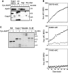
Figure 1
Figure 1
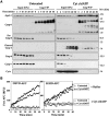
One-step isolation of native apoptosomes using a differential immunoprecipitation strategy. (A) Jurkat cell-free extracts were incubated in the presence or absence of 50 μg/ml cytochrome c/1 mM dATP for 10 min at 37°C. Reactions were adjusted to 50 mM NaCl and 0.3% CHAPS and precleared by incubating with protein A/G agarose beads. Protein complexes immunoprecipitated with a monoclonal anti-caspase-9 antibody (Upstate Biotechnology) were analyzed for the presence of Apaf-1 and caspase-9 by immunoblotting. Equivalent amounts of the input (6.5 μl of the reaction), the IP supernatant (Sup) and protein binding to the preclearing beads (preclear) were loaded to aid comparison. (B) Jurkat cell-free extracts were incubated for 15 min at 37°C in the presence (filled circles) or absence (open circles) of 50 μg/ml cytochrome c/1 mM dATP. Caspase-9 complexes isolated from 100 μl reactions were washed extensively and then incubated for 1 h at 37°C with 50 μM of the indicated fluorogenic peptides. (C) Caspase-9 complexes, isolated from Jurkat cell-free extract incubated for 15 min at 37°C in the presence or absence of 50 μg/ml cytochrome c/1 mM dATP, were incubated with in vitro translated Bid, caspase-3, vimentin or pro-interleukin-1β for 2 h at 37°C. Proteolysis of radiolabeled substrate proteins was analyzed by SDS—PAGE, followed by autoradiography.
Figure 2
Figure 2
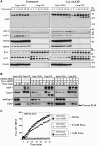
Cytochrome c triggers the rapid and stable association of Apaf-1, caspase-9 and caspase-3. (A) Jurkat cell-free extracts were incubated at 37°C in the presence or absence of 50 μg/ml cytochrome c/1 mM dATP for the times indicated and then brought to 4°C. Caspase-9 was then immunoprecipitated from each reaction as described in the legend to Figure 1A. Caspase-9 immunocomplexes (Casp-9 IP) or 10% of the input reactions (input) were then probed for the presence of Apaf-1, caspase-3, -6, -7 and cytochrome c by immunoblotting. Bands corresponding to immunoglobulin heavy and light chains are indicated by asterisks. (B) Caspase-9 complexes were immunoprecipitated from Jurkat cell-free extracts incubated for 15 min at 37°C in the presence (filled symbols) or absence (open symbols) of 50 μg/ml cytochrome c/1 mM dATP. Caspase-9 immunocomplexes were then washed extensively, as described in Materials and methods, and peptide hydrolysis assays were then performed in the presence (triangles) or absence (circles) of 50 μg/ml cytochrome c/1 mM dATP. Note that for DEVD-AFC hydrolysis reactions complexes were prepared from 50 μl cell-free reactions, whereas for LEHD-AFC hydrolysis assays 150 μl cell-free reactions were required.
Figure 3
Figure 3
XIAP is a constituent of native apoptosomes. (A) Jurkat cell-free extracts were incubated at 37°C in the presence or absence of 50 μg/ml cytochrome c/1 mM dATP for the times indicated and then brought to 4°C. Caspase-9 was then immunoprecipitated from each reaction as described in the legend to Figure 1A. Caspase-9 immunocomplexes (Casp-9 IP) or 10% of the input reactions (Input) were analyzed for the presence of IAPs, HSP90, HSP70, Bcl-2, Bcl-XL and PHAPI by immunoblotting. Bands corresponding to immunoglobulin light chain are indicated by asterisks. (B) Apoptosome assembly was initiated in Jurkat cell-free extracts in the presence or absence of 1 μM Ac-DEVD-CHO, as indicated. Caspase-9 was then immunoprecipitated from each reaction as described in the legend to Figure 1A. Caspase-9 complexes (Casp-9 IP) or 10% of the input reactions (Input) were analyzed for the presence of Apaf-1, caspase-9, -3 and XIAP by immunoblotting. Cleaved XIAP is indicated with an arrow. (C) Caspase-9 complexes were immunoprecipitated from Jurkat cell-free extracts incubated for 15 min at 37°C in the presence (filled symbols) or absence (open symbols) of 50 μg/ml cytochrome c/1 mM dATP. Peptide hydrolysis assays were subsequently performed in the presence of buffer (circles), 0.5 μM (triangles) or 5 μM (squares) recombinant Smac Δ1–55.
Figure 4
Figure 4
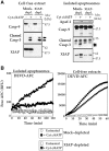
XIAP acts as a tether for recruitment of caspase-3 to the apoptosome. (A) Left panel, mock-depleted or XIAP-immunodepleted cell-free extracts were treated with 50 μg/ml cytochrome c/1 mM dATP for 15 min at 37°C, followed by immunoblotting and probing for the indicated proteins. Right panel, mock-depleted or XIAP-immunodepleted cell-free extracts were treated with 50 μg/ml cytochrome c/1 mM dATP for 15 min at 37°C. Reactions were then brought to 4°C and caspase-9 immunocomplexes were prepared, followed by immunoblotting for the indicated proteins. Arrows indicate mature caspase-3. Asterisks denotes immunoglobulin heavy and light chains. (B) Caspase-9 complexes (left panel) were immunoprecipitated from mock-depleted (circles) or XIAP-depleted (squares) Jurkat cell-free extracts incubated for 15 min at 37°C in the presence (filled symbols) or absence (open symbols) of 50 μg/ml cytochrome c/1 mM dATP. Hydrolysis of DEVD-AFC (50 μM) was monitored for 2 h at 37°C. Cell-free reactions (right panel) from the same extracts were incubated in the presence or absence of 50 μg/ml cytochrome c/1 mM dATP for 30 min at 37°C, and assayed for DEVD-AFC hydrolysis for 60 min at 37°C.
Figure 5
Figure 5
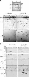
Analysis of native Apaf-1 apoptosomes by 2D-PAGE. (A) Caspase-9 complexes, isolated from 2 ml Jurkat cell-free reactions incubated for 15 min at 37°C in the presence or absence of 50 μg/ml cytochrome c/1 mM dATP, were analyzed by 2D gel electrophoresis (first dimension: pH 5–8, second dimension: 12% SDS–PAGE). Top: A representative silver-stained preparative gel is shown. Heavy and light chains of the immunoprecipitating antibody are indicated. Bottom: enlarged areas containing proteins that differentially immunoprecipitate with caspase-9 (areas I and II in the gel above) are shown. (B) Caspase-9 complexes were isolated from Jurkat cell-free extracts incubated for 15 min at 37°C in the presence or absence of 50 μg/ml cytochrome c/1 mM dATP. Proteins co-precipitating with caspase-9 were analyzed by 2D gel electrophoresis (pH 5–8 first dimension and 12% SDS–PAGE second dimension), followed by immunoblotting with the indicated antibodies. Spots corresponding to immunoglobulin heavy and light chains are indicated by asterisks.
Figure 6
Figure 6
Analysis of apoptosome constituents from Jurkat, BJAB and U937 cells. Cell-free extracts derived from Jurkat, BJAB or U937 cells were incubated at 37°C in the presence or absence of 50 μg/ml cytochrome c/1 mM dATP for the times indicated and then brought to 4°C. Caspase-9 was then immunoprecipitated from each reaction as described in the legend to Figure 1A. Caspase-9 complexes (CASP-9 IP) or 10% of the input reactions (input) were then immunoblotted for Apaf-1, caspase-3, caspase-9, RhoGDI2 and XIAP.
Figure 7
Figure 7
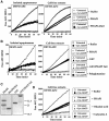
PHAPI enhances the catalytic activity of apoptosomes. (A) Caspase-9 apoptosome complexes (left) and Jurkat cell-free extracts (right), incubated for 15 min at 37°C in the presence (filled symbols) or absence (open symbols) of 50 μg/ml cytochrome c/1 mM dATP, were assayed for DEVDase activity in the presence of 0.5 μM recombinant PHAPI (triangles), PHAPI-ΔTail (squares) or buffer alone (circles). (B) Caspase-9 apoptosome complexes (left) and Jurkat cell-free extracts (right), incubated for 15 min at 37°C in the presence (filled symbols) or absence (open symbols) of cytochrome c/dATP, were assayed for DEVDase activity in the presence of 0.5 μM recombinant PHAPI (small circles), GST (large triangles), GST-PHAPI-Tail (small triangles), 15–50 kDa polyglutamine polymers (squares) or buffer alone (large circles). The results shown are representative of three separate experiments. (C) Aliquots (750 ng) of purified bacterially expressed PHAPI, PHAPI ΔTail, GST and GST-PHAP-Tail proteins were separated by SDS–PAGE, and visualized by coomassie blue staining. A degradation product of full-length PHAPI is indicated by an asterisk. (D) Jurkat cell-free extracts, incubated for 30 min at 37°C in the presence (filled symbols) or absence (open symbols) of 2 μg/ml cytochrome c/1 mM dATP, were assayed for DEVDase activity in the presence of 1 μM recombinant PHAPI (small circles), 5 μM okadaic acid (triangles), 1 μM calyculin A (squares) or buffer alone (large circles).
Figure 8
Figure 8
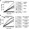
PHAPI and Smac enhance apoptosome activity in distinct ways. (A) Caspase-9 complexes, prepared from Jurkat cell-free extracts incubated for 15 min at 37°C in the presence (filled symbols) or absence (open symbols) of 50 μg/ml cytochrome c/1 mM dATP, were assayed for DEVDase activity in the presence of 1 μM recombinant PHAPI (triangles), 1 μM recombinant Smac (squares), 1 μM recombinant PHAPI plus 1 μM recombinant Smac (small circles) or buffer alone (large circles). (B) Jurkat cell-free extracts, either mock-depleted (small and large circles) or depleted of XIAP using anti-XIAP antibody (triangles and squares), were incubated for 30 min at 37°C in the presence (filled symbols) or absence (open symbols) of 50 μg/ml cytochrome c/1 mM dATP and then assayed for DEVDase activity in the presence or absence of 1 μM recombinant PHAPI, as indicated. Depletion of XIAP from extracts was confirmed by immunoblotting (inset).
Figure 9
Figure 9
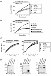
PHAPI specifically enhances the proteolytic activity of caspase-3. (A) The catalytic activity of purified recombinant caspase-3 (5 U/μl) was assayed in the presence or absence of 0.5 μM of the indicated recombinant proteins or BSA. Caspase-3 was allowed to pre-equilibrate with the proteins for 10 min at 4°C prior to addition of Ac-DEVD-AFC substrate. The result shown is representative of four separate experiments. (B) The catalytic activity of purified recombinant caspase-3 (5 U/μl) was assayed in the presence or absence of 0.5 μM of the indicated recombinant proteins. Caspase-3 was allowed to pre-equilibrate with the proteins for 10 min at 4°C prior to addition of Ac-DEVD-AFC substrate. The result shown is representative of three independent experiments. (C) The effect of PHAPI, Smac and control proteins on the catalytic activity of recombinant caspase-7 (0.02 U/μl, left panel) or recombinant caspase-9 (0.05 U/μl; right panel) was assayed as described above. The caspases were allowed to pre-equilibrate with the indicated proteins for 10 min at 4°C prior to addition of Ac-DEVD-AFC or Ac-LEHD substrates. The results shown are representative of three independent experiments. (D) The recombinant caspases used in the above assays were confirmed to be fully processed by immunoblot analysis. In all, 200 U of recombinant caspase-3, 1 U of recombinant caspase-7 and 0.0165 U of recombinant caspase-9 were analyzed by Western blotting, as indicated. As controls, 20 μg of control or activated (treated with 50 μg/ml cytochrome c, 1 mM dATP for 2 h at 37°C) Jurkat cell-free extracts were also run. Blots were probed with a mixture of antibodies that detect the pro-forms as well as the processed forms of caspase-3, -7 and -9. The asterisks denote nonspecific bands recognized by the antibodies.

Similar articles

Cited by

References

    1. Acehan D, Jiang X, Morgan DG, Heuser JE, Wang X, Akey CW (2002) Three-dimensional structure of the apoptosome: implications for assembly, procaspase-9 binding, and activation. Mol Cell 9: 423–432 - PubMed
    1. Adrain C, Creagh EM, Martin SJ (2001) Apoptosis-associated release of Smac/DIABLO from mitochondria requires active caspases and is blocked by Bcl-2. EMBO J 20: 6627–6636 - PMC - PubMed
    1. Adrain C, Slee EA, Harte MT, Martin SJ (1999) Regulation of apoptotic protease activating factor-1 oligomerization and apoptosis by the WD-40 repeat region. J Biol Chem 274: 20855–20860 - PubMed
    1. Beere HM, Wolf BB, Cain K, Mosser DD, Mahboubi A, Kuwana T, Tailor P, Morimoto RI, Cohen GM, Green DR (2000) Heat-shock protein 70 inhibits apoptosis by preventing recruitment of procaspase-9 to the Apaf-1 apoptosome. Nat Cell Biol 2: 469–475 - PubMed
    1. Bratton SB, Walker G, Srinivasula SM, Sun XM, Butterworth M, Alnemri ES, Cohen GM (2001) Recruitment, activation and retention of caspases-9 and -3 by Apaf-1 apoptosome and associated XIAP complexes. EMBO J 20: 998–1009 - PMC - PubMed

Publication types

MeSH terms

Substances