An essential role for vesicular glutamate transporter 1 (VGLUT1) in postnatal development and control of quantal size
- PMID: 15103023
- PMCID: PMC406482
- DOI: 10.1073/pnas.0401764101
An essential role for vesicular glutamate transporter 1 (VGLUT1) in postnatal development and control of quantal size
Abstract
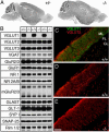
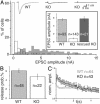
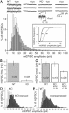
Quantal neurotransmitter release at excitatory synapses depends on glutamate import into synaptic vesicles by vesicular glutamate transporters (VGLUTs). Of the three known transporters, VGLUT1 and VGLUT2 are expressed prominently in the adult brain, but during the first two weeks of postnatal development, VGLUT2 expression predominates. Targeted deletion of VGLUT1 in mice causes lethality in the third postnatal week. Glutamatergic neurotransmission is drastically reduced in neurons from VGLUT1-deficient mice, with a specific reduction in quantal size. The remaining activity correlates with the expression of VGLUT2. This reduction in glutamatergic neurotransmission can be rescued and enhanced with overexpression of VGLUT1. These results show that the expression level of VGLUTs determines the amount of glutamate that is loaded into vesicles and released and thereby regulates the efficacy of neurotransmission.
Figures


Similar articles
-
Cellular localization of three vesicular glutamate transporter mRNAs and proteins in rat spinal cord and dorsal root ganglia.Synapse. 2003 Nov;50(2):117-29. doi: 10.1002/syn.10249. Synapse. 2003. PMID: 12923814
-
Expression of the vesicular glutamate transporters during development indicates the widespread corelease of multiple neurotransmitters.J Comp Neurol. 2004 Dec 13;480(3):264-80. doi: 10.1002/cne.20354. J Comp Neurol. 2004. PMID: 15515175
-
Most peptide-containing sensory neurons lack proteins for exocytotic release and vesicular transport of glutamate.J Comp Neurol. 2005 Feb 28;483(1):1-16. doi: 10.1002/cne.20399. J Comp Neurol. 2005. PMID: 15672399
-
Activity-dependent regulation of vesicular glutamate and GABA transporters: a means to scale quantal size.Neurochem Int. 2006 May-Jun;48(6-7):643-9. doi: 10.1016/j.neuint.2005.12.029. Epub 2006 Mar 20. Neurochem Int. 2006. PMID: 16546297 Review.
-
VGLUTs: 'exciting' times for glutamatergic research?Neurosci Res. 2006 Aug;55(4):343-51. doi: 10.1016/j.neures.2006.04.016. Epub 2006 Jun 9. Neurosci Res. 2006. PMID: 16765470 Review.
Cited by
-
SCAMP5 mediates activity-dependent enhancement of NHE6 recruitment to synaptic vesicles during synaptic plasticity.Mol Brain. 2021 Mar 4;14(1):47. doi: 10.1186/s13041-021-00763-0. Mol Brain. 2021. PMID: 33663553 Free PMC article.
-
Developmental up-regulation of vesicular glutamate transporter-1 promotes neocortical presynaptic terminal development.PLoS One. 2012;7(11):e50911. doi: 10.1371/journal.pone.0050911. Epub 2012 Nov 30. PLoS One. 2012. PMID: 23226425 Free PMC article.
-
Prefrontal cortical alterations of glutamate and GABA neurotransmission in schizophrenia: Insights for rational biomarker development.Biomark Neuropsychiatry. 2020 Dec;3:100015. doi: 10.1016/j.bionps.2020.100015. Epub 2020 May 18. Biomark Neuropsychiatry. 2020. PMID: 32656540 Free PMC article.
-
Inhibition of Vesicular Glutamate Transporters (VGLUTs) with Chicago Sky Blue 6B Before Focal Cerebral Ischemia Offers Neuroprotection.Mol Neurobiol. 2023 Jun;60(6):3130-3146. doi: 10.1007/s12035-023-03259-1. Epub 2023 Feb 18. Mol Neurobiol. 2023. PMID: 36802054 Free PMC article.
-
Deletion of the NR2A subunit prevents developmental changes of NMDA-mEPSCs in cultured mouse cerebellar granule neurones.J Physiol. 2005 Mar 15;563(Pt 3):867-81. doi: 10.1113/jphysiol.2004.079467. Epub 2005 Jan 13. J Physiol. 2005. PMID: 15649973 Free PMC article.
References
-
- Bellocchio, E. E., Reimer, R. J., Fremeau, R. T., Jr. & Edwards, R. H. (2000) Science 289, 957-960. - PubMed
-
- Takamori, S., Rhee, J. S., Rosenmund, C. & Jahn, R. (2000) Nature 407, 189-194. - PubMed
-
- Fremeau, R. T., Jr., Troyer, M. D., Pahner, I., Nygaard, G. O., Tran, C. H., Reimer, R. J., Bellocchio, E. E., Fortin, D., Storm-Mathisen, J. & Edwards, R. H. (2001) Neuron 31, 247-260. - PubMed
Publication types
MeSH terms
Substances
LinkOut - more resources
Full Text Sources
Other Literature Sources
Molecular Biology Databases

