. 2004 May 4;101(18):6975-80.
doi: 10.1073/pnas.0401833101. Epub 2004 Apr 20.

Essential role of GATA-4 in cell survival and drug-induced cardiotoxicity

Affiliations

Essential role of GATA-4 in cell survival and drug-induced cardiotoxicity

Anne Aries et al. Proc Natl Acad Sci U S A. .

Abstract

In recent years, significant progress has been made in understanding cardiomyocyte differentiation. However, little is known about the regulation of myocyte survival despite the fact that myocyte apoptosis is a leading cause of heart failure. Here we report that transcription factor GATA-4 is a survival factor for differentiated, postnatal cardiomyocytes and an upstream activator of the antiapoptotic gene Bcl-X. An early event in the cardiotoxic effect of the antitumor drug doxorubicin is GATA-4 depletion, which in turn causes cardiomyocyte apoptosis. Mouse heterozygotes for a null Gata4 allele have enhanced susceptibility to doxorubicin cardiotoxicity. Genetic or pharmacologic enhancement of GATA-4 prevents cardiomyocyte apoptosis and drug-induced cardiotoxicity. The results indicate that GATA-4 is an antiapoptotic factor required for the adaptive stress response of the adult heart. Modulation of survival/apoptosis genes by tissue-specific transcription factors may be a general paradigm that can be exploited effectively for cell-specific regulation of apoptosis in disease states.


Figures

Fig. 1.
Fig. 1.
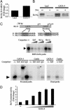
Identification of a Dox response element on the ANF gene. (A) Northern blot analyses of total RNA from primary cardiomyocyte cultures treated with Dox for the indicated time period. (B and C) Inhibition of ANF promoter activity by 12 h of treatment with Dox (19). The -700mutGATA construct contains point mutations in the three GATA-binding sites. The -700mutSRE construct contains point mutations in the proximal SRE. The results shown represent the mean ± SEM of at least six independent determinations. RSV, Rous sarcoma virus. (D) Gel-shift analyses with nuclear extracts prepared from control or Dox-treated cardiomyocytes and the GATA/SRE probe. Supershift (indicated by asterisks) with the serum response factor (αSRF) and GATA-4 (αG4) antibodies was used to confirm the identity of each complex.
Fig. 2.
Fig. 2.
Rapid depletion of GATA-4 transcripts and proteins after Dox treatment in vitro (A, B, and E) and in vivo (C and D). Total RNA (A) and nuclear extracts (B) were prepared from primary cardiomyocyte cultures treated for the indicated times with Dox and subjected to Northern and Western blot analyses, respectively. The arrowhead indicates GATA-4. Note how GATA-4 mRNA is already depleted after 3 h of Dox treatment. (C and D) Seven days after treatment with or without Dox, total RNA or tissue sections were prepared from the heart and assessed by semiquantitative RT-PCR (C) and immunohistochemistry (D). Similar decreases in GATA-4 mRNA or proteins were systematically observed in all animals treated with Dox (six to eight per group). (E) Northern blot analysis using total RNA from cardiomyocytes treated with vehicle (Veh), 5 mg/liter actinomycin D (ActD), or 10 μM cycloheximide (CHX) for 6 h. Dox-induced inhibition of GATA-4 mRNA is abrogated in the presence of actinomycin D but not cycloheximide.
Fig. 3.
Fig. 3.
GATA-4 protects cardiomyocytes against Dox cardiotoxicity. GATA-4 levels were manipulated by using adenovirus-mediated transfer of GATA-4 sense (G4S) or antisense (G4AS) transcripts (20). (A) Northern blot quantification of changes in ANF mRNA in cardiomyocytes infected with control (LacZ) or G4S adenovirus vectors. Cells were harvested after 12 h of treatment. The data represent the mean ± SEM (three in each group). (B) The contribution of GATA-4 to myocyte apoptosis was assessed 24 h after Dox treatment by using TUNEL assays. (Scale bar, 20 μm.) (C) Quantification of TUNEL assays in cardiomyocytes with altered GATA-4 levels. The data represent the mean ± SEM (three per group; *, P < 0.01 vs. LacZ).
Fig. 4.
Fig. 4.
A 50% reduction in GATA-4 levels exacerbates Dox cardiotoxicity in vivo. FS was determined by using echocardiography in GATA-4+/- mice and their wild-type (Wt) littermates before (Pre-Dox) and 1 week after (Post-Dox) Dox treatment. FS of the GATA-4+/- mice was reduced significantly after Dox treatment and as compared with the wild-type group. The data represent each individual mouse FS and mean ± SEM (three per group; *, P < 0.05 vs. pre-Dox wild type; †, P < 0.05 vs. pre-Dox GATA-4+/- mice).
Fig. 5.
Fig. 5.
Bcl-X is a GATA-4 target. (A and B) Quantitative PCR (A) and Western blots (B) were used to measure the level of Bcl-XL mRNA and protein in cardiomyocytes infected with adenoviruses expressing LacZ or GATA-4. The data in A are the mean ± SEM of four independent experiments, and the results in B are representative of two independent experiments. (C Top) Schematic representation of the proximal promoter of the Bcl-X gene showing the two conserved GATA elements. (C Middle) Gel-shift analyses using nuclear extracts prepared from NIH 3T3-overexpressing GATA-4 or cardiomyocytes (Cardio) and the BNP-GATA, Bcl-X proximal-GATA (Bcl-p), and Bcl-X distal-GATA (Bcl-d) probes. (C Bottom) Supershift (indicated by asterisks) with the GATA-4 (αG4) antibody was used to confirm the identity of the DNA-binding complex in cardiomyocytes. Cold competitor DNAs were used at 25- and 100-fold excess. Arrowheads indicate GATA-4 binding. (D) The Bcl-X luciferase reporter was cotransfected with increasing amounts of GATA-4 expression vector in NIH 3T3 cells. (E) Bcl-X promoter activity in cardiomyocytes is decreased 5-fold after Dox treatment. In D and E, the data represent the mean ± SEM of four independent determinations.
Fig. 6.
Fig. 6.
GATA-4 is essential for α1-adrenergic action. (A) Inhibition of Dox-induced apoptosis of cardiomyocytes by cotreatment with Phe for 12 h as determined by a TUNEL assay. The data are the mean ± SEM, (three to six per group; *, P < 0.01 vs. control; †, P < 0.01 vs. Dox). (B and C) Inhibition of Dox-induced change in gene expression. (B) Northern blot analyses after 12 h of Dox treatment in the presence or absence of Phe. (C Upper) Gel-shift analyses using cardiomyocyte nuclear extracts. (C Lower) Western blot showing Bcl-XL protein in the same extracts. (D) Quantification of TUNEL assays in cardiomyocytes infected with adenovirus expressing LacZ or antisense GATA-4 transcripts (ASG4); the protective effect of Phe is abrogated in cells lacking GATA-4. The data are the mean ± SEM of two independent experiments carried out in duplicate [*, P < 0.05, and **, P < 0.01 vs. respective control (Ctl); †, P < 0.05, and ††, P < 0.01 vs. respective Dox; and ‡, P < 0.01 vs. control LacZ].
Fig. 7.
Fig. 7.
Prevention of Dox cardiotoxicity by infusion of α1-adrenergic agonist in vivo.(A) GATA-4 protein levels (brown nuclei) were determined in the ventricles of the control (Ctl) and Dox- and Dox+Phe-treated mice by using immunohistochemistry. (Scale bar, 10 μm.) (B) Quantification of TUNEL assay in the ventricles of control (n = 3) and Dox-(n = 4) and Dox+Phe-(n = 4) treated mice. The data represent mean ± SEM (*, P < 0.05 vs. control; †, P < 0.05 vs. Dox). (C) Left ventricle tissue sections of control and Dox- and Dox+Phe-treated mice stained with hematoxylin and eosin. (Scale bar, 20 μm.) (D) Phe prevention of Dox cardiotoxicity. FS, frequency of CD, systolic blood pressure (SBP), and survival were determined in control and Dox- and Dox + Phe-treated mice. Data are mean ± SEM or frequency ratios of three separate experiments, with n = 8–22 in the 1-week follow-up group and n = 1–6 in the 2-week follow-up group (*, P < 0.05, and **, P < 0.01 vs. control; †, P < 0.05, and ††, P < 0.01 vs. Dox). (E) Phe prevention of chronic Dox cardiotoxicity. Heart rate (HR), FS, and frequency of CD were determined in control and Dox- and Dox+Phe-treated mice. Data are mean ± SEM or frequency ratios, with n = 3–8(*, P < 0.05 vs. control; †, P < 0.01 vs. Dox).

Similar articles

Cited by

References

    1. Olson, E. N. & Schneider, M. D. (2003) Genes Dev. 17, 1937-1956. - PubMed
    1. Regula, K. M., Ens, K. & Kirshenbaum, L. A. (2003) J. Mol. Cell Cardiol. 35, 559-567. - PubMed
    1. Yamamoto, S., Yang, G., Zablocki, D., Liu, J., Hong, C., Kim, S. J., Soler, S., Odashima, M., Thaisz, J., Yehia, G., et al. (2003) J. Clin. Invest. 111, 1463-1474. - PMC - PubMed
    1. Wencker, D., Chandra, M., Nguyen, K., Miao, W., Garantziotis, S., Factor, S. M., Shirani, J., Armstrong, R. C. & Kitsis, R. N. (2003) J. Clin. Invest. 111, 1497-1504. - PMC - PubMed
    1. Shi, Y. (2002) Mol. Cell 9, 459-470. - PubMed

Publication types

MeSH terms

LinkOut - more resources