. 2004 Mar 30;101(13):4419-24.
doi: 10.1073/pnas.0307700101. Epub 2004 Mar 22.

M-phase kinases induce phospho-dependent ubiquitination of somatic Wee1 by SCFbeta-TrCP

Affiliations

M-phase kinases induce phospho-dependent ubiquitination of somatic Wee1 by SCFbeta-TrCP

Nobumoto Watanabe et al. Proc Natl Acad Sci U S A. .

Abstract

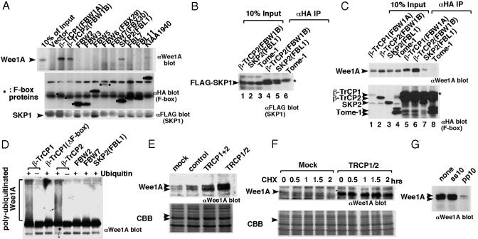
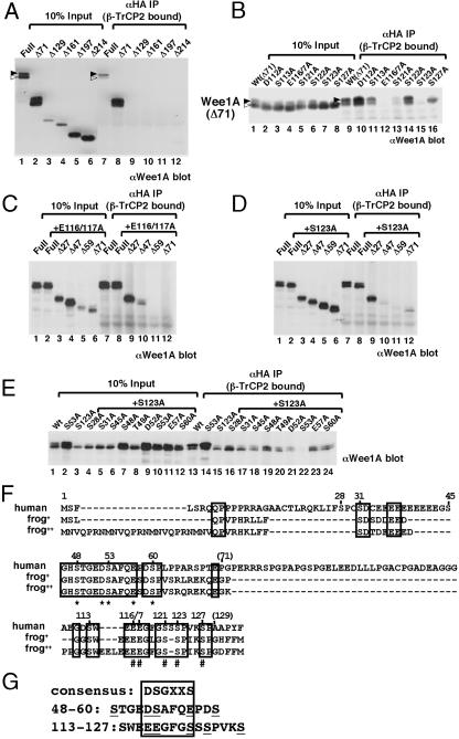
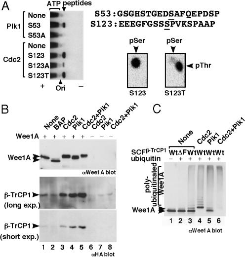
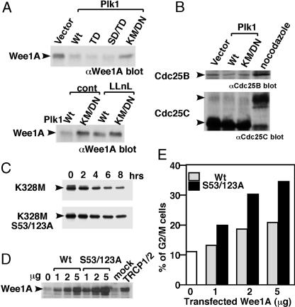
Wee1, the Cdc2 inhibitory kinase, needs to be down-regulated at the onset of mitosis to ensure rapid activation of Cdc2. Previously, we have shown that human somatic Wee1 (Wee1A) is down-regulated both by protein phosphorylation and degradation, but the underlying mechanisms had not been elucidated. In the present study, we have identified the beta-transducin repeat-containing protein 1/2 (beta-TrCP1/2) F-box protein-containing SKP1/Cul1/F-box protein (SCF) complex (SCF(beta-TrCP1/2)) as an E3 ubiquitin ligase for Wee1A ubiquitination. Although Wee1A lacks a consensus DS(p)GXXS(p) phospho-dependent binding motif for beta-TrCP, recognition of Wee1A by beta-TrCP depended on phosphorylation, and two serine residues in Wee1A, S53 and S123, were found to be the most important phosphorylation sites for beta-TrCP recognition. We have found also that the major M-phase kinases polo-like kinase 1 (Plk1) and Cdc2 are responsible for the phosphorylation of S53 and S123, respectively, and that in each case phosphorylation generates an unconventional phospho-degron (signal for degradation) that can be recognized by beta-TrCP. Phosphorylation of Wee1A by these kinases cooperatively stimulated the recognition and ubiquitination of Wee1A by SCF(beta-TrCP1/2) in vitro. Mutation of these residues or depletion of beta-TrCP by small-interfering RNA treatment increased the stability of Wee1A in HeLa cells. Moreover, our analysis indicates that beta-TrCP-dependent degradation of Wee1A is important for the normal onset of M-phase in vivo. These results also establish the existence of a feedback loop between Cdc2 and Wee1A in somatic cells that depends on ubiquitination and protein degradation and ensures the rapid activation of Cdc2 when cells are ready to divide.


Figures

Fig. 1.
Fig. 1.
Interaction between human Wee1A and SCF components. (A) Wee1A and FLAG-epitope tagged SKP1 were coexpressed with various HA epitope-tagged F-box proteins in HeLa-derived HtTA-1 cells. Anti-HA Ab immunoprecipitates from the transfected cell lysates were examined by immunoblotting with anti-Wee1A Ab (Top), anti-HA Ab (Middle); and anti-FLAG Ab (Bottom), respectively. Circles indicate F-box proteins. (B) Wee1A and FLAG epitope-tagged SKP1 were coexpressed with various HA epitope-tagged F-box proteins (β-TrCP2, SKP2, and Tome-1) in HeLa-derived HtTA-1 cells. Cell lysates (lanes 1–3) and anti-HA Ab immunoprecipitates (lanes 4–6) were examined by immunoblotting with anti-SKP1 Ab (Santa Cruz Biotechnology; SC-7163). *, signal derived from IgG light chain. (C) The nocodazole-treated transfected cell lysates as in B (lanes 1–4) or anti-HA Ab immunoprecipitates (lanes 5–8) from them were examined by immunoblotting with anti-Wee1A Abs (upper blot) and anti-HA mAb (lower blot), respectively. *, signal derived from IgG heavy chain. (D) GST-Wee1A (K328R) expressed, purified from insect cells and phosphorylated with cyclin B1/Cdc2 was ubiquitinated in vitro by using SCF complexes with various F-box proteins in the presence (+) or absence (–) of ubiquitin and analyzed by immunoblotting by using anti-Wee1A Ab. (E) HeLa cells were transfected with control unrelated siRNA, mixture of siRNAs that are specific for β-TrCP1 or β-TrCP2 siRNA (TRCP1+2) or siRNA that targets both of β-TrCP1 and β-TrCP2 (TRCP1/2). Wee1A levels were analyzed by immunoblotting at 2 days after transfection. Coomassie blue staining (CBB) of the filter is shown also (arrowheads indicate the position of Wee1A). (F) HeLa cells (105 cells per 6-cm dish) were transfected with siRNA (TRCP1/2). Two days later, cells were treated with nocodazole (0.3 μg/μl) for 21 h. CHX (25 μg/ml) was then added, and the stability of Wee1A was compared with that in mock-transfected cells at the times indicated. Wee1A in nocodazole-treated cells slowly migrated as a single band and was not detected in mock-transfected cells later than 0.5 h after the CHX addition. (G) Association of Wee1A to β-TrCP was abolished by the diphosphorylated IκBα peptide pp10 but not by its nonphosphorylated version, ss10 (29). Peptides were added at 100 μM before the addition of the anti-HA mAb.
Fig. 2.
Fig. 2.
Amino acid residues in Wee1A important for its recognition by β-TrCP. (AE) Wee1A (Full) and its N-terminal-deletion mutants (Δ71, Δ129, Δ161, Δ197 and Δ214) (A); Wee1A lacking 71 aa from the N terminus [Wt(Δ71)] and various point mutants as indicated (B); Wee1A (Full), E116/117A mutant (+E116/117A, Full), and its N-terminal-deletion mutants (+E116/117A, Δ27, Δ47, Δ59, and Δ71) (C); Wee1A (Full), S123A mutant (+S123A, Full), and its N-terminal-deletion mutants (+S123A, Δ27, Δ47, Δ59, and Δ71) (D); or Wee1A (Wt) and various mutants as indicated (E) were coexpressed with HA-tagged β-TrCP2 and SKP1 in HtTA-1 cells. Cell lysates (10% of total input is shown) and anti-HA immunoprecipitates of the lysates were analyzed by immunoblotting with anti-Wee1A Ab. Upper (hyperphosphorylated) and lower bands of Wee1A doublets are indicated by solid and open arrowheads, respectively, in A and B. (F) N-terminal region of human Wee1A [Wee1Hu; U10564; Top) (1) and two sequences of frog somatic, Wee1 (Wee2:AF358869; frog+; Wee1B:AB071983; frog++) (17, 30) were aligned. Identical amino acids among the three sequences are indicated by boxes. #, residues important for binding to β-TrCP in Δ71 Wee1A; *, residues important for binding to β-TrCP in S123A Wee1A. Positions of amino acids of human Wee1A are shown. (G) Phosphodegrons in Wee1A sequence are aligned with known consensus-recognition sequences for β-TrCP (boxed). The important amino acids shown in F are underlined.
Fig. 3.
Fig. 3.
Plk1 and Cdc2 cooperate to phosphorylate Wee1A and induce its binding to SCFβ-TrCP, ubiquitination, and proteasome-dependent degradation. (A) Synthetic peptides corresponding to sequences around S53 or S123 (and their derivatives) were phosphorylated by Plk1 or Cdc2. The weak phosphorylation of the S123A peptide by Cdc2 is presumably due to phosphorylation of S127, which is also succeeded by proline. Phosphorylation of S123 was also confirmed by the detection of phosphothreonine (pThr) in phosphoamino acid analysis of the S123T peptide (Right). Plk1 and Cdc2 did not phosphorylate S123 and S53, respectively (data not shown). (B) GST-Wee1A (K328R) expressed and purified from insect cells was treated with bacterial alkaline phosphatase (BAP) or the indicated protein kinases, mixed with cell lysates of 293T cells expressing HA-tagged β-TrCP1, and reisolated on glutathione agarose beads. Isolated GST-Wee1A (Top) and bound β-TrCP (Middle and Bottom) were detected by immunoblotting using anti-Wee1A and anti-HA Abs, respectively. No significant binding of β-TrCP1 was detected by using kinase reactions without GST-Wee1A(–). (C) GST-Wee1A (K328R) expressed and purified from insect cells was phosphorylated by the indicated protein kinases and ubiquitinated in vitro by using purified SCFβ-TrCP complexes together with E1, E2 (UbcH5a), and ATP in the presence (+) or absence (–) of ubiquitin, and it was analyzed by immunoblotting using anti-Wee1A Abs. No significant ubiquitination was detected when β-TrCP1 lacking the F-box domain (ΔF) was used.
Fig. 4.
Fig. 4.
Phosphorylation of two serines (S53 and S123) is important for ubiquitination and proteasome-dependent degradation of Wee1A in vivo, and it is required for the G2–M-phase transition. (A) Various Plk1 expression constructs [wild-type (Wt), constitutively active mutants T210D (TD) or S137D/T210D (SD/TD) and the kinase-negative mutant K82M/D194N (KM/DN)] were transfected into HeLa cells. Levels of endogenous Wee1A were examined by immunoblotting using anti-Wee1A Abs (Upper). In some experiments, transfected cells were treated with 50 μM N-acetyl-leucyl-leucyl-norleucinal (LLnL) for 6 h before harvesting (Lower). (B) Effect of Plk1 overexpression on endogenous Cdc25B and Cdc25C levels was analyzed by immunoblotting using anti-Cdc25B (upper) and anti Cdc25C (lower) Abs, respectively. An increased level of Cdc25B and a mobility retardation of Cdc25C were detected in nocodazole-treated M-phase cells. (C) Kinase negative mutant Wee1A (K328M) or the phosphorylation site mutant (K328M, S53/123A) was expressed in HeLa cells, and turnover was examined after inhibition of protein synthesis by using CHX (25 μg/ml) for the indicated times. (D and E) Various amounts of expression plasmids encoding wild-type or S53/123A Wee1A were transfected with GFP in HtTA1 cells. At 2 days after transfection, levels of Wee1A expression were compared with those in β-TrCP siRNA-treated cells (D), and cell-cycle distribution of GFP positive cells (≈100% of the transfected cells) was determined. The percentage of G2/M cells is shown in E. Because round-up mitotic cells were not increased in Wee1A-transfected cells under examination with microscope, we concluded that arrest at G2-phase rather than M-phase is responsible for the increased G2/M population (data not shown).

Similar articles

Cited by

References

    1. Watanabe, N., Broome, M. & Hunter, T. (1995) EMBO J. 14, 1878–1891. - PMC - PubMed
    1. Liu, F., Stanton, J. J., Wu, Z. & Piwnica-Worms, H. (1997) Mol. Cell. Biol. 17, 571–583. - PMC - PubMed
    1. Booher, R. N., Holman, P. S. & Fattaey, A. (1997) J. Biol. Chem. 272, 22300–22306. - PubMed
    1. Izumi, T., Walker, D. H. & Maller, J. L. (1992) Mol. Biol. Cell 3, 927–939. - PMC - PubMed
    1. Sia, R. A., Bardes, E. S. G. & Lew, D. J. (1998) EMBO J. 17, 6678–6688. - PMC - PubMed

MeSH terms