. 2004 Apr;24(7):2605-13.
doi: 10.1128/MCB.24.7.2605-2613.2004.

Histone fold protein Dls1p is required for Isw2-dependent chromatin remodeling in vivo

Affiliations

Histone fold protein Dls1p is required for Isw2-dependent chromatin remodeling in vivo

Audrey D McConnell et al. Mol Cell Biol. 2004 Apr.

Abstract

We report the identification of two new subunits of the Isw2 chromatin-remodeling complex in Saccharomyces cerevisiae. Both proteins, Dpb4p and Yjl065cp (named Dls1p), contain histone fold motifs and are homologous to the two smallest subunits of ISWI-containing CHRAC complexes in higher eukaryotes. Dpb4p is also a subunit of the DNA polymerase epsilon (polepsilon) complex, and Dls1p is homologous to another polepsilon subunit, Dpb3p. Therefore, these small histone fold proteins may fulfill functions that are required for both polepsilon and Isw2 complexes. We characterized the role of Dls1p in known roles of the Isw2 complex in vivo. Transcriptional analyses reveal that the Isw2 complex requires Dls1p to various degrees at a wide variety of loci in vivo. Consistent with this, Dls1p is required for Isw2-dependent chromatin remodeling in vivo, although the requirement for this protein varies among Isw2 targets. Dls1p is likely required for functions of the Isw2 complex at steps subsequent to its interaction with chromatin, since a dls1 mutation does not affect cross-linking of Isw2 with chromatin.


Figures

FIG. 1.
FIG. 1.
Identification of two previously unidentified subunits of the Isw2 complex. (A) Silver-stained gel of Isw2p purified by FLAG immunoaffinity chromatography followed by Source Q anionic-exchange chromatography. The identities of Isw2 complex subunits are shown on the right. The asterisk shows contaminants in the preparation. The numbers on the top indicate the fractions from the Source Q column. “Load” corresponds to the fraction from the FLAG immunoaffinity purification step, and “FT” represents the flowthrough fractions. (B) Silver-stained gel of Dls1p (Yjl065cp) purified by FLAG immunoaffinity chromatography followed by the Source Q column. The identities of Isw2 complex subunits are shown on the right. The numbers indicate the fractions from the Source Q column.
FIG. 2.
FIG. 2.
Quantitative analyses of interactions between small histone fold proteins in the Isw2 complex. (A) Coimmunoprecipitation assay of Dpb4p with both Pol2p and Isw2p. Dpb4p was immunoprecipitated from extracts prepared from YTT2186 (Dpb4p-6HA Pol2p-3FLAG Isw2p-13Myc) and YTT2133 (Pol2p-3FLAG Isw2p-13Myc) strains, and Dpb4p, Pol2p, and Isw2p were detected by Western blotting. To estimate the degree of depletion of epitope-tagged proteins from YTT2186 extract, serially diluted input materials were run in parallel. (B) Coimmunoprecipitation assay of Dpb2p with Isw2p. Dpb2p was immunoprecipitated from extracts prepared from YTT2756 (Dpb2-3FLAG) and YTT2757 (ISW2-13MYC DPB2-3FLAG) strains, and Isw2p and Dpb2p were detected by Western blotting. (C) Coimmunoprecipitation assay of Dls1p with both Itc1 and Isw2p. Dls1p was immunoprecipitated from extracts prepared from YTT2527 (Dls1p-3FLAG Itc1p-13Myc Isw2p-6HA) and YTT2529 (Itc1p-13Myc Isw2p-6HA) strains, and Dls1p, Itc1p, and Isw2p were detected by Western blotting.
FIG. 2.
FIG. 2.
Quantitative analyses of interactions between small histone fold proteins in the Isw2 complex. (A) Coimmunoprecipitation assay of Dpb4p with both Pol2p and Isw2p. Dpb4p was immunoprecipitated from extracts prepared from YTT2186 (Dpb4p-6HA Pol2p-3FLAG Isw2p-13Myc) and YTT2133 (Pol2p-3FLAG Isw2p-13Myc) strains, and Dpb4p, Pol2p, and Isw2p were detected by Western blotting. To estimate the degree of depletion of epitope-tagged proteins from YTT2186 extract, serially diluted input materials were run in parallel. (B) Coimmunoprecipitation assay of Dpb2p with Isw2p. Dpb2p was immunoprecipitated from extracts prepared from YTT2756 (Dpb2-3FLAG) and YTT2757 (ISW2-13MYC DPB2-3FLAG) strains, and Isw2p and Dpb2p were detected by Western blotting. (C) Coimmunoprecipitation assay of Dls1p with both Itc1 and Isw2p. Dls1p was immunoprecipitated from extracts prepared from YTT2527 (Dls1p-3FLAG Itc1p-13Myc Isw2p-6HA) and YTT2529 (Itc1p-13Myc Isw2p-6HA) strains, and Dls1p, Itc1p, and Isw2p were detected by Western blotting.
FIG. 3.
FIG. 3.
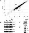
Requirement for Dls1p in Isw2-dependent effects on transcription. (A) Direct comparison of transcriptional profiles in isw2 rpd3 and dls1 rpd3 cells by DNA microarray analysis. Signals (pixels) normalized from six independent hybridizations were plotted. x axis, isw2 rpd3 mutant; y axis, dls1 rpd3 mutant. Note that the signals are represented by the number of pixels detected, rather than the ratio of expression to that of wild-type cells. The numbers of pixels detected are proportional to the strength of signals, hence the abundance of RNA molecules. The line in the center represents identical signals from each mutant. The two outside lines represent a twofold difference between the mutants. The two dots that are most distant from the diagonal line represent ISW2 and YJL064W genes. YJL064W is a dubious open reading frame that overlaps with the DLS1 gene (YJL065C). The DLS1 gene itself was eliminated from this analysis because of high local backgrounds in the batch of DNA microarrays used. (B) Northern blot analysis of Isw2 target genes. The genotypes of the cells used are indicated on the top. Listed vertically on the right of each blot is the gene being probed. The number under each band indicates the fold change in expression level relative to wild-type cells as determined with a phosphorimager. Signals were normalized for a loading control, ACT1. (C) Epistasis analysis of ISW2 and DLS1 genes by Northern blotting. (D) Northern blot analysis of Mata-specific genes STE2 and STE6 in Matα isw2 and dls1 mutants. As an internal positive control, the REC104 gene was also probed. WT, wild type.
FIG. 4.
FIG. 4.
Requirement for Dls1p in Isw2-dependent chromatin regulation. (A) Nucleosome mapping at the REC104 locus. The genotypes of cells used are indicated on the top. “N” stands for naked DNA control. The bracket on the right shows the region where differences in MNase sensitivities were observed between isw2 and dls1 mutants. (B) Nucleosome mapping at the POT1 locus. The arrowheads indicate the sites where a dls1 mutant exhibits a mixture of mutant and wild-type patterns. (C) Nucleosome mapping at the STE6 locus. The arrowheads indicate the sites where a dls1 mutant exhibits a mixture of mutant and wild-type patterns. The bracket shows the region where wild-type and dls1 cells have the same MNase digestion pattern, which is distinct from that of an isw2 mutant. WT, wild type.
FIG. 5.
FIG. 5.
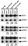
CHIP analysis of Isw2p and Dls1p. The relevant genotypes of the strains are shown on the top. The numbers indicate the averages and standard deviations of the CHIP signals at each locus relative to those at a control (YPL025C) locus. See Materials and Methods for the selection of the YPL025C locus as a control locus. As a control 100-fold (POT1 and STE6)- or 400-fold (REC104)-diluted input DNA from each strain was used.

Similar articles

Cited by

References

    1. Alen, C., N. A. Kent, H. S. Jones, J. O'Sullivan, A. Aranda, and N. J. Proudfoot. 2002. A role for chromatin remodeling in transcriptional termination by RNA polymerase II. Mol. Cell 10:1441-1452. - PubMed
    1. Araki, H., R. K. Hamatake, A. Morrison, A. L. Johnson, L. H. Johnston, and A. Sugino. 1991. Cloning DPB3, the gene encoding the third subunit of DNA polymerase II of Saccharomyces cerevisiae. Nucleic Acids Res. 19:4867-4872. - PMC - PubMed
    1. Badenhorst, P., M. Voas, I. Rebay, and C. Wu. 2002. Biological functions of the ISWI chromatin remodeling complex NURF. Genes Dev. 16:3186-3198. - PMC - PubMed
    1. Becker, P. B., and W. Horz. 2002. ATP-dependent nucleosome remodeling. Annu. Rev. Biochem. 71:247-273. - PubMed
    1. Bochar, D. A., J. Savard, W. Wang, D. W. Lafleur, P. Moore, J. Cote, and R. Shiekhattar. 2000. A family of chromatin remodeling factors related to Williams syndrome transcription factor. Proc. Natl. Acad. Sci. USA 97:1038-1043. - PMC - PubMed

Publication types

MeSH terms