

Neutralization assay using a modified vaccinia virus Ankara vector expressing the green fluorescent protein is a high-throughput method to monitor the humoral immune response against vaccinia virus
- PMID: 15013995
- PMCID: PMC371213
- DOI: 10.1128/cdli.11.2.406-410.2004
Neutralization assay using a modified vaccinia virus Ankara vector expressing the green fluorescent protein is a high-throughput method to monitor the humoral immune response against vaccinia virus
Abstract
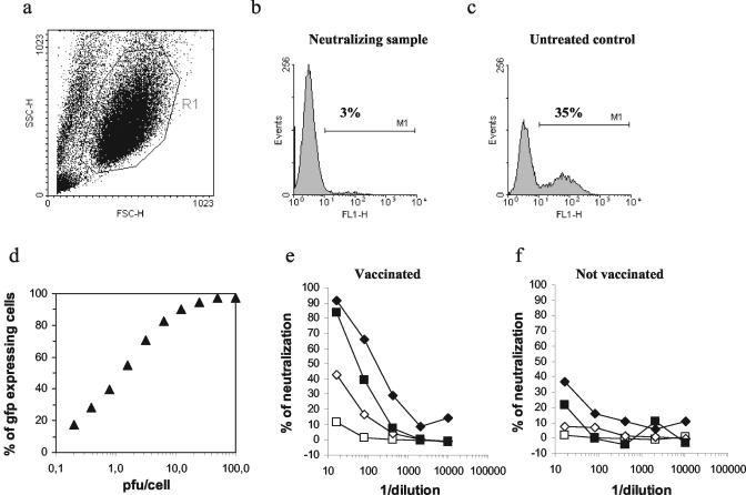
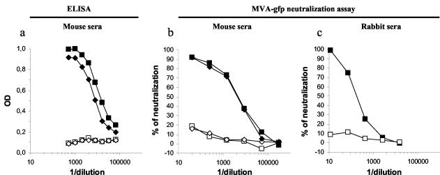
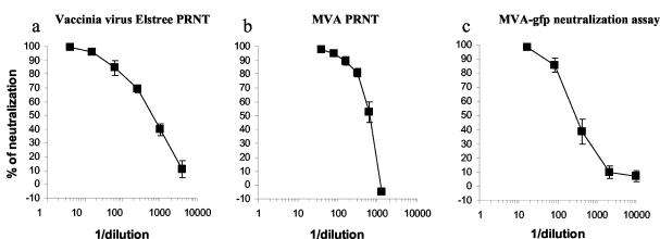
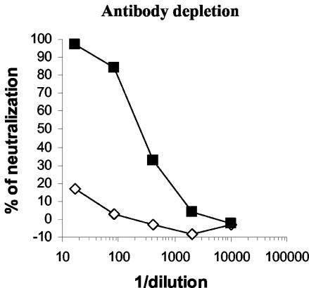
Vaccination against smallpox is again considered in order to face a possible bioterrorist threat, but the nature and the level of the immune response needed to protect a person from smallpox after vaccination are not totally understood. Therefore, simple, rapid, and accurate assays to evaluate the immune response to vaccinia virus need to be developed. Neutralization assays are usually considered good predictors of vaccine efficacy and more informative with regard to protection than binding assays. Currently, the presence of neutralizing antibodies to vaccinia virus is measured using a plaque reduction neutralization test, but this method is time-consuming and labor-intensive and has a subjective readout. Here, we describe an innovative neutralization assay based on a modified vaccinia virus Ankara (MVA) vector expressing the green fluorescent protein (MVA-gfp). This MVA-gfp neutralization assay is rapid and sensitive and has a high-throughput potential. Thus, it is suitable to monitor the immune response and eventually the efficacy of a large campaign of vaccination against smallpox and to study the vector-specific immune response in clinical trials that use genetically engineered vaccinia viruses. Most importantly, application of the highly attenuated MVA eliminates the safety concern in using the replication-competent vaccinia virus in the standard clinical laboratory.
Figures
Similar articles
-
A rapid, high-throughput vaccinia virus neutralization assay for testing smallpox vaccine efficacy based on detection of green fluorescent protein.J Virol Methods. 2008 Jun;150(1-2):14-20. doi: 10.1016/j.jviromet.2008.02.009. Epub 2008 Apr 2. J Virol Methods. 2008. PMID: 18387679
-
A novel high-throughput vaccinia virus neutralization assay and preexisting immunity in populations from different geographic regions in China.PLoS One. 2012;7(3):e33392. doi: 10.1371/journal.pone.0033392. Epub 2012 Mar 16. PLoS One. 2012. PMID: 22438922 Free PMC article.
-
Monitoring of human immunological responses to vaccinia virus.Methods Mol Biol. 2004;269:243-66. doi: 10.1385/1-59259-789-0:243. Methods Mol Biol. 2004. PMID: 15114020 Review.
-
Development of a novel vaccinia-neutralization assay based on reporter-gene expression.J Infect Dis. 2003 Aug 1;188(3):440-8. doi: 10.1086/376557. Epub 2003 Jul 16. J Infect Dis. 2003. PMID: 12870127
-
Clinical development of Modified Vaccinia virus Ankara vaccines.Vaccine. 2013 Sep 6;31(39):4241-6. doi: 10.1016/j.vaccine.2013.03.020. Epub 2013 Mar 21. Vaccine. 2013. PMID: 23523410 Review.
Cited by
-
Immune Profiles Identification by Vaccinomics After MVA Immunization in Randomized Clinical Study.Front Immunol. 2020 Nov 10;11:586124. doi: 10.3389/fimmu.2020.586124. eCollection 2020. Front Immunol. 2020. PMID: 33244316 Free PMC article. Clinical Trial.
-
Hepatitis B Virus DNA is a Substrate for the cGAS/STING Pathway but is not Sensed in Infected Hepatocytes.Viruses. 2020 May 29;12(6):592. doi: 10.3390/v12060592. Viruses. 2020. PMID: 32485908 Free PMC article.
-
Investigating the Effect of Encapsulation Processing Parameters on the Viability of Therapeutic Viruses in Electrospraying.Pharmaceutics. 2020 Apr 24;12(4):388. doi: 10.3390/pharmaceutics12040388. Pharmaceutics. 2020. PMID: 32344667 Free PMC article.
-
A Sensitive and High-Throughput Flow Cytometry-Based Assay for Measuring Antibody Neutralization of Human Adenovirus Type 3.Virol Sin. 2021 Jun;36(3):537-544. doi: 10.1007/s12250-020-00295-2. Epub 2020 Sep 29. Virol Sin. 2021. PMID: 32990935 Free PMC article.
-
Humoral Immunity to Primary Smallpox Vaccination: Impact of Childhood versus Adult Immunization on Vaccinia Vector Vaccine Development in Military Populations.PLoS One. 2017 Jan 3;12(1):e0169247. doi: 10.1371/journal.pone.0169247. eCollection 2017. PLoS One. 2017. PMID: 28046039 Free PMC article.
References
-
- Cosma, A., R. Nagaraj, S. Bühler, J. Hinkula, D. H. Busch, G. Sutter, F. D. Goebel, and V. Erfle. 2003. Therapeutic vaccination with MVA-HIV-1 nef elicits Nef-specific T-helper cell responses in chronically HIV-1 infected individuals. Vaccine 22:21-29. - PubMed
-
- Drexler, I., E. Antunes, M. Schmitz, T. Wolfel, C. Huber, V. Erfle, P. Rieber, M. Theobald, and G. Sutter. 1999. Modified vaccinia virus Ankara for delivery of human tyrosinase as melanoma-associated antigen: induction of tyrosinase- and melanoma-specific human leukocyte antigen A*0201-restricted cytotoxic T cells in vitro and in vivo. Cancer Res. 59:4955-4963. - PubMed
-
- Drexler, I., K. Heller, B. Wahren, V. Erfle, and G. Sutter. 1998. Highly attenuated modified vaccinia virus Ankara replicates in baby hamster kidney cells, a potential host for virus propagation, but not in various human transformed and primary cells. J. Gen. Virol. 79:347-352. - PubMed
-
- Geretti, A. M., M. E. Dings, C. A. van Els, C. A. van Baalen, F. J. Wijnholds, J. C. Borleffs, and A. D. Osterhaus. 1996. Human immunodeficiency virus type 1 (HIV-1)-and Epstein-Barr virus-specific cytotoxic T lymphocyte precursors exhibit different kinetics in HIV-1-infected persons. J. Infect. Dis. 174:34-45. - PubMed
-
- Goldstein, J. A., J. M. Neff, J. M. Lane, and J. P. Koplan. 1975. Smallpox vaccination reactions, prophylaxis, and therapy of complications. Pediatrics 55:342-347. - PubMed
MeSH terms
Substances
LinkOut - more resources
Full Text Sources
Other Literature Sources
