. 2004 Mar 15;164(6):819-29.
doi: 10.1083/jcb.200310077. Epub 2004 Mar 8.

The cohesion protein ORD is required for homologue bias during meiotic recombination

Affiliations

The cohesion protein ORD is required for homologue bias during meiotic recombination

Hayley A Webber et al. J Cell Biol. .

Abstract

During meiosis, sister chromatid cohesion is required for normal levels of homologous recombination, although how cohesion regulates exchange is not understood. Null mutations in orientation disruptor (ord) ablate arm and centromeric cohesion during Drosophila meiosis and severely reduce homologous crossovers in mutant oocytes. We show that ORD protein localizes along oocyte chromosomes during the stages in which recombination occurs. Although synaptonemal complex (SC) components initially associate with synapsed homologues in ord mutants, their localization is severely disrupted during pachytene progression, and normal tripartite SC is not visible by electron microscopy. In ord germaria, meiotic double strand breaks appear and disappear with frequency and timing indistinguishable from wild type. However, Ring chromosome recovery is dramatically reduced in ord oocytes compared with wild type, which is consistent with the model that defects in meiotic cohesion remove the constraints that normally limit recombination between sisters. We conclude that ORD activity suppresses sister chromatid exchange and stimulates inter-homologue crossovers, thereby promoting homologue bias during meiotic recombination in Drosophila.


Figures

Figure 1.
Figure 1.
Localization of ORD protein in wild-type germaria. (A) Schematic depiction of germline cysts and SC in different regions of the germarium. (B–D) Full projection of deconvolved z-series shows ORD localization in single germarium. Bar, 10 μm. (B) Linear stretches of ORD signal are visible in region 3 oocyte nucleus (arrowhead). (C) ORD foci and whole cyst staining are visible in region 1 before C(3)G localization (arrow). Threadlike ORD staining colocalizes with C(3)G in the oocyte nucleus (arrowhead in B and C). (D) ORD foci in a four-cell cyst and an early eight-cell cyst (arrowheads). In addition to bright ORD foci, diffuse ORD signal becomes visible throughout an older 8-cell cyst (arrow) and persists during the development of 16-cell germarial cysts. (E–G) Chromosome spread of semi-intact region 2A cyst showing single optical section from a deconvolved z-series. Bar, 10 μm. (E) Bright foci and linear stretches of ORD are visible within all 16 nuclei of the cyst. (F) Threadlike ORD staining (green) is more pronounced within nuclei that also stain for C(3)G (magenta). (G) DAPI-stained DNA. (H–J) Enlarged view from G (arrow) shows ORD (green) concentrated along the center of the bivalent (blue). Bar, 500 nm. (K–M) Extensive colocalization of ORD and C(3)G in a single nucleus. (N–P) Bright focus of ORD staining overlaps with CID signal. (K–P) Each image represents a single optical section from a deconvolved z-series of a chromosome spread. Bar, 2 μm.
Figure 2.
Figure 2.
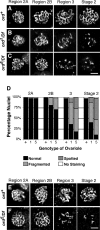
ord mutations disrupt C(3)G and C(2)M localization. (A–C) C(3)G localization within a single nucleus is shown for each region of the germarium and a vitellarial stage 2 egg chamber. Each image represents a full projection from a deconvolved z-series. Staining was performed on intact ovarioles (not spreads). ORB staining (not depicted) was used to identify oocytes in ord mutants. (A) In ord + germaria, extensive ribbonlike C(3)G is visible at each stage. (B) In region 2A of ord 1/Df mutant germaria, normal ribbonlike C(3)G staining is detected, but C(3)G staining becomes fragmented in the majority of ord 1/Df oocytes by region 3. (C) ord 5/Df mutant germaria also contain normal C(3)G staining in region 2A. However, the C(3)G signal is severely fragmented and spotted by region 3. Bar, 3 μm. (D) Quantification of the C(3)G defects in ord mutants shows the percentage of nuclei with normal, fragmented, spotted, or no C(3)G staining. Over 170 cells corresponding to each stage were examined. (E) Localization of C(2)M in ord + ovarioles. (F) C(2)M localization defects in ord 5/Df mutant germaria are similar to those observed for C(3)G. Although normal C(2)M signal is visible in region 2A, only fragmented and spotty staining is visible in region 3. Bar, 3 μm.
Figure 3.
Figure 3.
Abnormal SC ultrastructure in ord mutants. Transmission electron micrographs of germaria from ord + and ord 5/Df females. (A–C) Single sections showing examples of normal tripartite SC observed in each region of an ord + germarium. Lateral elements (le), transverse filaments (tf), and central elements (ce) are well defined and clearly visible in wild type. (D) Lower magnification of the entire region 3 nucleus showing multiple stretches of SC (arrowheads). The SC shown in C is indicated by the arrow. (E–G) Examples of abnormal SC-like structures from each region of ord 5/Df germaria. In some instances, (E and F) a central element (ce) is visible without obvious lateral elements. (H) Lower magnification of the entire region 3 nucleus with the area shown in G indicated by the arrow. Bars: (A–C and E–G) 200 nm; (D and H) 1 μm.
Figure 4.
Figure 4.
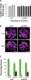
Homologues synapse in the absence of ORD activity. (A) Measured length of C(3)G staining in chromosome spread preparations from region 2B of wild-type and ord 5/Df mutant germaria. Mean C(3)G length is 114.6 ± 8.4 μm (n = 4) in ord + and 107.5 ± 3.2 μm (n = 8) in ord 5/Df. (B) Examples of chromosome spreads stained for C(3)G (magenta) and hybridized with probes (green) corresponding to pericentromeric heterochromatin or single copy sequences on the X chromosome arm (green). Bars, 2 μm. (C) Quantification of FISH analysis shows percentage of nuclei containing a single hybridization signal (green) or two to four signals (black) separated by <1 μm. n values are noted for each bar.
Figure 5.
Figure 5.
Normal numbers of DSBs appear and disappear in ord oocytes. (A–C) Whole germaria costained for C(3)G (magenta) and γ-H2Av (green). Images represent a full projection of a deconvolved z-series. Bars, 10 μm. Region 2B and region 3 cysts are outlined in white. (A) No γ-H2Av foci are visible in a mei-W68 germarium. (B) In wild type, γ-H2Av foci appear in region 2A (asterisk) and disappear in the oocyte by region 3 (arrow). (C) In ord 10/Df, γ-H2Av foci appear and disappear with normal kinetics. Asterisk marks region 2A and arrow points to region 3 cyst. (D and E) Examples of region 2B nuclei used to generate the graph in F. Bar, 3 μm. (F) Scatter plot representing the number of γ-H2Av foci per C(3)G staining nucleus in region 2B. Mean number of γ-H2Av foci in ord + is 7.0 (n = 25) and for ord 10/Df mutant is 7.2 (n = 25). Late 2B cysts in which γ-H2Av foci were absent in both pro-oocytes were not included in this analysis.
Figure 6.
Figure 6.
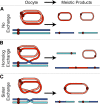
Possible meiotic outcomes for females carrying one Ring X chromosome and one normal Rod X chromosome. (A) Absence of inter-homologue or inter-sister exchange should yield equal numbers of meiotic products/progeny carrying either the Ring X or the Rod X chromatid. (B) Although a single crossover between homologous chromosomes will generate a dicentric chromosome that will not be transmitted, the number of progeny inheriting a nonrecombinant Ring or Rod chromosome should still be relatively equal. (C) Sister chromatid exchange between Rod X chromosomes should not affect their segregation. In contrast, a single crossover between the two Ring X sister chromatids will yield a large dicentric Ring chromosome that will not be transmitted to progeny. Therefore, high levels of sister chromatid exchange should cause Rod-containing progeny to greatly outnumber the Ring-containing progeny.
Figure 7.
Figure 7.
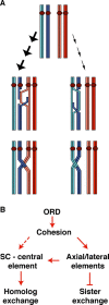
Homologue bias during meiotic recombination. (A) After premeiotic S phase, the oocyte enters meiotic prophase with two homologous chromosomes (blue and orange) each composed of a pair of sister chromatids (top). Different hues are used for each sister chromatid and both strands of DNA are shown. After the formation of a DSB, strand invasion can occur between homologues (middle left) or between sisters (middle right). Only a crossover between homologous chromosomes can generate a stable chiasma and the preferred pathway for strand invasion and recombination is between homologues (large arrows). Sister chromatid strand invasion and exchange occur much less frequently (small arrows). (B) Model for how ORD promotes homologue bias. ORD maintains meiotic sister chromatid cohesion, which is required for normal AE/LEs and stabilization of the CE of the SC. AE/LEs are required to suppress recombination between sister chromatids and stabilize CEs. In Drosophila, the SC is required for crossovers between homologous chromosomes. High levels of sister chromatid exchange occur in ord mutants because DSBs are preferentially repaired by strand invasion into the sister chromatid. ORD promotes homologue bias both by limiting crossovers between sister chromatids and by promoting exchange between homologous chromosomes.

Similar articles

Cited by

References

    1. Balicky, E.M., M.W. Endres, C. Lai, and S.E. Bickel. 2002. Meiotic cohesion requires accumulation of ORD on chromosomes prior to condensation. Mol. Biol. Cell. 21:3890–3900. - PMC - PubMed
    1. Bickel, S.E., D.W. Wyman, W.Y. Miyazaki, D.P. Moore, and T.L. Orr-Weaver. 1996. Identification of ORD, a Drosophila protein essential for sister-chromatid cohesion. EMBO J. 15:1451–1459. - PMC - PubMed
    1. Bickel, S.E., D.W. Wyman, and T.L. Orr-Weaver. 1997. Mutational analysis of the Drosophila sister-chromatid cohesion protein ORD and its role in the maintenance of centromeric cohesion. Genetics. 146:1319–1331. - PMC - PubMed
    1. Bickel, S.E., T. Orr-Weaver, and E.M. Balicky. 2002. The sister-chromatid cohesion protein ORD is required for chiasma maintenance in Drosophila oocytes. Curr. Biol. 12:925–929. - PubMed
    1. Blower, M.D., and G.H. Karpen. 2001. The role of Drosophila CID in kinetochore formation, cell-cycle progression and heterochromatin interactions. Nat. Cell Biol. 3:730–739. - PMC - PubMed

Publication types

MeSH terms