. 2004 Mar 3;24(9):2236-46.
doi: 10.1523/JNEUROSCI.4464-03.2004.

Macrophage-derived tumor necrosis factor alpha, an early developmental signal for motoneuron death

Affiliations

Macrophage-derived tumor necrosis factor alpha, an early developmental signal for motoneuron death

Frédéric Sedel et al. J Neurosci. .

Abstract

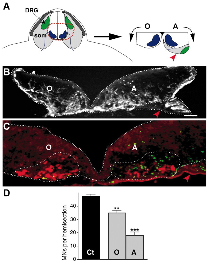
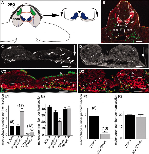
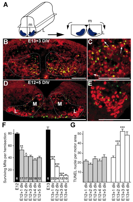
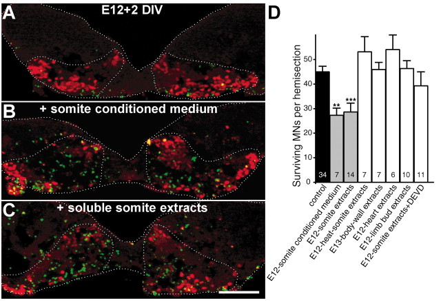
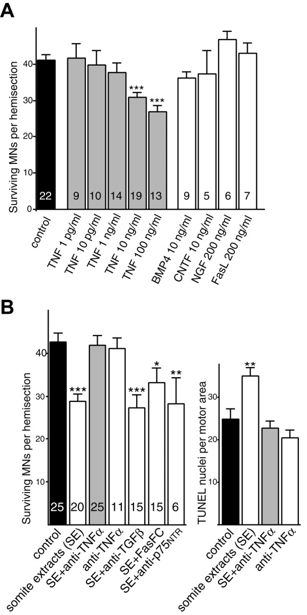
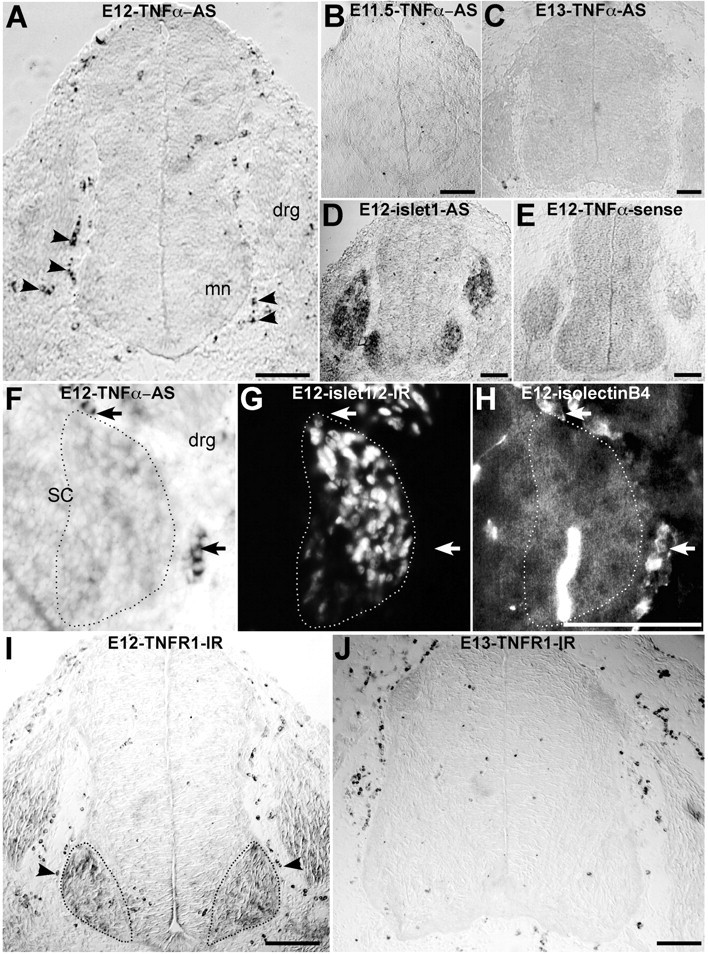
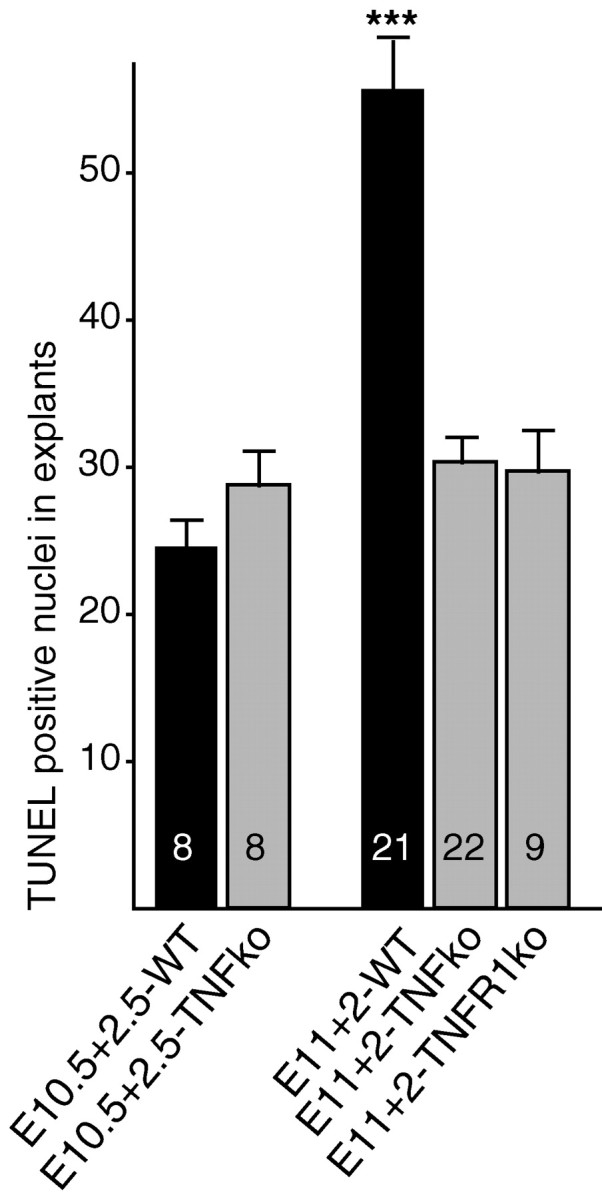
Mechanisms inducing neuronal death at defined times during embryogenesis remain enigmatic. We show in explants that a developmental switch occurs between embryonic day 12 (E12) and E13 in rats that is 72-48 hr before programmed cell death. Half the motoneurons isolated from peripheral tissues at E12 escape programmed cell death, whereas 90% of motoneurons isolated at E13 enter a death program. The surrounding somite commits E12 motoneurons to death. This effect requires macrophage cells, is mimicked by tumor necrosis factor alpha (TNFalpha), and is inhibited by anti-TNFalpha antibodies. In vivo, TNFalpha is detected within somite macrophages, and TNF receptor 1 (TNFR1) is detected within motoneurons precisely between E12 and E13. Although motoneuron cell death occurs normally in TNFalpha-/- mice, this process is significantly reduced in explants from TNFalpha-/- and TNFR1-/- mice. Thus, embryonic motoneurons acquire the competence to die, before the onset of programmed cell death, from extrinsic signals such as macrophage-derived TNFalpha


Figures

Figure 2.
Figure 2.
The proximal somite induces motoneuron death in spinal explants. A, Dissection of an E12 asymmetric explant. som, Somite; DRG, dorsal root ganglia; O, ventral horn opposite to the somite; A, ventral horn adjacent to the somite (red arrowhead). B, Transverse section of an E12 asymmetric explant cultured for 2 d in vitro. The explant section is immunolabeled with an anti-neurofilament antibody to show the position of the somite (red arrowhead). C, The section adjacent to that shown in B was double-labeled with TUNEL (green) and islet1/2 immunostaining (red). Note that there are numerous TUNEL- and fewer islet1/2-stained nuclei in the motor area (dotted) close to the somite (red arrowhead). Although some apoptotic nuclei are observed outside the motor area, most of them appear confined to the motor region (dotted). D, Quantification of surviving motoneurons (MNs) opposite (O) and adjacent (A) to the somite compared with control (Ct) E12 explants cultured for 2 d in vitro (8 explants quantified in each condition). **p < 0.01; ***p < 0.0001 (O and A vs Ct), Student's t test. Scale bars, 100 μm.
Figure 4.
Figure 4.
Macrophages provide the death signal to E12 but not to E13 motoneurons. A, Scheme of dissection of an asymmetric explant in which the most proximal part of the somite was left in contact with one ventral horn (same abbreviations and representation as in Fig. 2A). B, Macrophages and motoneurons simultaneously labeled with anti-Iba1 (green) and anti-islet1/2 (red) antibodies on a 36-somite (E12) rat embryo section passing through the brachial region. Motoneuron areas (mn), somites (som), and dorsal root ganglia (drg, only visible on one side) are outlined (dotted line). Note that numerous macrophages are within the somite, and some of them (arrows) are also detected in the proximal somite and occasionally within the neural tube (asterisks). C1-D2, Asymmetric explants cultured for 2 d without (C1, C2) or with (D1, D2) the macrophage-specific toxin IB4sap. Triple labeling identifying Iba1 (C1, D1), islet1/2 (C2, D2, red) immunoreactivities and TUNEL (C2, D2, green) are shown. The position of the somite is indicated by a large arrow, and Iba1-positive macrophages are indicated by small arrows. E, F, Quantification of macrophages (E1, F1) and motoneurons (E2, F2) on same sections from E12 (E1, E2) and E13 (F1, F2) explants cultured for 2 d. E12-ct, Explants cultured without somite; ct-asym-O, ct-asym-A, asymmetric explants cultured without IB4sap, ventral horn opposite (ct-asym-O) and adjacent (ct-asym-A) to the somite; IB4sap-O, IB4sap-A, asymmetric explants cultured with IB4sap, ventral horn opposite (IB4sap-O) and adjacent (OB4sap-A) to the somite. Note that in E12 explants, the somite prodeath effect is abolished in IB4sap-treated explants, which contain almost no macrophages. In E13 explants, the elimination of macrophages has no effect on motoneuron survival. ***p < 0.0001, Student's t test. Numbers of explants quantified in each condition are indicated on the histograms. Scale bars: B, 250 μm; C1-D2, 100 μm.
Figure 1.
Figure 1.
Motoneuron cell death in E12 and E13 ventral horn explants cultured in vitro. A, Dissection of an E12 ventral horn explant. Motoneuron areas are in blue. The relative positions of medial (m) and lateral (L) motoneurons are indicated. B-E, Explant sections were colabeled with TUNEL staining of apoptotic nuclei (green) and islet1/2 immunolabeling of motoneuron nuclei (red). B, C, Transverse sections of an E13 explant cultured for 3 d in vitro (DIV) at low (10×; B) and high (25×; C) magnifications. Arrows indicate some surviving motoneurons, e.g., intact nuclei positive for islet1/2 but negative for TUNEL. D, E, E12 explant cultured for 5 DIV at low (D) and high (E) magnification. Note that more surviving motoneurons are observed in E12+5 DIV explants compared with E13+3 div explants. Note also that in E12 explants the localization of apoptotic nuclei and of motoneuron loss corresponds to the medial part of the motor area. The observed number of apoptotic nuclei in explants is higher than in vivo at similar embryonic stages (Kablar and Rudnicki, 1999), suggesting that, in contrast to the in vivo situation, TUNEL-positive cells are not cleared by phagocytes in explant cultures. F, Absolute numbers of surviving motoneurons (MNs) in E12 and E13 explants cultured for the indicated numbers of days (+n) in vitro. The number of explants in each condition is marked inside the bars of histograms. G, Quantification of TUNEL-positive nuclei in same explants as in F. For both E12 and E13 explants, each value obtained for a given time of culture was compared with the value from the previous day. *p < 0.05; **p < 0.01; ***p < 0.0001, Student's t test. Scale bars, 100 μm.
Figure 3.
Figure 3.
Induction of motoneuron death by a somite-derived soluble protein. A-C, Double islet1/2-TUNEL labeling of E12 explants cultured for 2 d with culture medium alone (A), somite conditioned medium (B), or soluble somite extracts (C). Numerous apoptotic nuclei (green) and few surviving motoneurons (red) are present in the motor area of somite-treated explants. D, Numbers of surviving motoneurons (MNs) in E12 explants cultured for 2 d with control medium or medium supplemented with various soluble extracts as indicated. The number of explants quantified is indicated inside the bars of the histogram. **p < 0.01; ***p < 0.0001, Student's t test. Scale bar, 100 μm.
Figure 5.
Figure 5.
TNFα activates a death program in E12 motoneurons, and the somite prodeath effect is specifically inhibited by anti-TNFα blocking antibodies. A, E12 explants were treated for 2 d with increasing concentrations of TNFα or other proteic factors known to trigger neuronal death. B, E12 explants were treated for 2 d with somite extracts alone, somite extracts preincubated with anti-TNFα antibodies, or other blocking antibodies as indicated. An analysis of double islet1/2-TUNEL labeling experiments is shown. Note that anti-TNFα reverses the somite prodeath effect. Values corresponding to each test condition are compared with the control condition (black bar). **p < 0.01; ***p < 0.0001, Student's t test.
Figure 6.
Figure 6.
Transient TNFα mRNA and TNFR1 protein expression in spinal cord region during embryonic development in rats. A-C, In situ hybridization to detect TNFα mRNA with digoxigenin-labeled TNFα antisense (AS) probe. A, E12 (38 somites); B, E11.5 (31 somites); C, E13 spinal cord sections at the brachial level. Note that at E12, numerous round-labeled cells (arrowheads) are located close to the neural tube in the somite. Labeling is absent at E11.5 and E13. D, E, In situ hybridization of an E12 section labeled with islet-1 antisense (AS) probe, which labels motoneurons and dorsal root ganglia (D) and TNFα sense probe (E) as a negative control. F-H, E12 section at the brachial level triple-stained with TNFα antisense probe (F), Islet1/2 immunolabeling (G), and isolectin B4 (H). TNFα-expressing cells (arrows) are located in the somite close to motoneurons (G, dotted) and are stained with the macrophage marker isolectin B4 (H). SC, Spinal cord; DRG, dorsal root ganglia. I, J, TNFR1 immunoperoxidase of E12 (I) and E13 (J) spinal cord sections at the brachial level. On E12 (I), immunoreactivity (IR) is detected in the motor area (dotted), in the dorsal root ganglia, and in macrophage cells (arrowheads), whereas on E13 (J), immunoreactivity is only detected within macrophages. Scale bars, 100 μm.
Figure 7.
Figure 7.
Motoneuron death in TNFα-/- and TNFR1 -/- mouse explants. A comparison of apoptotic nuclei numbers per motor area in E10.5 explants cultured for 2.5 d in vitro (E10.5+2.5) or in E11 explants cultured for 2 d (E11+2) is shown. Note that in explants from wild-type mice (WT), there are twice as many apoptotic nuclei in E11+2 explants than in E10.5+2.5 explants. In contrast, no difference in apoptotic nuclei numbers is observed between E11+2 and E10.5+2.5 explants from both TNFα-/- and TNFR1 -/- mice. ***p < 0.0001, Student's t test. ko, Knock-out.

Similar articles

Cited by

References

    1. Altman J, Bayer SA (1984) The development of the rat spinal cord. Adv Anat Embryol Cell Biol 85: 1-164. - PubMed
    1. Barker V, Middleton G, Davey F, Davies AM (2001) TNFalpha contributes to the death of NGF-dependent neurons during development. Nat Neurosci 4: 1194-1198. - PubMed
    1. Boka G, Anglade P, Wallach D, Javoy-Agid F, Agid Y, Hirsch EC (1994) Immunocytochemical analysis of tumor necrosis factor and its receptors in Parkinson's disease. Neurosci Lett 172: 151-154. - PubMed
    1. Butler H, Juurlink BHJ (1987) An atlas for staging mammalian and chick embryos. Boca Raton, FL: CRC.
    1. Cuadros MA, Navascues J (1998) The origin and differentiation of microglial cells during development. Prog Neurobiol 56: 173-189. - PubMed

Publication types

MeSH terms

Substances

LinkOut - more resources