

STAT1 is essential for antimicrobial effector function but dispensable for gamma interferon production during Toxoplasma gondii infection
- PMID: 14977926
- PMCID: PMC356043
- DOI: 10.1128/IAI.72.3.1257-1264.2004
STAT1 is essential for antimicrobial effector function but dispensable for gamma interferon production during Toxoplasma gondii infection
Abstract
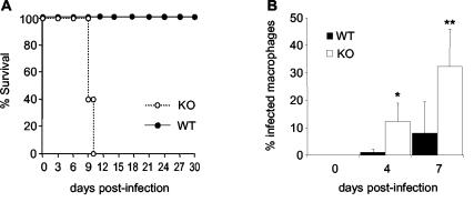
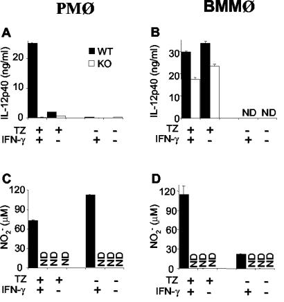
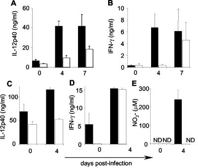
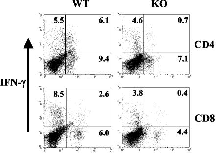
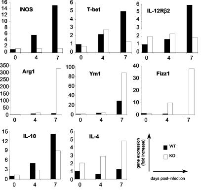
The opportunistic protozoan Toxoplasma gondii is a prototypic Th1-inducing pathogen inducing strong gamma interferon (IFN-gamma) cytokine responses that are required to survive infection. Intracellular signaling intermediate STAT1 mediates many effects of IFN-gamma and is implicated in activation of T-bet, a master regulator of Th1 differentiation. Here, we show that T. gondii-infected STAT1-null mice fail to upregulate the IFN-gamma-dependent effector molecules inducible nitric oxide synthase (iNOS), IGTP, and LRG-47, which are required for mice to survive infection. Both T-bet and interleukin-12 receptor beta2 (IL-12Rbeta2) failed to undergo normal upregulation in response to T. gondii. Development of IFN-gamma-producing CD4(+) and CD8(+) T lymphocytes was severely curtailed in the absence of STAT1, but a substantial level of STAT1-independent non-T-cell-derived IFN-gamma was induced. Absence of STAT1 also resulted in increased IL-4, Arg1, Ym1, and Fizz1, markers of Th2 differentiation and alternative macrophage activation. Together, the results show that T. gondii induces STAT1-dependent T-lymphocyte and STAT1-independent non-T-cell IFN-gamma production, but that effector functions of this type 1 cytokine cannot operate in the absence of STAT1, resulting in extreme susceptibility to acute infection.
Figures

Similar articles
-
STAT1 plays a critical role in the regulation of antimicrobial effector mechanisms, but not in the development of Th1-type responses during toxoplasmosis.J Immunol. 2004 Jan 1;172(1):457-63. doi: 10.4049/jimmunol.172.1.457. J Immunol. 2004. PMID: 14688355
-
The function of gamma interferon-inducible GTP-binding protein IGTP in host resistance to Toxoplasma gondii is Stat1 dependent and requires expression in both hematopoietic and nonhematopoietic cellular compartments.Infect Immun. 2002 Dec;70(12):6933-9. doi: 10.1128/IAI.70.12.6933-6939.2002. Infect Immun. 2002. PMID: 12438372 Free PMC article.
-
Identification of STAT4-dependent and independent mechanisms of resistance to Toxoplasma gondii.J Immunol. 2000 Sep 1;165(5):2619-27. doi: 10.4049/jimmunol.165.5.2619. J Immunol. 2000. PMID: 10946290
-
Cell-mediated immunity to Toxoplasma gondii: initiation, regulation and effector function.Immunobiology. 1999 Dec;201(2):240-7. doi: 10.1016/S0171-2985(99)80064-3. Immunobiology. 1999. PMID: 10631573 Review.
-
From cells to signaling cascades: manipulation of innate immunity by Toxoplasma gondii.FEMS Immunol Med Microbiol. 2003 Dec 5;39(3):193-203. doi: 10.1016/S0928-8244(03)00279-7. FEMS Immunol Med Microbiol. 2003. PMID: 14642303 Review.
Cited by
-
STAT1-Dependent Recruitment of Ly6ChiCCR2+ Inflammatory Monocytes and M2 Macrophages in a Helminth Infection.Pathogens. 2021 Oct 6;10(10):1287. doi: 10.3390/pathogens10101287. Pathogens. 2021. PMID: 34684235 Free PMC article.
-
Transcriptomic Profiling of Mouse Brain During Acute and Chronic Infections by Toxoplasma gondii Oocysts.Front Microbiol. 2020 Oct 19;11:570903. doi: 10.3389/fmicb.2020.570903. eCollection 2020. Front Microbiol. 2020. PMID: 33193165 Free PMC article.
-
Overview of Poultry Eimeria Life Cycle and Host-Parasite Interactions.Front Vet Sci. 2020 Jul 3;7:384. doi: 10.3389/fvets.2020.00384. eCollection 2020. Front Vet Sci. 2020. PMID: 32714951 Free PMC article. Review.
-
Th2 signals induce epithelial injury in mice and are compatible with the biliary atresia phenotype.J Clin Invest. 2011 Nov;121(11):4244-56. doi: 10.1172/JCI57728. Epub 2011 Oct 17. J Clin Invest. 2011. PMID: 22005305 Free PMC article.
-
STAT1 Signaling in Astrocytes Is Essential for Control of Infection in the Central Nervous System.mBio. 2016 Nov 8;7(6):e01881-16. doi: 10.1128/mBio.01881-16. mBio. 2016. PMID: 27834206 Free PMC article.
References
-
- Afkarian, M., J. R. Sedy, J. Yang, N. G. Jacobson, N. Cereb, S. Y. Yang, T. L. Murphy, and K. M. Murphy. 2002. T-bet is a STAT1-induced regulator of IL-12R expression in naive CD4+ T cells. Nat. Immunol. 6:549-557. - PubMed
-
- Bliss, S. K., A. J. Marshall, Y. Zhang, and E. Y. Denkers. 1999. Human polymorphonuclear leukocytes produce IL-12, TNF-α, and the chemokines macrophage-inflammatory protein-1α and -1β in response to Toxoplasma gondii antigens. J. Immunol. 162:7369-7375. - PubMed
-
- Bliss, S. K., Y. Zhang, and E. Y. Denkers. 1999. Murine neutrophil stimulation by Toxoplasma gondii antigen drives high level production of IFN-γ-independent IL-12. J. Immunol. 163:2081-2088. - PubMed
-
- Chang, N. C., S. I. Hung, K. Y. Hwa, I. Kato, J. E. Chen, C. H. Liu, and A. C. Chang. 2001. A macrophage protein, Ym1, transiently expressed during inflammation is a novel mammalian lectin. J. Biol. Chem. 276:17497-17506. - PubMed
-
- Collazo, C. M., G. S. Yap, S. Hieny, P. Caspar, C. G. Feng, G. A. Taylor, and A. Sher. 2002. The function of gamma interferon-inducible GTP-binding protein IGTP in host resistance to Toxoplasma gondii is Stat1 dependent and requires expression in both hematopoietic and nonhematopoietic cellular compartments. Infect. Immun. 70:6933-6939. - PMC - PubMed
Publication types
MeSH terms
Substances
Grants and funding
LinkOut - more resources
Full Text Sources
Molecular Biology Databases
Research Materials
Miscellaneous
