. 2004 Mar 10;23(5):1155-65.
doi: 10.1038/sj.emboj.7600069. Epub 2004 Feb 19.

Integration of TGF-beta/Smad and Jagged1/Notch signalling in epithelial-to-mesenchymal transition

Affiliations

Integration of TGF-beta/Smad and Jagged1/Notch signalling in epithelial-to-mesenchymal transition

Jiri Zavadil et al. EMBO J. .

Abstract

Epithelial-to-mesenchymal transitions (EMTs) underlie cell plasticity required in embryonic development and frequently observed in advanced carcinogenesis. Transforming growth factor-beta (TGF-beta) induces EMT phenotypes in epithelial cells in vitro and has been associated with EMT in vivo. Here we report that expression of the hairy/enhancer-of-split-related transcriptional repressor Hey1, and the Notch-ligand Jagged1 (Jag1), was induced by TGF-beta at the onset of EMT in epithelial cells from mammary gland, kidney tubules, and epidermis. The HEY1 expression profile was biphasic, consisting of immediate-early Smad3-dependent, Jagged1/Notch-independent activation, followed by delayed, indirect Jagged1/Notch-dependent activation. TGF-beta-induced EMT was blocked by RNA silencing of HEY1 or JAG1, and by chemical inactivation of Notch. The EMT phenotype, biphasic activation of Hey1, and delayed expression of Jag1 were induced by TGF-beta in wild-type, but not in Smad3-deficient, primary mouse kidney tubular epithelial cells. Our findings identify a new mechanism for functional integration of Jagged1/Notch signalling and coordinated activation of the Hey1 transcriptional repressor controlled by TGF-beta/Smad3, and demonstrate functional roles for Smad3, Hey1, and Jagged1/Notch in mediating TGF-beta-induced EMT.


Figures

Figure 1
Figure 1
HEY1 is a direct target gene of TGF-β/Smad signalling. (A) Bar graphs show averages of relative HEY1 mRNA abundance determined by quantitative RT-PCR analysis in keratinocytes (HaCaT), mouse mammary gland epithelial cells (NMuMG), human kidney proximal tubular epithelial cells (HK-2), and canine kidney distal tubular epithelial cell line (MDCK). (B) Immunoblots show HEY1 and C-terminally phosphorylated Smad2 p-Smad2 proteins in keratinocytes stimulated with TGF-β. PARP, loading control. (C) Bars show average (N=3) fold induction by TGF-β of relative luciferase units (RLUs) in keratinocytes transfected with reporter constructs of human HEY1 promoter deletions (full-length FL, M7 to M1). (▿) positions of SCRs; (*) positions of NICD-responsive CBEs. (D) Bars indicate average (N=3) fold induction by TGF-β of luciferase activity (RLU) in keratinocytes cotransfected with luciferase reporter constructs (FL-HEY1, M7 HEY1, or 3TPLux) and plasmids expressing dominant-negative mutants of type II TGF-β receptor (dnTbRII), Smad2, 3 and 4 (dn Smad2, 3 and 4), or NICD. p3TPlux, positive control for TGF-β/Smad; NICD, positive control for activation of CBE sites.
Figure 2
Figure 2
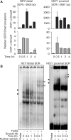
TGF-β induces rapid in vivo binding of endogenous Smad3/Smad4 protein complexes with SCR elements in the HEY1 promoter. (A) Bar graphs show the relative amount of distal HEY1 SCR (−3005 bp), or proximal SCR (−2681 bp), respectively, determined by quantitative, sequence-specific PCR that is bound with immunoprecipitated Smad3 (black bars) or Smad4 (gray bars) in untreated keratinocytes (time 0 h; baseline unit set at 1) and TGF-β-treated keratinocytes. (B) EMSAs demonstrate binding of oligonucleotide probes containing the distal (−3005) SCR or proximal (−2681) SCR, respectively, and nuclear protein extracts from keratinocytes left untreated (−) or treated with TGF-β (+) for 0.5 and 1 h. Specific DNA-binding protein complexes are depicted by block arrowheads, and are supershifted by preincubation with anti-Smad3 antibody (open arrowhead).
Figure 3
Figure 3
Activation of the Notch receptor is not involved in TGF-β induction of HEY1. (A) Histograms show the relative abundance of HEY1 mRNA expression as determined by quantitative RT-PCR in keratinocytes treated with TGF-β in the absence or presence of γ-secretase inhibitor GSI. (B) Immunoblot showing NICD in nuclear and cytoplasmic protein fractions of keratinocytes treated with TGF-β. PARP (poly-α-ADP-ribosyltransferase) and TUBB (tubulin β) demonstrate purity of cytoplasmic and nuclear protein lysate fractions, respectively. (C) EMSA demonstrates binding of CBF-1 protein complex with oligonucleotide probe containing the distal CBE of the HEY1 promoter (lanes 1–5). Lanes 6 and 7: EMSA using mutated CBE oligonucleotide probe (CBEmut) with single bp exchange (cgtggGaaa → cgtggCaaa). Lanes 8 and 9: EMSA using probe containing the distal (−3005 bp) HEY1 SCR sequence (HEY1 distal SCR) demonstrates TGF-β-inducible Smad protein complexes.
Figure 4
Figure 4
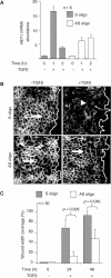
HEY1 is required for EMT and cell motility induced by TGF-β in keratinocytes. (A) Histograms show the relative abundance of HEY1 mRNA after treatment with TGF-β in keratinocytes transfected with the control S (sense) and AS (antisense) oligonucleotides, respectively. (B) In vitro scratch-wound assay of the cells transfected with S and AS oligonucleotide and stimulated by TGF-β for 24 h and immunostained for E-cadherin (white staining). The empty space right of the white line indicates the wound area. The arrow depicts E-cadherin-positive cell adherens junctions in untreated cells, and the arrowhead indicates loss of E-cadherin-positive adherens junctions in TGF-β-treated cells. (C) Histograms indicate the average coverage of scratch wound widths in % relative to baseline wound width (0 h) at 24 and 48 h after TGF-β treatment in keratinocyte cultures transfected with S (gray bars) or AS (open bars) oligonucleotides. Significance of the motility change was determined by Student's t-test. Wound width coverage at 24 and 48 h in untreated control cells was not significantly different from baseline (not shown).
Figure 5
Figure 5
Jagged1-induced Notch receptor activation initiates a second, delayed activation of HEY1. (A) Histograms show the mean of relative mRNA abundance determined by quantitative RT-PCR of Notch ligand JAGGED1 in response to TGF-β in epithelial cells (N=3). (B) Immunoblotting depicts JAGGED1 protein, intact 300 kDa transmembrane form (Notch1tm) of Notch in the absence or presence of γ-secretase inhibitor GSI, and nuclear accumulation of NICD (NICDnuc) in the absence or presence of GSI in TGF-β-treated keratinocytes. (C) Bar graphs indicate the relative abundance of HEY1 mRNA as determined by quantitative RT-PCR analysis in keratinocytes treated with TGF-β in the absence or presence of GSI. (D) Histogram shows RLUs in keratinocytes transfected with reporter constructs containing proximal human and mouse HEY1 promoter fragments, as indicated, after TGF-β stimulation (N=3). (E) Left histogram: Bars indicate a representative experiment showing the relative abundance of JAGGED1 mRNA determined by quantitative RT-PCR in keratinocytes, either control transfected (mock) or transfected with anti-JAGGED1 siRNA (anti-JAG1 siRNA). Right histogram: Bars show the mean relative abundance of HEY1 mRNA at 4 and 6 h after TGF-β treatment in untransfected keratinocytes (no TF), control-transfected keratinocytes (mock), control-transfected keratinocytes pretreated with GSI (GSI), or in anti-JAGGED1 siRNA-transfected keratinocytes (anti-JAG1 siRNA).
Figure 6
Figure 6
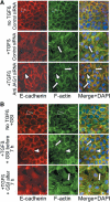
Jagged1 and Notch activation is required for TGF-β-induced EMT. (A) Immunofluorescence labelling for E-cadherin (red), F-actin (green), or both merged with nuclear DAPI staining, as indicated, of control-transfected, untreated (no TGF-β, control siRNA), or TGF-β-treated (+TGF-β, control siRNA) keratinocytes, and keratinocytes transfected with anti-JAGGED1 siRNA and treated with TGF-β (+TGF-β, anti-JAG1 siRNA). Immunostaining assays were performed 24 h after TGF-β induction. Open arrowheads show E-cadherin-positive adherens junctions and cortical actin bundles, respectively. White arrowheads depict widened E-cadherin-positive adherens junctions. Arrows denote actin stress fibers. (B) Experiments shown as in (A) in untransfected keratinocytes treated with TGF-β in the absence or presence of γ-secretase inhibitor (GSI) added either within 0–7 h of TGF-β treatment (+GSI before 7 h) or after 7 h (+GSI after 7 h).
Figure 7
Figure 7
Smad3 is required for induction of Hey1 and Jagged1, and for EMT, in primary mouse kidney tubular epithelial cells (mTEC). (A) Quantitative RT-PCR analysis of Smad3 in wild-type (mTEC WT) and Smad3-deficient (mTEC S3KO) primary tubular epithelial cells of the mouse kidney. (B, C) Quantitative RT-PCR analysis of Hey1 and Jagged1 inductions by TGF-β in wild-type (mTEC WT) and Smad3-deficient (mTEC S3KO) primary mTEC cultures. (D) Immunofluorescence labelling for E-cadherin (red), and phalloidin labelling for F-actin (green) in wild-type (mTEC WT) and Smad3-deficient (mTEC S3KO) primary, left untreated or treated with TGF-β for 24 h. Arrowheads indicate E-cadherin adherens junctions and cortical actin, respectively. Arrows depict actin stress fibers.
Figure 8
Figure 8
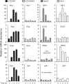
Histograms demonstrate the effects of TGF-β treatments, as indicated, on the relative abundance of Hey1/HEY1, Slug/SLUG, Snail/SNAIL, and Sip1/SIP1 mRNAs, respectively, in primary mouse kidney tubular epithelial cultures derived from wild-type control (mTEC WT) and Smad3 knockout (mTEC S3KO) mice, and in human keratinocytes (HaCaT) and human kidney tubular epithelial cells (HK-2). Graphs show representative results from triplicate experiments.

Similar articles

Cited by

References

    1. Bakin AV, Rinehart C, Tomlinson AK, Arteaga CL (2002) p38 mitogen-activated protein kinase is required for TGFbeta-mediated fibroblastic transdifferentiation and cell migration. J Cell Sci 115: 3193–3206 - PubMed
    1. Bakin AV, Tomlinson AK, Bhowmick NA, Moses HL, Arteaga CL (2000) Phosphatidylinositol-3 kinase function is required for TGFbeta-mediated epithelial to mesenchymal transition and cell migration. J Biol Chem 275: 36803–36810 - PubMed
    1. Bhowmick NA, Ghiassi M, Bakin A, Aakre M, Lundquist CA, Engel ME, Arteaga CL, Moses HL (2001) Transforming growth factor-beta1 mediates epithelial to mesenchymal transdifferentiation through a RhoA-dependent mechanism. Mol Biol Cell 12: 27–36 - PMC - PubMed
    1. Camenisch TD, Molin DG, Person A, Runyan RB, Gittenberger-De Groot AC, Mcdonald JA, Klewer SE (2002) Temporal and distinct TGFbeta ligand requirements during mouse and avian endocardial cushion morphogenesis. Dev Biol 248: 170–181 - PubMed
    1. Cano A, Perez-Moreno MA, Rodrigo I, Locascio A, Blanco MJ, Del Barrio MG, Portillo F, Nieto MA (2000) The transcription factor snail controls epithelial-mesenchymal transitions by repressing E-cadherin expression. Nat Cell Biol 2: 76–83 - PubMed

Publication types

MeSH terms