. 2004 Mar;24(5):2153-68.
doi: 10.1128/MCB.24.5.2153-2168.2004.

Direct interaction between nucleosome assembly protein 1 and the papillomavirus E2 proteins involved in activation of transcription

Affiliations

Direct interaction between nucleosome assembly protein 1 and the papillomavirus E2 proteins involved in activation of transcription

Manuela Rehtanz et al. Mol Cell Biol. 2004 Mar.

Abstract

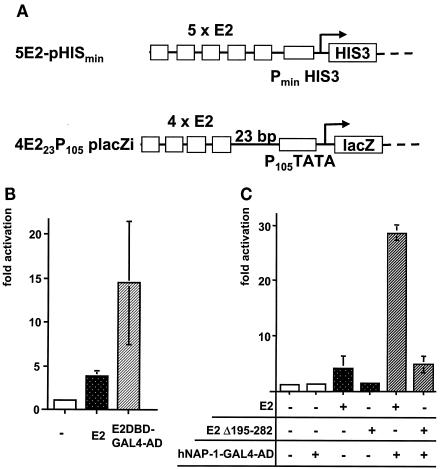
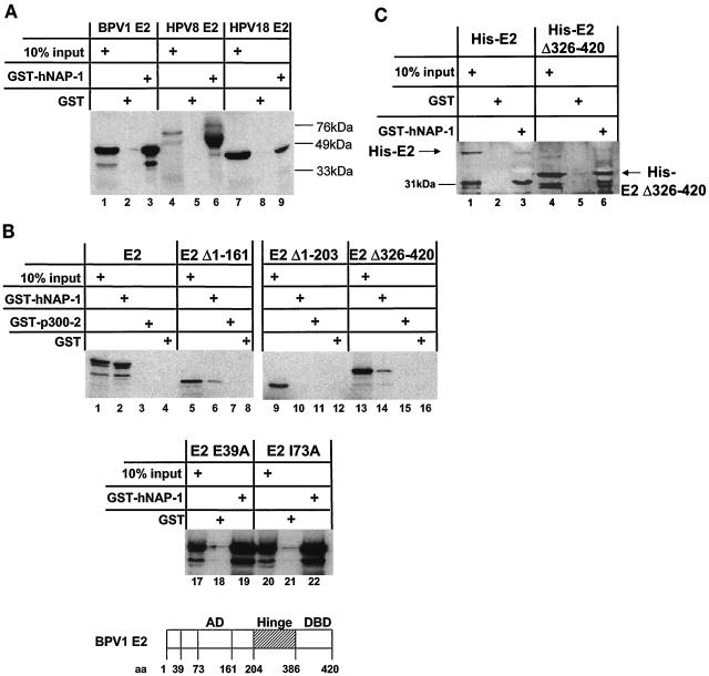
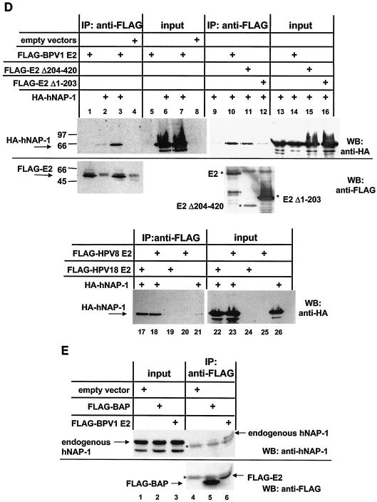
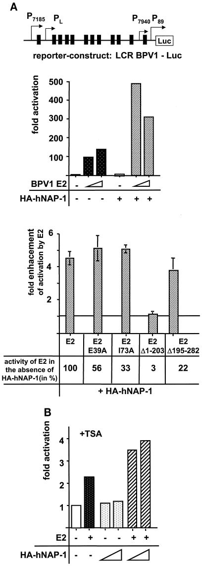
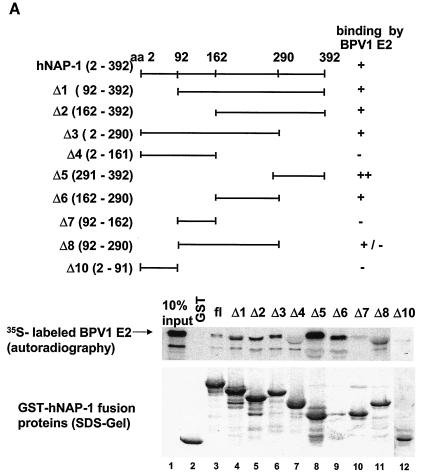
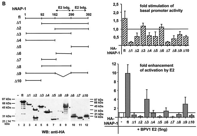
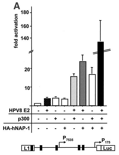
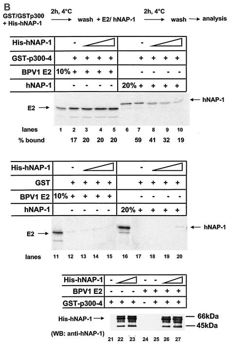
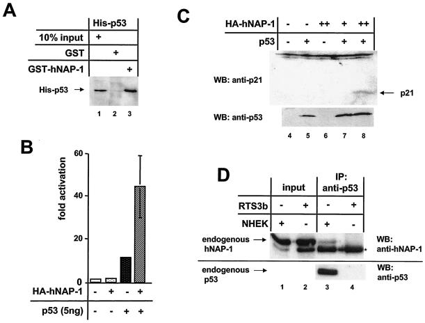
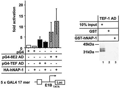
Using a yeast two-hybrid screen, we identified human nucleosome assembly protein 1 (hNAP-1) as a protein interacting with the activation domain of the transcriptional activator encoded by papillomaviruses (PVs), the E2 protein. We show that the interaction between E2 and hNAP-1 is direct and not merely mediated by the transcriptional coactivator p300, which is bound by both proteins. Coexpression of hNAP-1 strongly enhances activation by E2, indicating a functional interaction as well. E2 binds to at least two separate domains within hNAP-1, one within the C terminus and an internal domain. The binding of E2 to hNAP-1 is necessary for cooperativity between the factors. Moreover, the N-terminal 91 amino acids are crucial for the transcriptional activity of hNAP-1, since deletion mutants lacking this N-terminal portion fail to cooperate with E2. We provide evidence that hNAP-1, E2, and p300 can form a ternary complex efficient in the activation of transcription. We also show that p53 directly interacts with hNAP-1, indicating that transcriptional activators in addition to PV E2 interact with hNAP-1. These results suggest that the binding of sequence-specific DNA binding proteins to hNAP-1 may be an important step contributing to the activation of transcription.


Figures

FIG. 1.
FIG. 1.
Identification of hNAP-1 as an interaction partner for the PV E2 protein. (A) Schematic representation of two reporter constructs. 5E2-pHISmin expresses the yeast HIS3 gene under the control of its minimal promoter, pHISmin, and five E2 binding sites. 4E223P105placZi expresses the lacZ gene under the control of a synthetic promoter composed of the TATA box of the early promoter of HPV18 (P105) and four E2 binding sites located 23 bp further upstream. For the yeast two-hybrid system, both reporter constructs were integrated into the cellular genome of YM954. (B) Yeast expression vector pGBT9-E2, expressing the E2 protein of BPV1 (E2), or pGBT9-E2DBD-GAL4AD (E2DBD-GAL4-AD), expressing the AD of GAL4 fused to the DBD of BPV1 E2, or the vector alone (−) was transformed into the yeast strain harboring the two reporter constructs shown in panel A. β-Gal activities of the respective strains were determined. The activity in the presence of the vector alone (−) was assigned an arbitrary value of 1, and the fold activation was calculated. The columns represent the averages of three independent experiments performed with two different clones in each case; error bars are shown. (C) β-Gal activities of parental yeast strain YM954 transformed with a yeast expression vector for E2 or E2Δ195-282 (an E2 protein lacking the internal hinge region) and with plasmid pACT-c-ΔNAP-1, expressing hNAP-1 fused to the AD of GAL4, were determined. The latter was isolated by the yeast two-hybrid screen. The fold activation in relation to that of the strain transformed with the expression vector was calculated as described for panel B. The columns represent the averages for three independent clones; error bars are shown.
FIG. 2.
FIG. 2.
E2 proteins and hNAP-1 interact in vitro. (A) GST pull-down assays were carried out with purified GST, full-length hNAP-1 fused to GST (GST-hNAP-1), and 35S-labeled E2 proteins of BPV1, HPV8, and HPV18, obtained by in vitro translation with a rabbit reticulocyte lysate. Ten percent of the input is shown in lanes 1, 4, and 7. The positions of the marker proteins are indicated. (B) (Top panel) 35S-labeled full-length E2 of BPV1 (E2), two activation-deficient N-terminal deletion mutants (E2Δ1-161 and E2Δ1-203), and a C-terminal deletion mutant lacking the DBD (E2Δ326-420) were incubated with GST, GST-hNAP-1 or, as a negative control, GST-p300-2, expressing a fragment of p300 which was shown previously not to interact with E2 (49). Ten percent of the E2 protein and its derivatives used in one interaction assay is shown in the lanes labeled 10% input. (Middle panel) E2 proteins with point mutations in the AD, E2 E39A, which is replication deficient, and E2 I73A, which is impaired in transcription (14), were used in a GST pull-down assay. (Bottom panel) Structure of the E2 protein showing the positions of the amino acids (aa) in the various domains. (C) His-tagged, bacterially expressed, purified E2 protein of BPV1 (His-E2) or His-tagged E2Δ326-420 was incubated with purified GST or GST-hNAP-1. Bound E2 proteins were detected by Western blotting with an antibody directed against the His tag (Qiagen). The position of the 31-kDa marker protein is indicated. (D) (Upper panel) 293T cells were transfected with an expression vector expressing a FLAG-tagged full-length E2 protein of BPV1 (lanes 1, 3, 5, 7, 10, and 14), a FLAG-tagged BPV1 E2 mutant lacking the N-terminal AD (FLAG-E2Δ1-203; lanes 12 and 16), or FLAG-tagged AD (FLAG-E2Δ204-420; lanes 11 and 15) and an expression vector for HA-tagged hNAP-1 (lanes 2, 3, 6, 7, and 9 to 16). Cell extracts were incubated with the FLAG affinity gel, and bound hNAP-1 was detected with an antibody directed against the HA epitope (lanes 1 to 4 and lanes 9 to 12). In lanes 5 to 8 and lanes 13 to 16, 1/40 the input cellular extract was included, and the expression of HA-tagged hNAP-1 was analyzed with the HA antibody. The presence of FLAG-tagged E2 proteins was determined by reprobing of the blot shown in lanes 1 to 4 and lanes 10 to 12 with the FLAG M5 antibody. IP, immunoprecipitation; WB, Western blotting. (Lower panel) Western blot developed with the antibody directed against the HA epitope in a coimmunoprecipitation similar to that shown above but with cell extracts from 293T cells that had been transfected with an expression vector for FLAG-tagged HPV18 E2 (lanes 17, 19, 22, and 24) or HPV8 E2 (lanes 18, 20, 23, and 25) or an expression vector for HA-tagged hNAP-1 (lanes 17, 18, 21, 22, 23, and 26). (E) (Upper panel) Cell extracts of 293T cells that had been transfected with an expression vector for FLAG-tagged BPV1 E2 (lanes 3 and 6) or FLAG-tagged bacterial alkaline phosphatase (BAP) as a negative control (lanes 2 and 5) or with an empty vector (lanes 1 and 4) were incubated with the FLAG antibody coupled to Sepharose (lanes 4 to 6). Bound, endogenous hNAP-1 was detected by Western blotting with an antibody directed against hNAP-1. In lanes 1 to 3, 1/160 the cellular extract used for immunoprecipitation was loaded as an input control. (Lower panel) Part of the blot in the upper panel was reprobed with the FLAG antibody to detect the presence of FLAG-tagged proteins. A cellular protein cross-reacting with both antibodies is indicated by an asterisk.
FIG. 2.
FIG. 2.
E2 proteins and hNAP-1 interact in vitro. (A) GST pull-down assays were carried out with purified GST, full-length hNAP-1 fused to GST (GST-hNAP-1), and 35S-labeled E2 proteins of BPV1, HPV8, and HPV18, obtained by in vitro translation with a rabbit reticulocyte lysate. Ten percent of the input is shown in lanes 1, 4, and 7. The positions of the marker proteins are indicated. (B) (Top panel) 35S-labeled full-length E2 of BPV1 (E2), two activation-deficient N-terminal deletion mutants (E2Δ1-161 and E2Δ1-203), and a C-terminal deletion mutant lacking the DBD (E2Δ326-420) were incubated with GST, GST-hNAP-1 or, as a negative control, GST-p300-2, expressing a fragment of p300 which was shown previously not to interact with E2 (49). Ten percent of the E2 protein and its derivatives used in one interaction assay is shown in the lanes labeled 10% input. (Middle panel) E2 proteins with point mutations in the AD, E2 E39A, which is replication deficient, and E2 I73A, which is impaired in transcription (14), were used in a GST pull-down assay. (Bottom panel) Structure of the E2 protein showing the positions of the amino acids (aa) in the various domains. (C) His-tagged, bacterially expressed, purified E2 protein of BPV1 (His-E2) or His-tagged E2Δ326-420 was incubated with purified GST or GST-hNAP-1. Bound E2 proteins were detected by Western blotting with an antibody directed against the His tag (Qiagen). The position of the 31-kDa marker protein is indicated. (D) (Upper panel) 293T cells were transfected with an expression vector expressing a FLAG-tagged full-length E2 protein of BPV1 (lanes 1, 3, 5, 7, 10, and 14), a FLAG-tagged BPV1 E2 mutant lacking the N-terminal AD (FLAG-E2Δ1-203; lanes 12 and 16), or FLAG-tagged AD (FLAG-E2Δ204-420; lanes 11 and 15) and an expression vector for HA-tagged hNAP-1 (lanes 2, 3, 6, 7, and 9 to 16). Cell extracts were incubated with the FLAG affinity gel, and bound hNAP-1 was detected with an antibody directed against the HA epitope (lanes 1 to 4 and lanes 9 to 12). In lanes 5 to 8 and lanes 13 to 16, 1/40 the input cellular extract was included, and the expression of HA-tagged hNAP-1 was analyzed with the HA antibody. The presence of FLAG-tagged E2 proteins was determined by reprobing of the blot shown in lanes 1 to 4 and lanes 10 to 12 with the FLAG M5 antibody. IP, immunoprecipitation; WB, Western blotting. (Lower panel) Western blot developed with the antibody directed against the HA epitope in a coimmunoprecipitation similar to that shown above but with cell extracts from 293T cells that had been transfected with an expression vector for FLAG-tagged HPV18 E2 (lanes 17, 19, 22, and 24) or HPV8 E2 (lanes 18, 20, 23, and 25) or an expression vector for HA-tagged hNAP-1 (lanes 17, 18, 21, 22, 23, and 26). (E) (Upper panel) Cell extracts of 293T cells that had been transfected with an expression vector for FLAG-tagged BPV1 E2 (lanes 3 and 6) or FLAG-tagged bacterial alkaline phosphatase (BAP) as a negative control (lanes 2 and 5) or with an empty vector (lanes 1 and 4) were incubated with the FLAG antibody coupled to Sepharose (lanes 4 to 6). Bound, endogenous hNAP-1 was detected by Western blotting with an antibody directed against hNAP-1. In lanes 1 to 3, 1/160 the cellular extract used for immunoprecipitation was loaded as an input control. (Lower panel) Part of the blot in the upper panel was reprobed with the FLAG antibody to detect the presence of FLAG-tagged proteins. A cellular protein cross-reacting with both antibodies is indicated by an asterisk.
FIG. 3.
FIG. 3.
E2 and hNAP-1 cooperate in the activation of gene expression. (A) (Top panel) RTS3b cells, immortalized skin keratinocyte cells (57), were transfected with a luciferase reporter construct containing the regulatory region of BPV1 called the LCR, the structure of which is shown. The different promoters are indicated, and the 12 E2 binding sites are depicted as black boxes. (Middle panel) Either 5 or 20 ng of expression vector for BPV1 E2, under the control of the SV40 promoter, was cotransfected together with 400 ng of expression vector for HA-tagged hNAP-1. The graph shows the results of one representative experiment. (Bottom panel) Transient transfection experiments with the BPV1 LCR-Luc reporter plasmid, an expression vector for HA-tagged hNAP-1, and expression vectors for full-length E2 (E2), E2 E39A (the replication-deficient mutant), E2 I73A (the mutant impaired in transcription), E2Δ1-203 (lacking the AD), and E2Δ195- 282 (lacking the internal hinge region). The activity of each of the E2 proteins in the absence of coexpressed HA-tagged hNAP-1 was arbitrary defined as 1. The fold enhancement of E2-mediated activation by hNAP-1 was calculated. The graph represents the averages of at least three independent experiments; error bars are shown. The activation of each E2 mutant protein in the absence of coexpressed hNAP-1, compared to that of the wild-type protein, which was set at 100%, is given below the graph. (B) RTS3b cells, containing the BPV1 LCR-Luc reporter construct integrated into the cellular genome, were cotransfected with expression vectors for HA-tagged hNAP-1 and for E2. At 24 h after transfection, TSA was added to the medium for another 24 h.
FIG. 4.
FIG. 4.
The binding of E2 to hNAP-1 is necessary but not sufficient for the stimulation of E2-mediated activation. (A) Different regions of hNAP-1 were fused to GST. Bacterially expressed, purified GST- hNAP-1 fusion proteins (shown in the sodium dodecyl sulfate [SDS] gel at the bottom, stained by Coomassie blue) were incubated with 35S-labeled BPV1 E2. Bound proteins were detected by autoradiography. The binding of E2 to hNAP-1 is summarized (−, no binding; +/−, weak binding; +, binding; ++, strong binding). In some cases, the presence of GST- hNAP-1 fusion proteins in the gel might have affected the migration of the E2 protein slightly. aa, amino acids; fl, full length. (B) (Upper left panel) RTS3b cells were cotransfected with the BPV1 LCR reporter construct and an expression vector for HA-tagged hNAP-1 or HA-tagged deletion mutants of hNAP-1, either alone or together with 5 ng of the E2 expression vector. (Upper right panel) Effects of the various hNAP-1 mutants on BPV1 promoter activity in the absence of E2. The activity of the BPV1 LCR in the absence of hNAP-1 was arbitrarily defined as 1. (Lower right panel) Effects of the hNAP-1 mutants on activation by E2. Here, the activity in the presence of E2 without hNAP-1 (lane −) was set at 1, and the effects of the various hNAP-1 mutants were calculated. Both graphs represent the averages of at least three independent experiments with two different plasmid DNA preparations; error bars are shown. (Lower left panel) Levels of expression of the various HA-tagged hNAP-1 deletion mutants analyzed by Western blotting (WB) with extracts of cells that had been transiently transfected with the corresponding expression vectors and a high-affinity antibody against the HA epitope. Asterisks indicate the positions of the respective hNAP-1 mutants. Lanes 1 to 9, 10% polyacrylamide gel; lanes 10 to 12, 15% polyacrylamide gel.
FIG. 4.
FIG. 4.
The binding of E2 to hNAP-1 is necessary but not sufficient for the stimulation of E2-mediated activation. (A) Different regions of hNAP-1 were fused to GST. Bacterially expressed, purified GST- hNAP-1 fusion proteins (shown in the sodium dodecyl sulfate [SDS] gel at the bottom, stained by Coomassie blue) were incubated with 35S-labeled BPV1 E2. Bound proteins were detected by autoradiography. The binding of E2 to hNAP-1 is summarized (−, no binding; +/−, weak binding; +, binding; ++, strong binding). In some cases, the presence of GST- hNAP-1 fusion proteins in the gel might have affected the migration of the E2 protein slightly. aa, amino acids; fl, full length. (B) (Upper left panel) RTS3b cells were cotransfected with the BPV1 LCR reporter construct and an expression vector for HA-tagged hNAP-1 or HA-tagged deletion mutants of hNAP-1, either alone or together with 5 ng of the E2 expression vector. (Upper right panel) Effects of the various hNAP-1 mutants on BPV1 promoter activity in the absence of E2. The activity of the BPV1 LCR in the absence of hNAP-1 was arbitrarily defined as 1. (Lower right panel) Effects of the hNAP-1 mutants on activation by E2. Here, the activity in the presence of E2 without hNAP-1 (lane −) was set at 1, and the effects of the various hNAP-1 mutants were calculated. Both graphs represent the averages of at least three independent experiments with two different plasmid DNA preparations; error bars are shown. (Lower left panel) Levels of expression of the various HA-tagged hNAP-1 deletion mutants analyzed by Western blotting (WB) with extracts of cells that had been transiently transfected with the corresponding expression vectors and a high-affinity antibody against the HA epitope. Asterisks indicate the positions of the respective hNAP-1 mutants. Lanes 1 to 9, 10% polyacrylamide gel; lanes 10 to 12, 15% polyacrylamide gel.
FIG. 5.
FIG. 5.
hNAP-1 and E2 can form a ternary complex with p300. (A) (Top panel) A reporter construct expressing the luciferase gene under the control of the regulatory region of HPV8 was cotransfected with expression vectors for HPV8 E2, p300, and HA-tagged hNAP-1, either alone or in combination. The luciferase activity in the presence of the empty expression vector was set at 1, and the fold activation of any of the proteins was calculated. The graph represents the averages of five independent experiments; error bars are shown. (Bottom panel) Structure of the regulatory region of HPV8, including the two promoters and the E2 binding sites (black boxes). (B) GST or GST-p300-4 was incubated with increasing amounts of His-tagged, bacterially expressed, purified hNAP-1 for 2 h at 4°C. After several washes to remove unbound His-tagged hNAP-1, incubation with 35S-labeled BPV1 E2 or hNAP-1 for another 2 h followed. The binding of radiolabeled E2 or hNAP-1 was analyzed by autoradiography. The radioactive signals were quantified with a PhosphorImager; the percentages bound to GST-p300-4 were calculated. Ten percent of the input of 35S-labeled E2 or 20% of the input of 35S-labeled hNAP-1 was loaded as an input control. (Bottom panel) The Western blot (WB) reveals the binding of hNAP-1 to GST-p300-4 in the absence (lanes 22 and 23) or presence (lanes 26 and 27) of 35S-labeled BPV1 E2. (C) Glycerol density sedimentation analysis performed as described previously (61) with 600 ng of His-tagged hNAP-1, 200 ng of His-tagged E2Δ326-420 (E2), and 800 ng of p300 1195 to 1761. Samples were subjected to 7.5 to 30% glycerol gradient centrifugation, and a total of 36 fractions were collected, starting from the top of the gradient. Since an initial analysis revealed that all proteins fractionated in fractions 10 to 31, only these fractions are shown here after analysis by Western blotting with an antibody against the His tag. The presence of E2, hNAP-1, and p300 in the various fractions is indicated on the right.
FIG. 5.
FIG. 5.
hNAP-1 and E2 can form a ternary complex with p300. (A) (Top panel) A reporter construct expressing the luciferase gene under the control of the regulatory region of HPV8 was cotransfected with expression vectors for HPV8 E2, p300, and HA-tagged hNAP-1, either alone or in combination. The luciferase activity in the presence of the empty expression vector was set at 1, and the fold activation of any of the proteins was calculated. The graph represents the averages of five independent experiments; error bars are shown. (Bottom panel) Structure of the regulatory region of HPV8, including the two promoters and the E2 binding sites (black boxes). (B) GST or GST-p300-4 was incubated with increasing amounts of His-tagged, bacterially expressed, purified hNAP-1 for 2 h at 4°C. After several washes to remove unbound His-tagged hNAP-1, incubation with 35S-labeled BPV1 E2 or hNAP-1 for another 2 h followed. The binding of radiolabeled E2 or hNAP-1 was analyzed by autoradiography. The radioactive signals were quantified with a PhosphorImager; the percentages bound to GST-p300-4 were calculated. Ten percent of the input of 35S-labeled E2 or 20% of the input of 35S-labeled hNAP-1 was loaded as an input control. (Bottom panel) The Western blot (WB) reveals the binding of hNAP-1 to GST-p300-4 in the absence (lanes 22 and 23) or presence (lanes 26 and 27) of 35S-labeled BPV1 E2. (C) Glycerol density sedimentation analysis performed as described previously (61) with 600 ng of His-tagged hNAP-1, 200 ng of His-tagged E2Δ326-420 (E2), and 800 ng of p300 1195 to 1761. Samples were subjected to 7.5 to 30% glycerol gradient centrifugation, and a total of 36 fractions were collected, starting from the top of the gradient. Since an initial analysis revealed that all proteins fractionated in fractions 10 to 31, only these fractions are shown here after analysis by Western blotting with an antibody against the His tag. The presence of E2, hNAP-1, and p300 in the various fractions is indicated on the right.
FIG. 6.
FIG. 6.
p53 interacts directly with hNAP-1. (A) The GST- hNAP-1 fusion protein or GST was incubated with His-tagged, bacterially expressed, purified p53. After the beads were washed with a buffer containing 200 mM KCl, bound p53 was revealed by an antibody directed against the His tag. (B) The p53-negative cell line RTS3b was cotransfected with a synthetic p53-responsive luciferase reporter construct, 5 ng of an expression vector for p53, and 400 ng of the vector for HA-tagged hNAP-1. The luciferase activity in the presence of the vector only was set at 1. Error bars are shown. (C) Extracts (90 μg) from RTS3b cells transiently transfected with an expression vector for p53 (lanes 5, 7, and 8) or for HA-tagged hNAP-1 in two different amounts (lanes 6 to 8) were used for Western blotting (WB) to detect endogenous p21. The blot was reprobed with an antibody against p53 to detect p53 protein levels. (D) Nuclear extracts from p53-negative RTS3b cells (lanes 2 and 4) and from neonatal human epidermal keratinocytes (NHEK) (lanes 1 and 3), which express wild-type p53, were subjected to immunoprecipitation (IP) with a p53 antibody. Bound endogenous hNAP-1 was analyzed by Western blotting with a monoclonal antibody against hNAP-1 (lanes 3 and 4). In lanes 1 and 2, one-sixth of the input of nuclear extracts was loaded to detect the level of expression of hNAP-1 in both types of cells. Lanes 3 and 4 were reprobed with the p53 antibody to confirm the pattern of expression of p53. A cross-reacting cellular protein is indicated by an asterisk.
FIG. 7.
FIG. 7.
TEF-1 does not interact with hNAP-1. (Left panel) An expression vector for the DBD of GAL4 fused to either the AD of TEF-1 (pG4-TEF AD) (68) or the AD of HPV8 E2 (pG4-8E2 AD) was cotransfected either alone or together with the expression vector for HA-tagged hNAP-1 and the GAL4-responsive reporter construct, which is shown schematically below the graph. The data represent the averages of four experiments, and error bars indicate standard deviations. (Right panel) 35S-labeled TEF-1 AD was incubated with GST or GST-hNAP-1. After the samples were washed with a buffer containing 100 mM KCl, the reaction was analyzed by autoradiography. Ten percent of the input is shown in lane 1.

Similar articles

Cited by

References

    1. Altman, R., and D. R. Kellogg. 1997. Control of mitotic events by NAP1 and the Gin4 kinase. J. Cell Biol. 138:119-130. - PMC - PubMed
    1. Armstrong, J. A., J. J. Bieker, and B. M. Emerson. 1998. A SWI/SNF-related chromatin remodeling complex, E-RC1 is required for tissue specific transcriptional regulation by EKLF in vitro. Cell 95:93-104. - PubMed
    1. Asahara, H., B. Santoso, E. Guzman, K. Du, P. A. Cole, I. Davidson, and M. Montminy. 2001. Chromatin-dependent cooperativity between constitutive and inducible activation domains in CREB. Mol. Cell. Biol. 21:7892-7900. - PMC - PubMed
    1. Asahara, H., S. Tartare-Deckert, T. Nakagawa, T. Ikehara, F. Hirose, T. Hunter, T. Ito, and M. Montminy. 2002. Dual roles of p300 in chromatin assembly and transcriptional activation in cooperation with nucleosome assembly protein 1 in vitro. Mol. Cell. Biol. 22:2974-2982. - PMC - PubMed
    1. Avantaggiati, M. L., V. V. Ogryzko, K. Gardner, A. Giordano, S. A. Levine, and K. Kelly. 1997. Recruitment of p300/CBP in p53 dependent signal pathways. Cell 89:1175-1184. - PubMed

Publication types

MeSH terms