Comparative Study
. 2004 Feb;14(2):287-95.
doi: 10.1101/gr.2012304.

High-resolution analysis of DNA copy number using oligonucleotide microarrays

Affiliations
Comparative Study

High-resolution analysis of DNA copy number using oligonucleotide microarrays

Graham R Bignell et al. Genome Res. 2004 Feb.

Abstract

Genomic copy number alterations are a feature of many human diseases including cancer. We have evaluated the effectiveness of an oligonucleotide array, originally designed to detect single-nucleotide polymorphisms, to assess DNA copy number. We first showed that fluorescent signal from the oligonucleotide array varies in proportion to both decreases and increases in copy number. Subsequently we applied the system to a series of 20 cancer cell lines. All of the putative homozygous deletions (10) and high-level amplifications (12; putative copy number >4) tested were confirmed by PCR (either qPCR or normal PCR) analysis. Low-level copy number changes for two of the lines under analysis were compared with BAC array CGH; 77% (n = 44) of the autosomal chromosomes used in the comparison showed consistent patterns of LOH (loss of heterozygosity) and low-level amplification. Of the remaining 10 comparisons that were discordant, eight were caused by low SNP densities and failed in both lines. The studies demonstrate that combining the genotype and copy number analyses gives greater insight into the underlying genetic alterations in cancer cells with identification of complex events including loss and reduplication of loci.


Figures

Figure 1
Figure 1
Plot of log(copy number) against log(intensity) for the spiking experiment in which 18 aliquots of the same DNA were spiked with varying concentrations of 42 SNPs from twofold up to 1000-fold. The black dots indicate the results for the individual SNPs across the 12 spiking concentrations (spiked with an extra 0, 2, 5, 10, 25, 50, 75, 100, 250, 500, 750, and 1000 copies); the green dots and line show the mean for all 42 SNPs. Two SNPs did not report increased fluorescence with increased copy number, and these are highlighted in red.
Figure 2
Figure 2
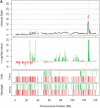
Examples of amplification and deletions together with genotyping data generated by the p501 array compared with BAC CGH data. (Top panel) The fluorescence ratio plot from the p501 array showing the smoothed fluorescent intensity data of the sample divided by the figure from the reference sample. (Second panel) The p-value plot for the individual SNPs calculated by comparison to the mean and standard deviation of 29 normal DNAs with deletions represented by red lines and amplifications in green. (Third and fourth panels) The genotypes for the tumor and matched normal samples, respectively; homozygous SNPs are represented by a red line below the center point whereas heterozygous markers are represented by a green line above the center point. (Bottom panel in C and D only) The results for BAC-array-based CGH (where available). (A) Genomic amplification at the C-MYC locus (arrow) on Chromosome 8 in the prostate line COR-L96-CAR. (B) A homozygous deletion (arrow) at the p16/INK4 locus on Chromosome 9 in the renal cell carcinoma cell line LB1047. (C) Chromosome 18 from the breast cancer cell line HCC1937. (i) This chromosome has a copy number of 2 (intensity ratio of 1) from the pter until 18q12.1. (ii) The copy number drops to 1 (intensity ratio of 0.5) from 18q12.1 until the 18qter, although the genotyping data indicate that this line is heterozygous across the full length of Chromosome 18. (D) Chromosome 5 from the small cell lung cancer line NCI-H209. This chromosome shows a complex pattern with partial amplification of the p-arm (i), followed by a drop in fluorescent intensity to 0.5 with corresponding LOH determined from the genotyping data (ii), until 5q23.2 (iii), where the intensity ratio recovers to 1 (copy number of 2); however, this region still represents LOH as determined by the genotyping data and therefore represents duplication of a single parental chromosome. There is also a homozygous deletion at 5q14.3 (arrow). This was not detected with the BAC array, as the array does not have a clone that covers this region.
Figure 2
Figure 2
Examples of amplification and deletions together with genotyping data generated by the p501 array compared with BAC CGH data. (Top panel) The fluorescence ratio plot from the p501 array showing the smoothed fluorescent intensity data of the sample divided by the figure from the reference sample. (Second panel) The p-value plot for the individual SNPs calculated by comparison to the mean and standard deviation of 29 normal DNAs with deletions represented by red lines and amplifications in green. (Third and fourth panels) The genotypes for the tumor and matched normal samples, respectively; homozygous SNPs are represented by a red line below the center point whereas heterozygous markers are represented by a green line above the center point. (Bottom panel in C and D only) The results for BAC-array-based CGH (where available). (A) Genomic amplification at the C-MYC locus (arrow) on Chromosome 8 in the prostate line COR-L96-CAR. (B) A homozygous deletion (arrow) at the p16/INK4 locus on Chromosome 9 in the renal cell carcinoma cell line LB1047. (C) Chromosome 18 from the breast cancer cell line HCC1937. (i) This chromosome has a copy number of 2 (intensity ratio of 1) from the pter until 18q12.1. (ii) The copy number drops to 1 (intensity ratio of 0.5) from 18q12.1 until the 18qter, although the genotyping data indicate that this line is heterozygous across the full length of Chromosome 18. (D) Chromosome 5 from the small cell lung cancer line NCI-H209. This chromosome shows a complex pattern with partial amplification of the p-arm (i), followed by a drop in fluorescent intensity to 0.5 with corresponding LOH determined from the genotyping data (ii), until 5q23.2 (iii), where the intensity ratio recovers to 1 (copy number of 2); however, this region still represents LOH as determined by the genotyping data and therefore represents duplication of a single parental chromosome. There is also a homozygous deletion at 5q14.3 (arrow). This was not detected with the BAC array, as the array does not have a clone that covers this region.
Figure 3
Figure 3
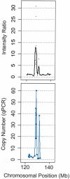
Comparison of copy number estimation for the amplification of the c-MYC proto-oncogene in COR-L96-CAR using the p501 array (A; where copy number equals two times the intensity ratio) and qPCR (B).

Similar articles

Cited by

References

    1. Albertson, D.G., Ylstra, B., Segraves, R., Collins, C., Dairkee, S.H., Kowbel, D., Kuo, W.L., Gray, J.W., and Pinkel, D. 2000. Quantitative mapping of amplicon structure by array CGH identifies CYP24 as a candidate oncogene. Nat. Genet. 25: 144-146. - PubMed
    1. Dumur, C.I., Dechsukhum, C., Ware, J.L., Cofield, S.S., Best, A.M., Wilkinson, D.S., Garrett, C.T., and Ferreira-Gonzalez, A. 2003. Genome-wide detection of LOH in prostate cancer using human SNP microarray technology. Genomics 81: 260-269. - PubMed
    1. Ghaffari, S.R., Boyd, E., Tolmie, J.L., Crow, Y.J., Trainer, A.H., and Connor, J.M. 1998. A new strategy for cryptic telomeric translocation screening in patients with idiopathic mental retardation. J. Med. Genet. 35: 225-233. - PMC - PubMed
    1. Hodgson, G., Hager, J.H., Volik, S., Hariono, S., Wernick, M., Moore, D., Nowak, N., Albertson, D.G., Pinkel, D., Collins, C., et al. 2001. Genome scanning with array CGH delineates regional alterations in mouse islet carcinomas. Nat. Genet. 29: 459-464. - PubMed
    1. Jain, A.N., Chin, K., Borresen-Dale, A.L., Erikstein, B.K., Eynstein Lonning, P., Kaaresen, R., and Gray, J.W. 2001. Quantitative analysis of chromosomal CGH in human breast tumors associates copy number abnormalities with p53 status and patient survival. Proc. Natl. Acad. Sci. 98: 7952-7957. - PMC - PubMed