. 2004 Feb 11;23(3):659-69.
doi: 10.1038/sj.emboj.7600081. Epub 2004 Jan 29.

Molecular clearance of ataxin-3 is regulated by a mammalian E4

Affiliations

Molecular clearance of ataxin-3 is regulated by a mammalian E4

Masaki Matsumoto et al. EMBO J. .

Abstract

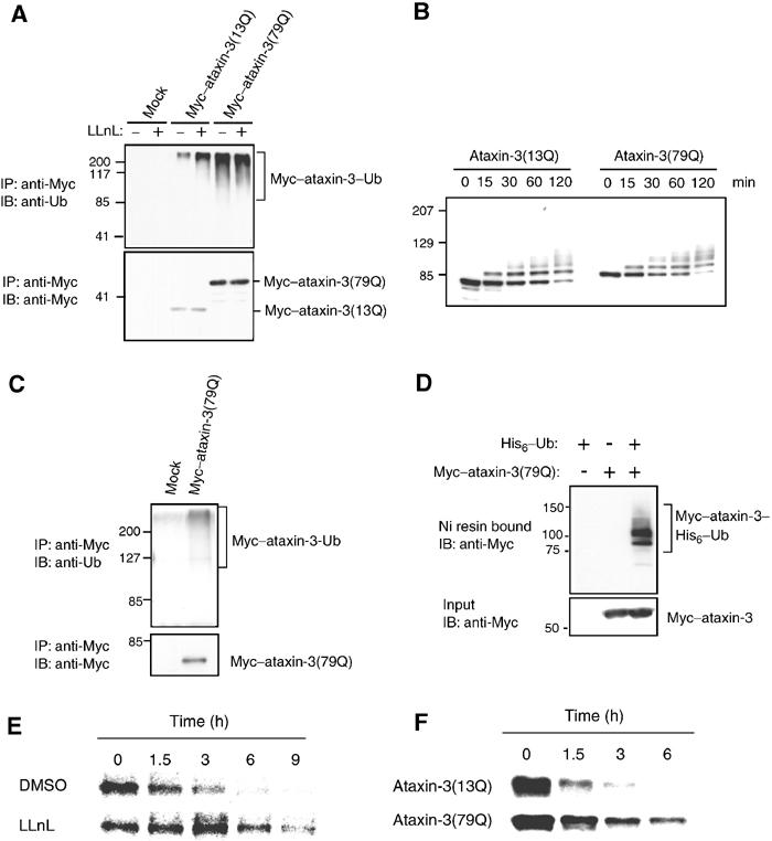
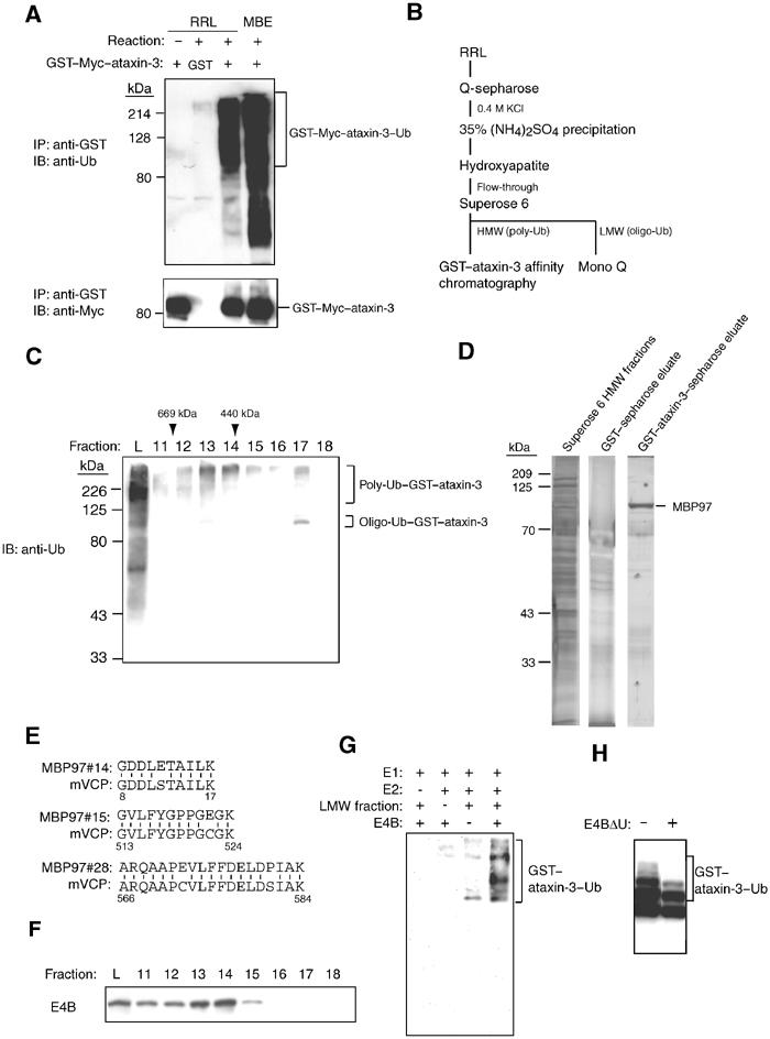
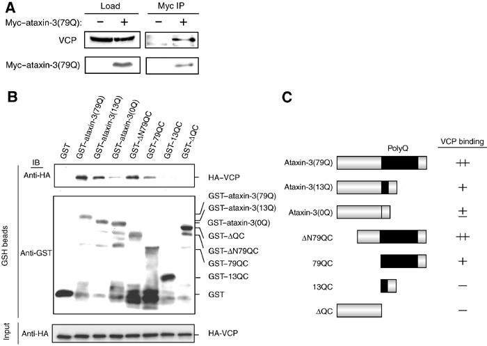
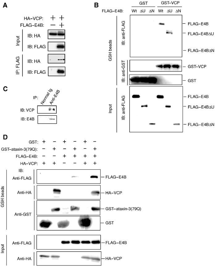
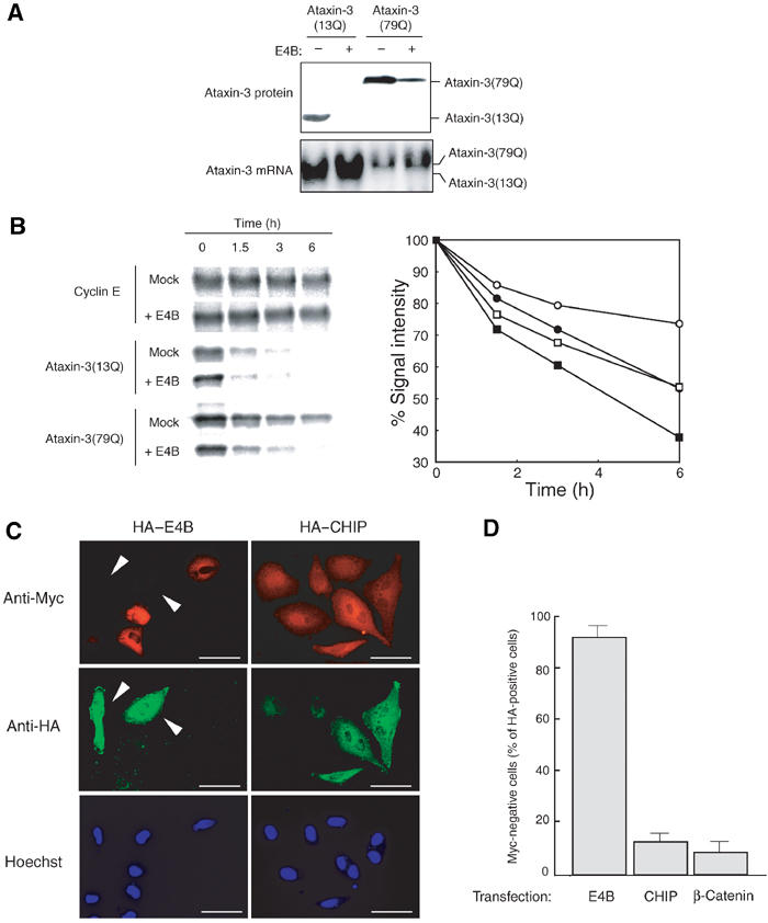
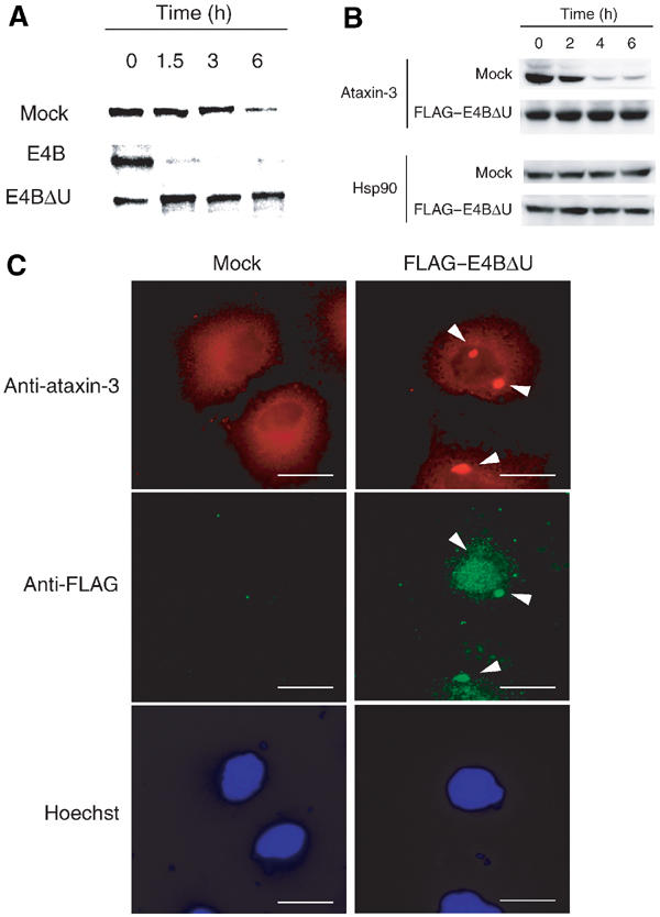
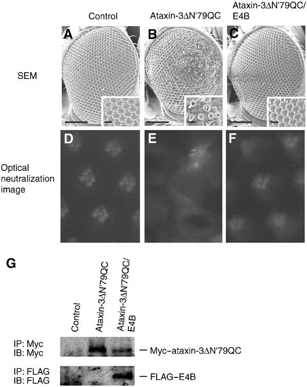
Insoluble aggregates of polyglutamine-containing proteins are usually conjugated with ubiquitin in neurons of individuals with polyglutamine diseases. We now show that ataxin-3, in which the abnormal expansion of a polyglutamine tract is responsible for spinocerebellar ataxia type 3 (SCA3), undergoes ubiquitylation and degradation by the proteasome. Mammalian E4B (UFD2a), a ubiquitin chain assembly factor (E4), copurified with the polyubiquitylation activity for ataxin-3. E4B interacted with, and thereby mediated polyubiquitylation of, ataxin-3. Expression of E4B promoted degradation of a pathological form of ataxin-3. In contrast, a dominant-negative mutant of E4B inhibited degradation of this form of ataxin-3, resulting in the formation of intracellular aggregates. In a Drosophila model of SCA3, expression of E4B suppressed the neurodegeneration induced by an ataxin-3 mutant. These observations suggest that E4 is a rate-limiting factor in the degradation of pathological forms of ataxin-3, and that targeted expression of E4B is a potential gene therapy for SCA3.


Figures

Figure 1
Figure 1
Degradation of ataxin-3 by the ubiquitin–proteasome pathway. (A) HEK293T cells were transiently transfected with an expression vector encoding Myc–ataxin-3(13Q) or Myc–ataxin-3(79Q), or with the empty vector as a control (mock), and then cultured in the absence or presence of 10 μM LLnL for 6 h. Cell lysates were then subjected to immunoprecipitation (IP) with anti-Myc, and the resulting precipitates were subjected to immunoblot (IB) analysis with either anti-ubiquitin (anti-Ub) or anti-Myc (upper and lower panels, respectively). (B) An in vitro ubiquitylation assay was performed with rabbit reticulocyte lysate and either GST–ataxin-3(13Q) or GST–ataxin-3(79Q). The reaction was terminated at the indicated times by the addition of SDS sample buffer, and the reaction mixture was subjected to IB analysis with anti-GST. (C) Extracts of control (mock) or Myc–ataxin-3(79Q)-expressing HEK293T cells were boiled in the presence of 1% SDS for 5 min. After dilution to 0.2% SDS, the extracts were subjected to IP with anti-Myc and the resulting precipitates were subjected to IB analysis with either anti-ubiquitin or anti-Myc. (D) HEK293T cells were transiently transfected with vectors for Myc–ataxin-3(79Q) and His6–ubiquitin (His6–Ub), as indicated. Ubiquitylated proteins were purified from cell lysates with a Ni-based resin in the presence of 4 M urea and then subjected to IB analysis with anti-Myc (upper panel). A portion (10%) of the input lysates was also subjected directly to IB analysis (lower panel). (E) HEK293T cells were transfected with the Myc–ataxin-3(13Q) vector and then labeled with [35S]methionine for 1 h. The cells were then washed, cultured for the indicated times in the presence of dimethyl sulfoxide (DMSO, vehicle control) or 100 μM LLnL, and lysed. Cell lysates were subjected to IP with anti-Myc, and the resulting precipitates were subjected to SDS–PAGE and autoradiography. (F) HEK293T cells expressing Myc–ataxin-3(13Q) or Myc–ataxin-3(79Q) were subjected to pulse-chase analysis (without LLnL) as described in (E).
Figure 2
Figure 2
Copurification of VCP and E4B with polyubiquitylation activity for ataxin-3. (A) Recombinant GST or GST–Myc–ataxin-3(79Q) was incubated for 0 or 1 h (reaction: (–) and (+), respectively) with rabbit reticulocyte lysate (RRL) or mouse brain extract (MBE) in the presence of an ATP-regenerating system. The reaction mixture was then subjected to immunoprecipitation with anti-GST, and the resulting precipitates were subjected to immunoblot analysis either with anti-ubiquitin (upper panel) or with anti-Myc (lower panel). The ubiquitylated material smaller than the full-length substrate corresponds to ubiquitylated degradation products of Myc–ataxin-3(79Q). (B) Protocol for purification of polyubiquitylation activity from rabbit reticulocyte lysate. (C) Rabbit reticulocyte lysate fractions from the Superose 6 gel filtration column were assayed for polyubiquitylation activity with GST–ataxin-3 as in (A). The positions of polyubiquitlyated (poly-Ub) and oligo-ubiquitylated (oligo-Ub) GST–ataxin-3 are indicated on the right. The elution of molecular size standards from the column is indicated by arrowheads. L, sample loaded onto the Superose 6 column. (D) The HMW fractions from the Superose 6 column were subjected to chromatography on a GST or GST–ataxin-3(79Q) affinity column. Portions of the applied sample and the eluates were analyzed by SDS–PAGE and silver staining. The position of a 97-kDa ataxin-3-binding protein (MBP97) is indicated. (E) Comparison of the amino-acid sequences of three peptides derived from MBP97 with internal sequences of mouse VCP. (F) Superose 6 column fractions were subjected to immunoblot analysis with anti-E4B. (G) The in vitro ubiquitylation assay was performed with GST–ataxin-3(79Q) as substrate and the indicated combinations of purified enzymes (E1, E2, and E4B) and the LMW fraction from the Superose 6 column as a crude source of E3. (H) The in vitro ubiquitylation assay was performed with GST–ataxin-3(79Q) and the HMW fractions from the Superose 6 column in the absence or presence of recombinant E4BΔU (1 μg).
Figure 3
Figure 3
Interaction between VCP and ataxin-3 in vivo and in vitro. (A) HEK293T cells were transiently transfected with an expression vector for Myc–ataxin-3(79Q) or with the empty vector as a control. Cell lysates were subjected to immunoprecipitation with anti-Myc, and the resulting precipitates as well as the original lysates (load) were subjected to immunoblot analysis with anti-VCP or anti-Myc. (B) GST, GST–ataxin-3(0Q, 13Q, or 79Q), or GST-fusion proteins of the indicated ataxin-3 deletion mutants were incubated for 30 min at room temperature with recombinant HA–VCP. GST or the GST-fusion proteins were then precipitated with glutathione–sepharose beads and subjected to immunoblot analysis with anti-HA (top panel) or anti-GST (middle panel). A portion (10%) of the input binding mixture was also subjected directly to immunoblot analysis with anti-HA (bottom panel). (C) Schematic representation of the ataxin-3 deletion mutants and summary of the data obtained from the in vitro assay of the binding of ataxin-3 derivatives to VCP.
Figure 4
Figure 4
Interaction of E4B with VCP and ataxin-3. (A) HEK293T cells transiently expressing HA–VCP and FLAG–E4B were subjected to immunoprecipitation with anti-FLAG, and the resulting precipitates were subjected to immunoblot analysis with anti-HA or anti-FLAG. A portion (10%) of the input lysates was also subjected directly to immunoblot analysis with anti-HA and anti-FLAG. (B) Recombinant GST or GST–VCP was mixed with recombinant FLAG-tagged wild-type (Wt) E4B, a deletion mutant lacking the U-box domain (ΔU), or an NH2-terminal deletion mutant (ΔN). Proteins precipitated with glutathione–sepharose beads were then subjected to immunoblot analysis with anti-FLAG (top panel) or anti-GST (middle panels). A portion (10%) of the input binding mixture was also subjected directly to immunoblot analysis with anti-FLAG (bottom panel). (C) An HMW fraction (Superose 6, fraction 13) of rabbit reticulocyte lysate was subjected to immunoprecipitation with anti-E4B or normal immunoglobulin, and the resulting precipitates were subjected to immunoblot analysis with anti-VCP and anti-E4B. (D) GST or GST–ataxin-3(79Q) was mixed with FLAG–E4B and HA–VCP, as indicated, and then precipitated with glutathione–sepharose beads. The precipitated proteins were subjected to immunoblot analysis with anti-FLAG, anti-HA, or anti-GST.
Figure 5
Figure 5
Promotion by E4B of ataxin-3 degradation. (A) HEK293T cells were transiently transfected with vectors for Myc–ataxin-3(13Q) (30 μg) or Myc–ataxin-3(79Q) (10 μg), in the absence or presence of a vector encoding FLAG–E4B (10 μg). The cells were subsequently lysed and subjected either to immunoblot analysis with anti-Myc (upper panel) or to Northern blot analysis with an ataxin-3 cDNA probe (lower panel). (B) HEK293T cells were transiently transfected with expression plasmids encoding Myc-cyclin E, Myc–ataxin-3(13Q), or Myc–ataxin-3(79Q), in the absence (mock) or presence of a vector for FLAG–E4B. The cells were labeled with [35S]methionine for 1 h, washed, and cultured for the indicated times before lysis. Cell lysates were subjected to immunoprecipitation with anti-Myc, and the resulting precipitates were subjected to SDS–PAGE and autoradiography (left panel). The Myc–ataxin-3 signals from two independent experiments were quantitated (right panel; ataxin-3(13Q) (squares) and ataxin-3(79Q) (circles) in the absence or presence of E4B are represented by open and closed symbols, respectively. (C) HeLa cells stably expressing Myc–ataxin-3(79Q) were transiently transfected with vectors encoding either HA–E4B (left panels) or HA–CHIP (right panels). The cells were then subjected to immunofluorescence analysis with anti-Myc (top panels) or anti-HA (middle panels) and were stained with Hoechst 33258 dye (bottom panels). Myc–ataxin-3(79Q) was not apparent in cells expressing HA–E4B (arrowheads), but was observed in cells expressing HA–CHIP. Scale bars, 50 μm. (D) The number of HA-positive cells in which Myc immunoreactivity was not detected was expressed as a percentage of all HA-positive cells in experiments similar to that shown in (C); in some instances, HA-β-catenin was used as the control protein instead of HA–CHIP. Data are means±SD of values from three independent experiments (200 HA-positive cells counted in each).
Figure 6
Figure 6
Stabilization and aggregation of ataxin-3 induced by inhibition of E4B function. (A) HEK293T cells were transiently transfected with an expression vector for Myc–ataxin-3(79Q) together with either expression plasmids for wild-type E4B or the mutant E4BΔU or the empty vector alone (mock). Cells were labeled with [35S]methionine for 1 h, washed, and cultured for the indicated times before lysis. Lysates were subjected to immunoprecipitation with anti-Myc, and the resulting precipitates were analyzed by SDS–PAGE and autoradiography. (B) Neuro2A cells were infected either with a control retrovirus (mock) or with a retrovirus encoding FLAG–E4BΔU, and were then incubated with 50 μg/ml cycloheximide for 0–6 h. Cell lysates were then subjected to immunoblot analysis with anti-ataxin-3 and anti-Hsp90 (control). (C) HeLa cells were first infected with a control retrovirus (mock, left panels) or a retrovirus encoding FLAG–E4BΔU (right panels), and were then transiently transfected with a vector for Myc–ataxin-3(79Q). The cells were then stained with Hoechst 33258 (bottom panels) and subjected to immunofluorescence analysis with anti-ataxin-3 (top panels) and anti-FLAG (middle panels). Arrowheads indicate ataxin-3 aggregates in or adjacent to the nucleus. Scale bars, 25 μm. The percentage of inclusion-positive cells was estimated by scoring 200 cells in each of two independent experiments.
Figure 7
Figure 7
Suppression of ataxin-3-induced neurodegeneration by E4B in Drosophila. The compound eyes of transgenic flies expressing either GAL4 alone (sev–Gal4/+) (A, D), Myc–ataxin-3ΔN′79QC (UAS–ataxin-3ΔN′79QC/sev–Gal4) (B, E), or both Myc–ataxin-3ΔN′79QC and FLAG–E4B (UAS–ataxin-3ΔN′79QC/sev–Gal4; UAS–E4B/+) (C, F) were examined by SEM at low and high (insets) magnification (A–C) and by the optical neutralization method (D–F). Scale bars: 100 μm (entire eye) or 10 μm (insets). (G) Immunoblot analysis for the expression of transgenes. Lane 1: GAL4 (GMR–Gal4/+); lane 2: Myc–ataxin-3ΔN′79QC (UAS–ataxin-3ΔN′79QC/GMR–Gal4); lane 3: Myc–ataxin-3ΔN′79QC; FLAG–E4B (UAS–ataxin-3ΔN′79QC/GMR–Gal4; UAS–E4B/+).

Similar articles

Cited by

References

    1. Alves-Rodrigues A, Gregori L, Figueiredo-Pereira ME (1998) Ubiquitin, cellular inclusions and their role in neurodegeneration. Trends Neurosci 21: 516–520 - PubMed
    1. Aravind L, Koonin EV (2000) The U box is a modified RING finger—a common domain in ubiquitination. Curr Biol 10: R132–R134 - PubMed
    1. Banerjee U, Renfranz PJ, Pollock JA, Benzer S (1987) Molecular characterization and expression of sevenless, a gene involved in neuronal pattern formation in the Drosophila eye. Cell 49: 281–291 - PubMed
    1. Brand AH, Perrimon N (1993) Targeted gene expression as a means of altering cell fates and generating dominant phenotypes. Development 118: 401–415 - PubMed
    1. Braun S, Matuschewski K, Rape M, Thoms S, Jentsch S (2002) Role of the ubiquitin-selective CDC48(UFD1/NPL4) chaperone (segregase) in ERAD of OLE1 and other substrates. EMBO J 21: 615–621 - PMC - PubMed

Publication types

LinkOut - more resources