The herpes simplex virus ICP0 RING finger domain inhibits IRF3- and IRF7-mediated activation of interferon-stimulated genes
- PMID: 14747533
- PMCID: PMC369457
- DOI: 10.1128/jvi.78.4.1675-1684.2004
The herpes simplex virus ICP0 RING finger domain inhibits IRF3- and IRF7-mediated activation of interferon-stimulated genes
Abstract
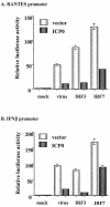
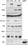
Virus infection induces a rapid cellular response in cells characterized by the induction of interferon. While interferon itself does not induce an antiviral response, it activates a number of interferon-stimulated genes that collectively function to inhibit virus replication and spread. Previously, we and others reported that herpes simplex virus type 1 (HSV-1) induces an interferon -independent antiviral response in the absence of virus replication. Here, we report that the HSV-1 proteins ICP0 and vhs function in concert to disable the host antiviral response. In particular, we show that ICP0 blocks interferon regulatory factor IRF3- and IRF7-mediated activation of interferon-stimulated genes and that the RING finger domain of ICP0 is essential for this activity. Furthermore, we demonstrate that HSV-1 modifies the IRF3 pathway in a manner different from that of the small RNA viruses most commonly studied.
Figures


Similar articles
-
Novel roles of cytoplasmic ICP0: proteasome-independent functions of the RING finger are required to block interferon-stimulated gene production but not to promote viral replication.J Virol. 2014 Jul;88(14):8091-101. doi: 10.1128/JVI.00944-14. Epub 2014 May 7. J Virol. 2014. PMID: 24807717 Free PMC article.
-
Expression of herpes simplex virus ICP0 inhibits the induction of interferon-stimulated genes by viral infection.J Virol. 2002 Mar;76(5):2180-91. doi: 10.1128/jvi.76.5.2180-2191.2002. J Virol. 2002. PMID: 11836395 Free PMC article.
-
Herpes simplex virus type 1 regulatory protein ICP0 aids infection in cells with a preinduced interferon response but does not impede interferon-induced gene induction.J Virol. 2009 May;83(10):4978-83. doi: 10.1128/JVI.02595-08. Epub 2009 Mar 4. J Virol. 2009. PMID: 19264774 Free PMC article.
-
The HSV-1 ubiquitin ligase ICP0: Modifying the cellular proteome to promote infection.Virus Res. 2020 Aug;285:198015. doi: 10.1016/j.virusres.2020.198015. Epub 2020 May 13. Virus Res. 2020. PMID: 32416261 Free PMC article. Review.
-
ICP0, a regulator of herpes simplex virus during lytic and latent infection.Bioessays. 2000 Aug;22(8):761-70. doi: 10.1002/1521-1878(200008)22:8<761::AID-BIES10>3.0.CO;2-A. Bioessays. 2000. PMID: 10918307 Review.
Cited by
-
Models of Herpes Simplex Virus Latency.Viruses. 2024 May 8;16(5):747. doi: 10.3390/v16050747. Viruses. 2024. PMID: 38793628 Free PMC article. Review.
-
Herpes simplex virus infected cell protein 8 is required for viral inhibition of the cGAS pathway.Virology. 2023 Aug;585:34-41. doi: 10.1016/j.virol.2023.05.002. Epub 2023 May 25. Virology. 2023. PMID: 37271042 Free PMC article.
-
Recent advances in ZBP1-derived PANoptosis against viral infections.Front Immunol. 2023 May 16;14:1148727. doi: 10.3389/fimmu.2023.1148727. eCollection 2023. Front Immunol. 2023. PMID: 37261341 Free PMC article. Review.
-
A Novel Recognition by the E3 Ubiquitin Ligase of HSV-1 ICP0 Enhances the Degradation of PML Isoform I to Prevent ND10 Reformation in Late Infection.Viruses. 2023 Apr 27;15(5):1070. doi: 10.3390/v15051070. Viruses. 2023. PMID: 37243155 Free PMC article.
-
Inborn Errors of Immunity Predisposing to Herpes Simplex Virus Infections of the Central Nervous System.Pathogens. 2023 Feb 13;12(2):310. doi: 10.3390/pathogens12020310. Pathogens. 2023. PMID: 36839582 Free PMC article. Review.
References
-
- Bandyopadhyay, S. K., G. T. Leonaard, T. Bandyopadhyay, G. R. Stark, and G. C. Sen. 1995. Transcriptional induction by double-stranded RNA is mediated by interferon-stimulated response elements without activation of interferon-stimulated gene factor 3. J. Biol. Chem. 270:19624-19629. - PubMed
-
- Barlow, P. N., B. Luisi, A. Milner, M. Elliott, and R. Everett. 1994. Structure of the C3HC4 domain by 1H-nuclear magnetic resonance spectroscopy. A new structural class of zinc-finger. J. Mol. Biol. 237:201-211. - PubMed
Publication types
MeSH terms
Substances
LinkOut - more resources
Full Text Sources
Molecular Biology Databases

