Herpes simplex virus 1 interaction with Toll-like receptor 2 contributes to lethal encephalitis
- PMID: 14739339
- PMCID: PMC337050
- DOI: 10.1073/pnas.0308057100
Herpes simplex virus 1 interaction with Toll-like receptor 2 contributes to lethal encephalitis
Abstract
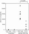
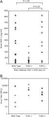
Human neonates infected with herpes simplex virus 1 (HSV-1) develop one of three distinct patterns of infection: (i) infection limited to the skin, eye or mouth; (ii) infection of the CNS; or (iii) disseminated infection. The disseminated form usually involves the liver, adrenal gland, and lung, and resembles the clinical picture of bacterial sepsis. This spectrum of symptoms in HSV-1-infected neonates suggests that inflammatory cytokines play a significant role in the pathogenesis of the disease. Recent studies suggest that the Toll-like receptors (TLRs) may play an important role in the induction of inflammatory cytokines in response to viruses. TLRs are mammalian homologues of Toll, a Drosophila protein that is essential for host defense against infection. Engagement of TLRs by bacterial, viral, or fungal components leads to the production and release of cytokines and other antimicrobial products. Here, we demonstrate that TLR2 mediates the inflammatory cytokine response to HSV-1 by using both transfected cell lines and knockout mice. Studies of infected mice revealed that HSV-1 induced a blunted cytokine response in TLR2(-/-) mice. Brain levels of monocyte chemoattractant protein 1 chemokine were significantly lower in TLR2(-/-) mice than in either wild-type or TLR4(-/-) mice. TLR2(-/-) mice had reduced mortality compared with wild-type mice. The differences between TLR2(-/-) mice and both wild-type and TLR4(-/-) mice in the induction of monocyte chemoattractant protein 1, brain inflammation, or mortality could not be accounted for on the basis of virus levels. Thus, these studies suggest the TLR2-mediated cytokine response to HSV-1 is detrimental to the host.
Figures


Similar articles
-
The role of toll-like receptors in herpes simplex infection in neonates.J Infect Dis. 2005 Mar 1;191(5):746-8. doi: 10.1086/427339. Epub 2005 Jan 18. J Infect Dis. 2005. PMID: 15688290
-
Toll-like receptor (TLR) 2 and TLR9 expressed in trigeminal ganglia are critical to viral control during herpes simplex virus 1 infection.Am J Pathol. 2010 Nov;177(5):2433-45. doi: 10.2353/ajpath.2010.100121. Epub 2010 Sep 23. Am J Pathol. 2010. PMID: 20864677 Free PMC article.
-
Lethal encephalitis in myeloid differentiation factor 88-deficient mice infected with herpes simplex virus 1.Am J Pathol. 2005 May;166(5):1419-26. doi: 10.1016/S0002-9440(10)62359-0. Am J Pathol. 2005. PMID: 15855642 Free PMC article.
-
Impact of Toll-Like Receptors (TLRs) and TLR Signaling Proteins in Trigeminal Ganglia Impairing Herpes Simplex Virus 1 (HSV-1) Progression to Encephalitis: Insights from Mouse Models.Front Biosci (Landmark Ed). 2024 Mar 14;29(3):102. doi: 10.31083/j.fbl2903102. Front Biosci (Landmark Ed). 2024. PMID: 38538263 Review.
-
Viruses and Toll-like receptors.Microbes Infect. 2004 Dec;6(15):1356-60. doi: 10.1016/j.micinf.2004.08.013. Microbes Infect. 2004. PMID: 15596120 Review.
Cited by
-
Viral miRNA regulation of host gene expression.Semin Cell Dev Biol. 2023 Sep 15;146:2-19. doi: 10.1016/j.semcdb.2022.11.007. Epub 2022 Nov 30. Semin Cell Dev Biol. 2023. PMID: 36463091 Free PMC article. Review.
-
The immunologic basis for severe neonatal herpes disease and potential strategies for therapeutic intervention.Clin Dev Immunol. 2013;2013:369172. doi: 10.1155/2013/369172. Epub 2013 Mar 31. Clin Dev Immunol. 2013. PMID: 23606868 Free PMC article. Review.
-
Toll-like receptor sensing of human herpesvirus infection.Front Cell Infect Microbiol. 2012 Oct 8;2:122. doi: 10.3389/fcimb.2012.00122. eCollection 2012. Front Cell Infect Microbiol. 2012. PMID: 23061052 Free PMC article. Review.
-
Viral encephalitis.J Neurol. 2005 Mar;252(3):268-72. doi: 10.1007/s00415-005-0770-7. Epub 2005 Mar 11. J Neurol. 2005. PMID: 15761675 Review.
-
Scratching the Surface Takes a Toll: Immune Recognition of Viral Proteins by Surface Toll-like Receptors.Viruses. 2022 Dec 24;15(1):52. doi: 10.3390/v15010052. Viruses. 2022. PMID: 36680092 Free PMC article. Review.
References
-
- Corey, L. (2001) in Harrison's Principles of Internal Medicine, eds. Braunwald, E., Fauci, A. S., Kasper, D. L., Hauser, S. L., Longo, D. L. & Jameson, J. L. (McGraw–Hill, New York), pp. 1100–1106.
-
- Whitley, R. J. (2001) in Fields' Virology, eds. Knipe, D. M., Howley, P. M., Griffin, D. E., Lamb, R. A., Martin, M. A., Roizman, B. & Straus, S. E. (Lippincott, Williams & Wilkins, Philadelphia), 4th. Ed., pp. 2461–2509.
-
- Whitley, R., Arvin, A., Prober, C., Corey, L., Burchett, S., Plotkin, S., Starr, S., Jacobs, R., Powell, D., Nahmias, A., et al. (1991) N. Engl. J. Med. 324, 450–454. - PubMed
-
- Takeda, K., Kaisho, T. & Akira, S. (2003) Annu. Rev. Immunol. 21, 335–376. - PubMed
-
- Imler, J. L. & Zheng, L. (July 15, 2003) J. Leukocyte Biol., 10.1189/jlb.0403160.
Publication types
MeSH terms
Substances
Grants and funding
LinkOut - more resources
Full Text Sources
Other Literature Sources
Medical
Molecular Biology Databases
Research Materials

