. 2004 Jan;2(1):E21.
doi: 10.1371/journal.pbio.0020021. Epub 2004 Jan 20.

Role of Saccharomyces single-stranded DNA-binding protein RPA in the strand invasion step of double-strand break repair

Affiliations

Role of Saccharomyces single-stranded DNA-binding protein RPA in the strand invasion step of double-strand break repair

Xuan Wang et al. PLoS Biol. 2004 Jan.

Abstract

The single-stranded DNA (ssDNA)-binding protein replication protein A (RPA) is essential for both DNA replication and recombination. Chromatin immunoprecipitation techniques were used to visualize the kinetics and extent of RPA binding following induction of a double-strand break (DSB) and during its repair by homologous recombination in yeast. RPA assembles at the HO endonuclease-cut MAT locus simultaneously with the appearance of the DSB, and binding spreads away from the DSB as 5' to 3' exonuclease activity creates more ssDNA. RPA binding precedes binding of the Rad51 recombination protein. The extent of RPA binding is greater when Rad51 is absent, supporting the idea that Rad51 displaces RPA from ssDNA. RPA plays an important role during RAD51-mediated strand invasion of the MAT ssDNA into the donor sequence HML. The replication-proficient but recombination-defective rfa1-t11 (K45E) mutation in the large subunit of RPA is normal in facilitating Rad51 filament formation on ssDNA, but is unable to achieve synapsis between MAT and HML. Thus, RPA appears to play a role in strand invasion as well as in facilitating Rad51 binding to ssDNA, possibly by stabilizing the displaced ssDNA.


Conflict of interest statement

The authors have declared that no conflicts of interest exist.

Figures
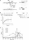
Figure 1
Figure 1. Recruitment of RPA to a DSB in the Absence of DNA Repair
A strain deleted for donors (yXW1), thus incapable of repairing a DSB by gene conversion, was pregrown in YP–lactate medium, and 2% galactose was added to the culture to induce a DSB at MAT. DNA was extracted at intervals after HO cutting, to which polyclonal antibody against Rfa1 was applied to immunoprecipitate RPA-bound chromatin. Another set of DNA samples were taken at the same time for Southern blot analysis. (A) Map of MAT showing the locations of the HO-cut site as well as the StyI restriction sites and the primers (P1 and P2), 189 bp to 483 bp distal to the DSB, used to PCR-amplify RPA-associated MAT DNA from the immunoprecipitated extract. Purified genomic DNA was digested with StyI, separated on a 1.4% native gel, and probed with a 32P-labeled MAT distal fragment to monitor the appearance of the HO-cut fragment (see Materials and Methods). The 1-h timepoint represents 1 h after galactose induction of the HO endonuclease. (B) PCR-amplified RPA-bound MAT DNA in a wild-type strain (yXW1). As controls, primers to an independent locus, ARG5,6 (see Materials and Methods), were used to amplify DNA from the immunoprecipitated chromatin. PCR samples were run on ethidium bromide-stained gels (reverse images are shown). Quantitated signals were graphed for the wild-type strain. IP represents ratio of the MAT IP signal to ARG5,6 IP signal. Error bars show one standard deviation. (C) RPA-bound chromatin was PCR-amplified from sites located proximal and distal to the DSB and then quantitated and graphed as described in (B). The DSB is shown at 0 bp. (D) Effect of formaldehyde cross-linking on RPA binding to ssDNA. In both the noncross-linked samples and the cross-linked samples, 4 ng of single-stranded heterologous β-lactamase (AMP) gene DNA was added during the extract preparation step of ChIP. The amount of input genomic and heterologous DNA was measured by PCR primers specific to the ARG5,6 locus and to the AMP sequence, respectively. RPA-associated ARG5,6 and AMP DNA were analyzed from the IP samples. PCR samples were run on ethidium bromide-stained gels (reverse images are shown).
Figure 2
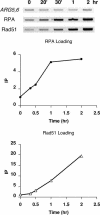
Figure 2. Timing of Recruitment of RPA versus Rad51 to the DSB
An unrepairable DSB was created in the wild-type strain (yXW1), and closer timepoints were harvested at 20 min and 30 min after the HO cut. DNA samples extracted at each timepoint were split. One half was applied with antibody against Rfa1 to immunoprecipitate RPA-associated DNA, while the other half was applied with anti-Rad51 antibody to immunoprecipitate Rad51-bound chromatin. RPA- or Rad51-associated MAT DNA was PCR-amplified and run on ethidium bromide-stained gels (reverse images are shown). DNA signals were quantitated and graphed as described in Figure 1 for RPA ChIP. PCR-amplified ARG5,6 signals from the input DNA were used as controls for quantitation and graphing for Rad51 ChIP (see Materials and Methods).
Figure 4
Figure 4. Localization of RPA and Rad51 to HML and MAT during DSB-Induced Gene Conversion
A strain carrying an HMLα donor (yXW2), thus able to repair the DSB at MAT by gene conversion, was treated with 2% galactose to induce HO endonuclease and then with 2% glucose after 1 h to repress further HO expression. DNA extracted at intervals after HO cutting was split. One half was applied with antibody against Rfa1 to immunoprecipitate RPA-associated DNA, while the other half was applied with anti-Rad51 antibody to immunoprecipitate Rad51-bound chromatin. Another set of DNA samples were taken at the same time for Southern blot analysis. (A) Diagram of MAT and HML showing the locations of the primers, 189 bp to 483 bp distal to the DSB at MAT (P1 and P2) and 189 bp to 467 bp from the uncleaved HO recognition site at HML (P1 and P3), used to PCR-amplify RPA- and Rad51-associated MAT and HML DNA from the immunoprecipitated extract. (B) Purified genomic DNA was digested with StyI, separated on a 1.4% native gel, and probed with a 32P-labeled MAT distal fragment to monitor the appearance of the HO-cut fragment and the repaired product Yα (see Figure 1A; see Materials and Methods). The arrowhead indicates the switched product Yα. RPA- and Rad51-bound MAT and HML DNA were PCR-amplified with primers P1 and P2 and with P1 and P3, respectively. Samples were run on ethidium bromide-stained gels. (C and D) Reverse images are shown for RPA ChIP (C) and Rad51 ChIP (D). DNA signals were quantitated and graphed as described in Figure 2. Error bars show one standard deviation.
Figure 3
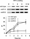
Figure 3. Effect of rad51Δ and rad52Δ on the Extent of RPA Binding to an Unrepairable DSB
An unrepairable DSB was created in wild-type (yXW1), rad51Δ (ySL306), and rad52Δ (ySL177) strains and RPA-bound chromatin was immunoprecipitated using anti-Rfa1 antibody. PCR-amplified DNA from the MAT locus was run on ethidium bromide-stained gels (reverse images are shown). DNA signals were quantitated and graphed as described in Figure 1. Error bars show one standard deviation.
Figure 6
Figure 6. rfa1-t11 Was Not Able to Associate with the Donor Sequence during Gene Conversion
The wild-type strain carrying the HMLα donor (yXW2) and an isogenic strain carrying the rfa1-t11 mutation (yXW3) were treated with 2% galactose to induce HO endonuclease and then with 2% glucose after 1 h to repress further HO expression. DNA extracted at intervals after HO cutting was split. One half was applied with antibody against Rfa1 to immunoprecipitate RPA-associated DNA, while the other half was applied with anti-Rad51 antibody to immunoprecipitate Rad51-bound chromatin. Another set of DNA samples was taken at the same time for Southern blot analysis. (A) Purified genomic DNA was digested with StyI, separated on a 1.4% native gel, and probed with a 32P-labeled MAT distal fragment to monitor the appearance of the HO-cut fragment and the repaired product Yα (see Figure 1A; see Materials and Methods). Arrowheads indicate the switched product Yα. (B) RPA-bound MAT and HML DNA was PCR-amplified with primers P1 and P2 and with P1 and P3, respectively (see Figure 4A). Samples were run on ethidium bromide-stained gels (reverse images are shown). DNA signals were quantitated and graphed as described in Figure 1.
Figure 5
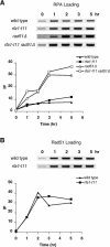
Figure 5. rfa1-t11 Mutation Does Not Affect the Recruitment of Itself or Rad51 to an Unrepairable DSB
(A) An unrepairable DSB was created in wild-type (yXW1), rfa1-t11 (ySL31), rad51Δ (ySL306), and rfa1-t11 rad51Δ (ySL351) strains, and half of the DNA sample was immunoprecipitated with anti-Rfa1 antibody to extract rfa1-t11-bound chromatin. (B) For wild-type (yXW1) and rfa1-t11 (ySL31) strains, the other half of the DNA sample was applied with anti-Rad51 antibody to extract Rad51-associated chromatin. PCR-amplified DNA from the MAT locus was run on ethidium bromide-stained gels (reverse images are shown). DNA signals were quantitated and graphed as described in Figure 2.
Figure 7
Figure 7. rfa1-t11 Mutants Are Defective in the Strand Invasion Step of Gene Conversion
(A) One half of the DNA extract collected from a typical timecourse experiment as described in Figure 6 was applied with anti-Rad51 antibody to immunoprecipitate Rad51-bound chromatin. Primers P1 and P2 and P1 and P3 were used to PCR-amplify Rad51-bound MAT and HML DNA, respectively (see Figure 4A). Samples were run on ethidium bromide-stained gels (reverse images are shown). DNA signals were quantitated and graphed as described in Figure 2. (B) Input DNA was used to PCR-amplify strand invasion product using a unique primer distal to MAT (pB) and a primer within the Yα sequence from HML (pA) (White and Haber 1990). PCR-amplified ARG5,6 signals from the input DNA were used as loading control.

Similar articles

Cited by

References

    1. Alani E, Thresher R, Griffith JD, Kolodner RD. Characterization of DNA-binding and strand-exchange stimulation properties of y-RPA, a yeast single-strand-DNA-binding protein. J Mol Biol. 1992;227:54–71. - PubMed
    1. Aparicio OM, Weinstein DM, Bell SP. Components and dynamics of DNA replication complexes in S. cerevisiae: Redistribution of MCM proteins and Cdc45p during S phase. Cell. 1997;91:59–69. - PubMed
    1. Bianco PR, Tracy RB, Kowalczykowski SC. DNA strand exchange proteins: A biochemical and physical comparison. Front Biosci. 1998;3:D570–D603. - PubMed
    1. Brill SJ, Stillman B. Replication factor-A from Saccharomyces cerevisiae is encoded by three essential genes coordinately expressed at S phase. Genes Dev. 1991;5:1589–1600. - PubMed
    1. Dedon PC, Soults JA, Allis CD, Gorovsky MA. A simplified formaldehyde fixation and immunoprecipitation technique for studying protein–DNA interactions. Anal Biochem. 1991;197:83–90. - PubMed

Publication types