. 2004 Feb;134(2):625-39.
doi: 10.1104/pp.103.030635. Epub 2004 Jan 15.

Identification of the protein storage vacuole and protein targeting to the vacuole in leaf cells of three plant species

Affiliations

Identification of the protein storage vacuole and protein targeting to the vacuole in leaf cells of three plant species

Misoon Park et al. Plant Physiol. 2004 Feb.

Abstract

Protein storage vacuoles (PSVs) are specialized vacuoles devoted to the accumulation of large amounts of protein in the storage tissues of plants. In this study, we investigated the presence of the storage vacuole and protein trafficking to the compartment in cells of tobacco (Nicotiana tabacum), common bean (Phaseolus vulgaris), and Arabidopsis leaf tissue. When we expressed phaseolin, the major storage protein of common bean, or an epitope-tagged version of alpha-tonoplast intrinsic protein (alpha-TIP, a tonoplast aquaporin of PSV), in protoplasts derived from leaf tissues, these proteins were targeted to a compartment ranging in size from 2 to 5 microm in all three plant species. Most Arabidopsis leaf cells have one of these organelles. In contrast, from one to five these organelles occurred in bean and tobacco leaf cells. Also, endogenous alpha-TIP is localized in a similar compartment in untransformed leaf cells of common bean and is colocalized with transiently expressed epitope-tagged alpha-TIP. In Arabidopsis, phaseolin contained N-glycans modified by Golgi enzymes and its traffic was sensitive to brefeldin A. However, trafficking of alpha-TIP was insensitive to brefeldin A treatment and was not affected by the dominant-negative mutant of AtRab1. In addition, a modified alpha-TIP with an insertion of an N-glycosylation site has the endoplasmic reticulum-type glycans. Finally, the early step of phaseolin traffic, from the endoplasmic reticulum to the Golgi complex, required the activity of the small GTPase Sar1p, a key component of coat protein complex II-coated vesicles, independent of the presence of the vacuolar sorting signal in phaseolin. Based on these results, we propose that the proteins we analyzed are targeted to the PSV or equivalent organelle in leaf cells and that proteins can be transported to the PSV by two different pathways, the Golgi-dependent and Golgi-independent pathways, depending on the individual cargo proteins.


Figures
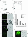
Figure 1.
Figure 1.
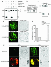
Expression and localization of phaseolin in leaf protoplasts of three plant species. A, Western-blot analysis of phaseolin in Arabidopsis. Protein extracts were prepared from protoplasts transformed with phaseolin (Pha) and untransformed protoplasts (Non) 24 h after transformation and were analyzed by western-blot analysis using an antiphaseolin antibody. Arrow and arrowheads indicate unprocessed and processed forms of phaseolin, respectively. B. Secretion of phaseolin Δ418. Protoplasts were transformed with phaseolin or phaseolin Δ418. At 24 to 36 h after transformation, protein extracts were prepared from the protoplasts (Cell) and medium (Med). The presence of phaseolin proteins in these protein extracts was detected by western-blot analysis using antiphaseolin antibody. C, Localization of phaseolin in protoplasts of Arabidopsis, tobacco, and bean. Protoplasts derived from leaf tissues of Arabidopsis (At), tobacco (Tb), and bean (Be) were transformed with the phaseolin construct and were fixed 24 to 48 h after transformation. The fixed cells were stained with antiphaseolin antibody followed by fluorescein isothiocyanate (FITC)-labeled anti-rabbit immunoglobulin (Ig) G antibody. As a control for immunostaining, the Arabidopsis protoplasts were treated exactly the same way except that the primary antibody was omitted in g and h. Bar = 20 μm. D, Quantification of the number of discs in a protoplast. The number of the disc as shown in C (a, c, and e) was counted from more than 50 immunostained protoplasts for each plant species. Three identical experiments were performed. E, Localization of phaseolin Δ418. Fixed Arabidopsis protoplasts transformed with phaseolin Δ418 was stained with antiphaseolin antibody followed by FITC-labeled anti-rabbit IgG antibody. Bar = 20 μm. At, Arabidopsis.
Figure 2.
Figure 2.
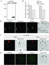
Golgi-dependent trafficking of phaseolin in Arabidopsis protoplasts. A, Western-blot analysis of phaseolin under various conditions. Arabidopsis protoplasts were transformed with phaseolin alone or together with AtSar1[H74L]. To inhibit glycosylation, tunicamycin (10 μg mL–1) was added to the incubation medium right after transformation and the protoplasts were incubated for 24 h. Protein extracts were prepared from the protoplasts and were treated with endo H. The migration patterns of phaseolin in these protein extracts were analyzed by western-blot analysis using an anti-phaseolin antibody. Bands I and II indicate glycosylated and unglycosylated phaseolin, respectively. Arrowheads indicate fragmented forms of phaseolin. B, Effect of AtSar1[H74L] on the secretion of phaseolin Δ418. Protoplasts were transformed with phaseolin Δ418 alone or together with AtSar1[H74L]. Protein extracts were prepared from the protoplasts (Cell) and medium (Med) and were analyzed for the presence of phaseolin Δ418 with the antiphaseolin antibody. C, The effect of BFA on the destiny of phaseolin. Protoplasts derived from leaf cells of Arabidopsis were transformed with phaseolin and were incubated in the presence (+BFA) or absence (–BFA) of BFA for 24 h. Protein extracts were prepared and analyzed for phaseolin by western-blot analysis. Arrow and arrowheads indicate intact and three fragmented forms of phaseolin, respectively. D, Localization of phaseolin in the presence of AtSar1[H74L]. Protoplasts were transformed with phaseolin and AtSar1[H74L] or phaseolin Δ418 and AtSar1[H74L] and localization of phaseolin proteins was detected with antiphaseolin antibody in fixed protoplasts. Bar = 20 μm. E, Localization of phaseolin in the presence of BFA. Arabidopsis protoplasts transformed with phaseolin or sialyltransferase (ST):green fluorescent protein (GFP) plus binding protein (BiP):red fluorescent protein (RFP) were incubated in the presence (+BFA) or absence (–BFA) of BFA for 6 to 24 h, and localization of phaseolin was detected by immunohistochemistry using the antiphaseolin antibody followed by FITC-labeled anti-rabbit IgG antibody. ST:GFP and BiP:RFP were detected directly by GFP and RFP signals, respectively, from intact protoplasts. Arrowheads indicate ST:GFP localized at the Golgi apparatus. CH, Chloroplasts. DsRed was used to generated BiP:RFP. Bar = 20 μm. F, Quantification of localization patterns. The number of protoplasts was counted based on the pattern shown in E (a and c) for phaseolin. More than 50 protoplasts were scored at each time and at least three independent experiments were performed. Disc and ER indicate the patterns shown in E (a and c, respectively).
Figure 3.
Figure 3.
Localization of α-TIP tagged with HA. A, Expression of α-TIP:HA. Arabidopsis protoplasts were transformed with α-TIP:HA and protein extracts were prepared from the transformed protoplasts 24 h after transformation. α-TIP:HA was detected by western-blot analysis using a monoclonal anti-HA antibody. Non, Protein extracts obtained from untransformed protoplasts. B, Localization of reporter proteins. Arabidopsis protoplasts were cotransformed with α-TIP:HA plus phaseolin and were fixed with paraformaldehyde 24 to 48 h after transformation. α-TIP:HA was treated with anti-HA antibodies followed by FITC-labeled anti-rat IgG antibody. Phaseolin was detected with antiphaseolin antibody followed by tetramethylrhodamine B isothiocyanate (TRITC)-labeled anti-rabbit IgG antibody. Arrows indicate untransformed cells used as controls. C, The effect of BFA on the localization of α-TIP:HA. Arabidopsis protoplasts transformed with α-TIP:HA were incubated in the presence (+BFA) or absence (–BFA) of BFA and were fixed 48 h after transformation. The localization of α-TIP:HA was detected using anti-HA antibody followed by TRITC-labeled anti-rat IgG antibody. Bar = 20 μm. D, Quantification of localization patterns. The numbers of protoplasts were counted based on the pattern of TRITC. Protoplasts (n = 50–100) were scored at each time and at least three independent experiments were performed. Disc indicates the protoplasts with the TRITC pattern shown in B (a). Nondisc indicates protoplasts with the TRITC pattern other than in B (a). The nondisc patterns include the ER pattern (network pattern), punctate staining pattern, and a minor portion of the lytic vacuolar pattern. E, Lack of colocalization of α-TIP:HA with ST:GFP. Protoplasts were transformed with α-TIP:HA plus ST:GFP. Protoplasts were fixed and stained with anti-HA antibody. GFP signals were directly observed on the fixed protoplasts. Bar = 20 μm.
Figure 4.
Figure 4.
α-TIP:gly:HA is transported to the PSV in protoplasts. A, No glycosylation of α-TIP:HA. Protein extracts were prepared from the protoplasts transformed with α-TIP:HA 36 h after transformation. The protein extracts were fractionated by SDS/PAGE without (Con) or with endo H treatment (Endo H). Also, protein extracts were prepared from protoplasts treated with tunicamycin after transformation (Tun). α-TIP:HA was detected with the anti-HA antibody. B, A scheme of α-TIP:gly: HA. The C-terminal region of phaseolin from amino acid positions 226 to 284, which contains an N-glycosylation site (denoted by dots), was inserted in the loop between transmembrane domains 5 and 6. Cyt, Cytosol. C, Localization of α-TIP:gly:HA. Protoplasts transformed with α-TIP:gly:HA were incubated in the presence and absence of BFA for 6 to 24 h and localization of α-TIP:gly:HA was examined by immunohistochemistry using anti-HA antibody. Bar = 20 μm. D, Western-blot analysis of α-TIP:gly:HA. Protoplasts transformed with α-TIP:HA or α-TIP:gly:HA were incubated in the presence and absence of tunicamycin (10 μg mL–1) for 24 h or in the presence and absence of BFA (25 μg mL–1) for 24 h. Protein extracts were prepared from protoplasts and were analyzed by western-blot analysis using anti-HA antibody. Also, protein extract treated with endo H was included in western-blot analysis. Bands I and II indicate glycosylated and unglycosylated forms, respectively, of α-TIP:gly:HA.
Figure 5.
Figure 5.
The dominant-negative mutant of AtRab1 inhibits trafficking of phaseolin but not α-TIP:HA to the PSV. A, Western-blot analysis of transiently expressed proteins. Protoplasts were transformed with the indicated amounts of each construct. Protein extracts were prepared from transformed protoplasts and transiently expressed proteins were detected by western-blot analysis using anti-GFP, antiphaseolin, or anti-HA antibodies. B, Localization of α-TIP and phaseolin. Protoplasts were transformed with the indicated constructs and were stained with anti-HA (α-TIP:HA) or antiphaseolin (phaseolin) antibodies. Bar = 20 μm. C, Quantification of targeting efficiency. Immunostained protoplasts were counted based on localization patterns of α-TIP and phaseolin. The disc pattern shown in a or c was taken to indicate targeting to the PSV. Three independent transformations were performed, and 150 to 200 immunostained cells were counted each time. Error bars indicates standard deviation.
Figure 6.
Figure 6.
Overexpressed T7:AtSec23 inhibits trafficking of phaseolin but not α-TIP:HA to the PSV. A, Western-blot analysis of transiently expressed proteins. Protoplasts were transformed with the indicated amounts of each construct. Protein extracts were prepared from transformed protoplasts and transiently expressed proteins were detected by western-blot analysis using anti-GFP, antiphaseolin, or anti-T7 antibodies. R6, An empty vector used as a control. B, Localization of α-TIP and phaseolin. Protoplasts were transformed with the indicated constructs and were stained with anti-HA (α-TIP:HA) or antiphaseolin (phaseolin) antibodies. Bar = 20 μm. C, Quantification of targeting efficiency. Immunostained protoplasts were counted based on localization patterns of α-TIP and phaseolin. The disc pattern shown in a or c was taken to indicate targeting to the PSV. Three independent transformations were performed, and 200 immunostained cells were counted each time. Error bars indicates standard deviation.
Figure 7.
Figure 7.
PSVs are present in untransformed leaf cells of common bean. A, Schemes of leaf tissues used in experiments. B, Western-blot analysis of endogenous α-TIP. Protein extracts were prepared from seeds (lane 1), cotyledons (lane 2), and leaf tissues (lanes 3–6) and were analyzed by western-blot analysis using anti-α-TIP antibody. The amount of total proteins loaded was 10 and 50 μg for top and bottom panels, respectively. The numbers are the same as in A. C, Localization of endogenous α-TIP. Protoplasts were prepared from the indicated bean tissues and were immunostained with anti-α-TIP antibody [α-TIP (endo)] after fixation. The numbers are the same as in A. Bar = 20 μm.
Figure 8.
Figure 8.
α-TIP is expressed in leaf tissues of Arabidopsis. Total RNA isolated from seeds or leaf tissues was used for RT-PCR analysis and PCR products were analyzed by an agarose gel. As a control, γ-TIP was included in the analysis. The products were sequenced to confirm the identity. The α- and γ-TIP-specific PCR fragments are indicated.
Figure 9.
Figure 9.
Colocalization of endogenous α-TIP with transiently expressed α-TIP:HA. Protoplasts prepared from leaf cells of common bean were transformed with α-TIP:HA and localization of proteins was examined at 24 h after transformation. Endogenous α-TIP [α-TIP (endo)] and transiently expressed α-TIP:HA (α-TIP:HA) were detected with anti-α-TIP and anti-HA antibodies, respectively. Bar = 20 μm.

Similar articles

Cited by

References

    1. Aubert S, Gout E, Bligny R, Marty-Mazars D, Barrieu F, Alabouvette J, Marty F, Douce R (1996) Ultrastructural and biochemical characterization of autophagy in higher plant cells subjected to carbon deprivation: control by the supply of mitochondria with respiratory substrates. J Cell Biol 133: 1251–1263 - PMC - PubMed
    1. Bagga S, Adams H, Kemp JD, Sengupta-Gopalan C (1995) Accumulation of the 15-kD zein in novel protein bodies in transgenic tobacco. Plant Physiol 107: 13–23 - PMC - PubMed
    1. Bassham DC, Raikhel NV (2000) Unique features of the plant vacuolar sorting machinery. Curr Opin Cell Biol 12: 491–495 - PubMed
    1. Batoko H, Zheng HQ, Hawes C, Moore I (2000) A rab1 GTPase is required for transport between the endoplasmic reticulum and Golgi apparatus and for normal Golgi movement in plants. Plant Cell 12: 2201–2218 - PMC - PubMed
    1. Boevink P, Martin B, Oparka K, Santa Cruz S, Hawes C (1999) Transport of virally expressed green fluorescent protein through the secretory pathway in tobacco leaves is inhibited by cold shock and brefeldin A. Planta 208: 392–400

Publication types

MeSH terms

LinkOut - more resources