. 2004 Jan 19;164(2):291-300.
doi: 10.1083/jcb.200307151. Epub 2004 Jan 12.

GADD34-PP1c recruited by Smad7 dephosphorylates TGFbeta type I receptor

Affiliations

GADD34-PP1c recruited by Smad7 dephosphorylates TGFbeta type I receptor

Weibin Shi et al. J Cell Biol. .

Abstract

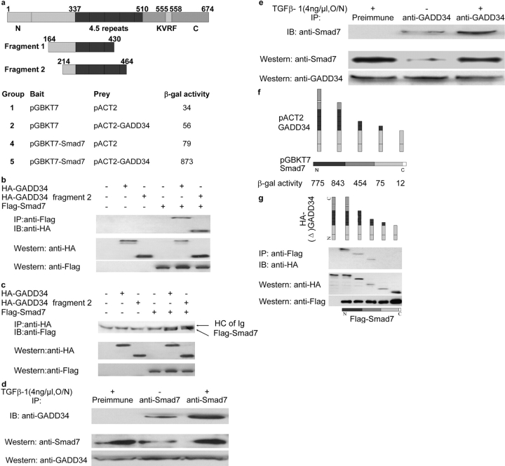
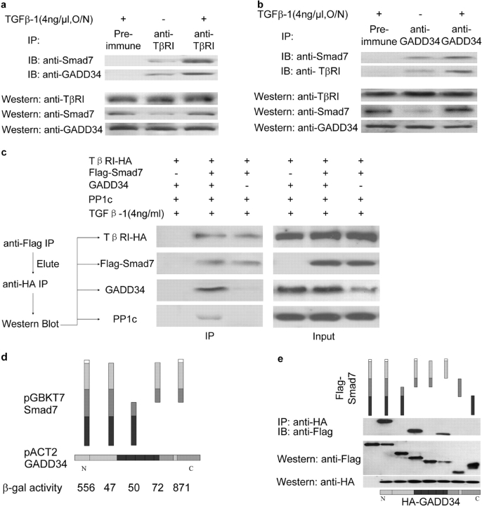
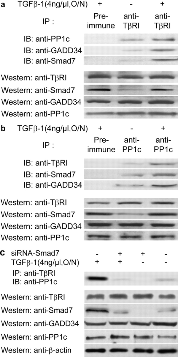
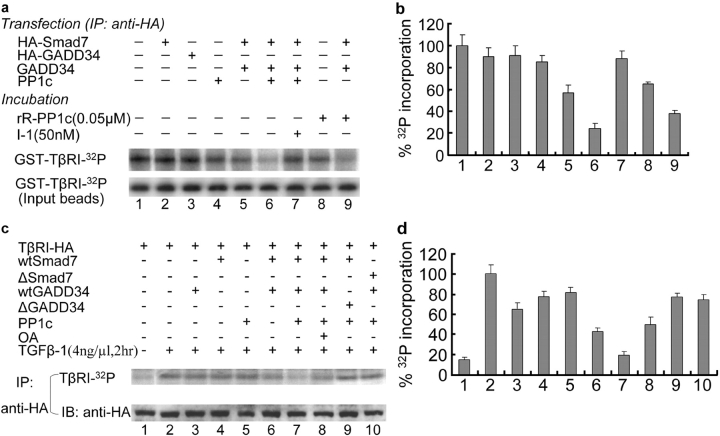
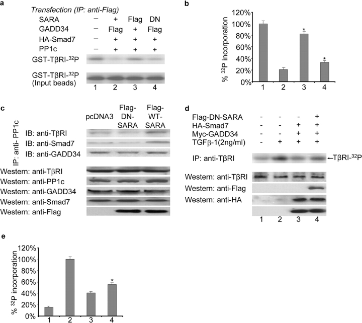
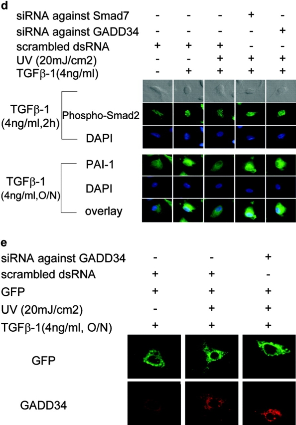
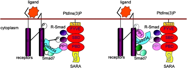
The cascade of phosphorylation is a pivotal event in transforming growth factor beta (TGFbeta) signaling. Reversible phosphorylation regulates fundamental aspects of cell activity. TGFbeta-induced Smad7 binds to type I receptor (TGFbeta type I receptor; TbetaRI) functioning as a receptor kinase antagonist. We found Smad7 interacts with growth arrest and DNA damage protein, GADD34, a regulatory subunit of the protein phosphatase 1 (PP1) holoenzyme, which subsequently recruits catalytic subunit of PP1 (PP1c) to dephosphorylate TbetaRI. Blocking Smad7 expression by RNA interference inhibits association of GADD34-PP1c complex with TbetaRI, indicating Smad7 acts as an adaptor protein in the formation of the PP1 holoenzyme that targets TbetaRI for dephosphorylation. SARA (Smad anchor for receptor activation) enhances the recruitment PP1c to the Smad7-GADD34 complex by controlling the specific subcellular localization of PP1c. Importantly, GADD34-PP1c recruited by Smad7 inhibits TGFbeta-induced cell cycle arrest and mediates TGFbeta resistance in responding to UV light irradiation. The dephosphorylation of TbetaRI mediated by Smad7 is an effective mechanism for governing negative feedback in TGFbeta signaling.


Figures

Figure 1.
Figure 1.
GADD34 as a Smad7-interacting protein through its central repeats. (a) GADD34 is shown schematically with conservative regions. Two fragments of GADD34 were isolated in a yeast two-hybrid screen using Smad7 as the bait. (b–e) Smad7 interacts with GADD34 in mammalian cells. (b and c) Immunoblots indicating the protein expression levels of the lysates of COS1 cells transfected with HA–GADD34 and Flag–Smad7 or HA–GADD34 fragment 2 are shown in the two lower panels. The upper panel shows anti-Flag immunoprecipitates probed with anti-HA antibody (b) or anti-HA immunoprecipitates probed with anti-Flag antibody (c). The bands at ∼50 kD close to Smad7 in immunoprecipitate lanes were due to the presence of cross-reacting Ig heavy chain. (d and e) Endogenous GADD34 immunoprecipitated from Mv1Lu cells coprecipitated Smad7 as determined by immunoblotting. Conversely, immunoprecipiated endogenous Smad7 coprecipitated GADD34. (f and g) Similar experiments were performed in both yeast (f) and mammalian (g) systems to map Smad7 binding to GADD34.
Figure 2.
Figure 2.
TGFβ regulates the formation of TβRI–Smad7–GADD34 complexes via Smad7. (a) Endogenous TβRI was immunoprecipitated from Mv1Lu cells with or without TGFβ-1 stimulation and precipitates were examined for the presence of GADD34 and Smad7 by immunoblotting. (b) Conversely, endogenous GADD34 was immunoprecipiated from Mv1Lu cells with or without TGFβ-1 stimulation and precipitates were examined for the presence of TβRI and Smad7. (c and d) Experiments were performed in both yeast (c) and mammalian (d) systems, as in Fig. 1, to map GADD34 binding to Smad7.
Figure 3.
Figure 3.
Smad7 mediates recruitment of PP1c to TβRI. (a and b) TGFβ regulates the interaction between TβRI and PP1c. Endogenous TβRI was immunoprecipitated from Mv1Lu cells with or without TGFβ-1 stimulation and precipitates were detected for the presence of PP1c, GADD34, and Smad7 by immunoblotting (a). Conversely, endogenous PP1 was immunoprecipitated and TβRI, Smad7, and GADD34 detected by immunoblotting (b). (c) Knockdown of Smad7 inhibits the formation of a GADD34–PP1c complex with TβRI. (c) Endogenous Smad7 in 293T cells was knocked down by using siRNA in the presence or absence of TGFβ-1. The cell lysates were immunoprecipitated with anti-TβRI and the presence of PP1c in the precipitate was detected by immunoblotting with anti-PP1.
Figure 4.
Figure 4.
Dephosphorylation of TβRI by Smad7-recruited PP1 complex. (a and b) In vitro dephosphorylation assay. (a) GST–TβRI was phosphorylated by an in vitro phosphorylation reaction. GST–TβRI–32P was incubated with different immunoprecipitates (anti-HA) from lysates of COS1 cells transfected with different combinations of genes, in the absence or presence of phosphatase inhibitor (I-1) or recombinant rabbit PP1c (rR-PP1c) as indicated. (b) The relative 32P phosphorylation level of the type I receptor in a, normalized to input of GST–TβRI–32P, is plotted as the mean ± SD from three experiments. (c and d) In vivo dephosphorylation assay. (c) Mv1Lu cells transfected with different combination of genes were labeled with [32P]orthophosphate in the presence or absence of TGFβ-1 as indicated. TβRI–HA was immunoprecipitated from lysates of treated cells and separated by 8.5% SDS-PAGE. Gels were dried and exposed to Biomax M r film (Eastman Kodak). ΔGADD34 is a mutant without the PP1c binding domain and ΔSmad7 is absent in its TβRI binding site. OA is an inhibitor for both PP1 and PP2. (d) The relative 32P phosphorylation level of TβRI in c was plotted as the mean ± SD from three experiments.
Figure 5.
Figure 5.
The regulation of PP1 intracellular localization and facilitation of TβRI dephosphorylation by SARA. (a and b) SARA–PP1c fails to dephosphorylate TβRI in vitro. (a) Similar in vitro dephosphorylation assay to that in Fig. 4 a was performed. GST–TβRI–32P was incubated with different immunoprecipitates (anti-Flag) from lysates of COS1 cells transfected with different combinations of genes as indicated. In lanes 2 and 4 Flag was tagged to GADD34 and in lane 3 Flag was tagged to SARA. In lane 4 SARA was replaced with DN–SARA (F728A). (b) The relative 32P phosphorylation level of the type I receptor in a, normalized to input of GST–TβRI–32P, is plotted as the mean ± SD from three experiments. *P < 0.05, compared with lane 2. (c) The availability of PP1 to the complex is mediated by SARA. COS1 cells were transfected with either Flag–WT–SARA or Flag–DN–SARA (F728A). The cell lysates were immunoprecipitated with anti-PP1c, and the TβRI, Smad7, and GADD34 in the precipitate were probed by immunoblotting. (d and e) In in vivo dephosphorylation assay, dominant-negative SARA with a mutation in the PP1c-binding domain (F728A) inhibits the dephosphorylation of TβRI by Smad7-recruited PP1 complex. (d) Similar experiments to that in Fig. 4 c were performed with dominant-negative SARA (F678A). (e) The relative 32P phosphorylation level of TβRI in panel d was plotted as the mean ± SD from three experiments. *P < 0.05, compared with lane 3.
Figure 6.
Figure 6.
GADD34–PP1c recruited by Smad7 inhibits TGFβ signaling by dephosphorylating TβRI and its induced cell cycle arrest and it contributes to UV light–induced TGFβ resistance. (a) Mv1Lu cells were transfected with 3TP–lux alone or together with the indicated amounts of expression constructs for different genes. Transfected cells were incubated in the presence (black bars) or absence (open bars) of TGFβ-1. Luciferase activity was normalized and plotted as the mean ± SD of triplicates from a representative experiment. (b) GADD34–PP1c recruited by Smad7 inhibits antiproliferative effect of TGFβ on Mv1Lu cells. Transfected cells with different combinations of expression constructs for different genes were incubated in the presence or absence of TGFβ-1, harvested 2 d after transfection, and then subjected to FACS®–DNA profiling assay. The percentage of cells in G1 phase was plotted as the mean ± SD of triplicates from a representative experiment. (c) Knockdown of Smad7 and GADD34 expression resensitizes TGFβ signaling in UV light–irradiated cells. The day before UV light irradiation, HepG2 cells were transfected with 3TP–lux together with the indicated siRNAs or scrambled dsRNA. 24 h after UV light irradiation, luciferase activity was assayed and normalized and plotted as the mean ± SD of triplicates from a representative experiment. *P < 0.05, compared with lane 2. (d) UV light irradiation inhibits Smad2 nuclear translocation and downstream gene expression, while blocking UV light–enhanced expression of Smad7 or GADD34 rescues it. HepG2 Cells were transfected with siRNA against Smad7 or GADD34 the day before UV light irradiation. The cells were stimulated by TGFβ for 2 h (Smad2 translocation) or 24 h (PAI-1 induction) after UV light irradiation. Immunostaining were then performed to visualize the intracellular translocation of Smad2 and expression of PAI-1. (e) GADD34 induced by UV light irradiation is knocked down by RNAi. GFP expression vector (one tenth the amount of siRNA) was cotransfected into HepG2 cells with siRNA against GADD34. The siRN-transfected cell (last column, showing GFP positive) showed less expression of GADD34, whereas the siRNA-untransfected cell (last column, showing GFP negative, at bottom of the panel) with GADD34 intact compared with siRNA-untransfected UV light–irradiated cell in middle column.
Figure 6.
Figure 6.
GADD34–PP1c recruited by Smad7 inhibits TGFβ signaling by dephosphorylating TβRI and its induced cell cycle arrest and it contributes to UV light–induced TGFβ resistance. (a) Mv1Lu cells were transfected with 3TP–lux alone or together with the indicated amounts of expression constructs for different genes. Transfected cells were incubated in the presence (black bars) or absence (open bars) of TGFβ-1. Luciferase activity was normalized and plotted as the mean ± SD of triplicates from a representative experiment. (b) GADD34–PP1c recruited by Smad7 inhibits antiproliferative effect of TGFβ on Mv1Lu cells. Transfected cells with different combinations of expression constructs for different genes were incubated in the presence or absence of TGFβ-1, harvested 2 d after transfection, and then subjected to FACS®–DNA profiling assay. The percentage of cells in G1 phase was plotted as the mean ± SD of triplicates from a representative experiment. (c) Knockdown of Smad7 and GADD34 expression resensitizes TGFβ signaling in UV light–irradiated cells. The day before UV light irradiation, HepG2 cells were transfected with 3TP–lux together with the indicated siRNAs or scrambled dsRNA. 24 h after UV light irradiation, luciferase activity was assayed and normalized and plotted as the mean ± SD of triplicates from a representative experiment. *P < 0.05, compared with lane 2. (d) UV light irradiation inhibits Smad2 nuclear translocation and downstream gene expression, while blocking UV light–enhanced expression of Smad7 or GADD34 rescues it. HepG2 Cells were transfected with siRNA against Smad7 or GADD34 the day before UV light irradiation. The cells were stimulated by TGFβ for 2 h (Smad2 translocation) or 24 h (PAI-1 induction) after UV light irradiation. Immunostaining were then performed to visualize the intracellular translocation of Smad2 and expression of PAI-1. (e) GADD34 induced by UV light irradiation is knocked down by RNAi. GFP expression vector (one tenth the amount of siRNA) was cotransfected into HepG2 cells with siRNA against GADD34. The siRN-transfected cell (last column, showing GFP positive) showed less expression of GADD34, whereas the siRNA-untransfected cell (last column, showing GFP negative, at bottom of the panel) with GADD34 intact compared with siRNA-untransfected UV light–irradiated cell in middle column.
Figure 7.
Figure 7.
A model for dephosphorylation of TβRI by Smad7-mediated PP1 holoenzyme in TGFβ signaling. See Discussion for details. SBD, Smad-binding domain; PBD, phosphatase-binding domain; CTD, COOH-terminal domain; NTD, NH2-terminal domain.

Similar articles

Cited by

References

    1. Abdollah, S., M. Macias-Silva, T. Tsukazaki, H. Hayashi, L. Attisano, and J.L. Wrana. 1997. TβRI phosphorylation of Smad2 on Ser465 and Ser467 is required for Smad2-Smad4 complex formation and signaling. J. Biol. Chem. 272:27678–27685. - PubMed
    1. Aggen, J.B., A.C. Nairn, and R. Chamberlin. 2000. Regulation of protein phosphatase-1. Chem. Biol. 7:R13–R23. - PubMed
    1. Attisano, L., and J.L. Wrana. 2002. Signal transduction by the TGF-β superfamily. Science. 296:1646–1647. - PubMed
    1. Bennett, D., and L. Alphey. 2002. PP1 binds Sara and negatively regulates Dpp signaling in Drosophila melanogaster. Nat. Genet. 31:419–423. - PubMed
    1. Bitzer, M., G. von Gersdorff, D. Liang, A. Dominguez-Rosales, A.A. Beg, M. Rojkind, and E.P. Bottinger. 2000. A mechanism of suppression of TGF-β/SMAD signaling by NF-kappa B/RelA. Genes Dev. 14:187–197. - PMC - PubMed

Publication types

MeSH terms