. 2004 Jan 9;32(1):287-97.
doi: 10.1093/nar/gkh161. Print 2004.

Characterization of cytosine methylated regions and 5-cytosine DNA methyltransferase (Ehmeth) in the protozoan parasite Entamoeba histolytica

Affiliations

Characterization of cytosine methylated regions and 5-cytosine DNA methyltransferase (Ehmeth) in the protozoan parasite Entamoeba histolytica

Ohad Fisher et al. Nucleic Acids Res. .

Abstract

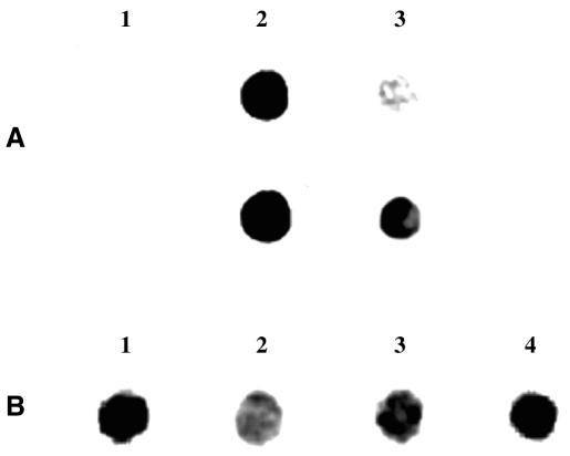
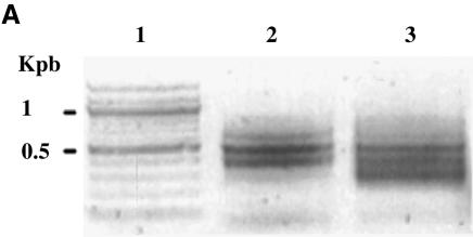
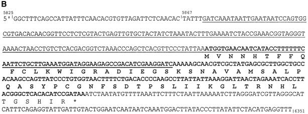
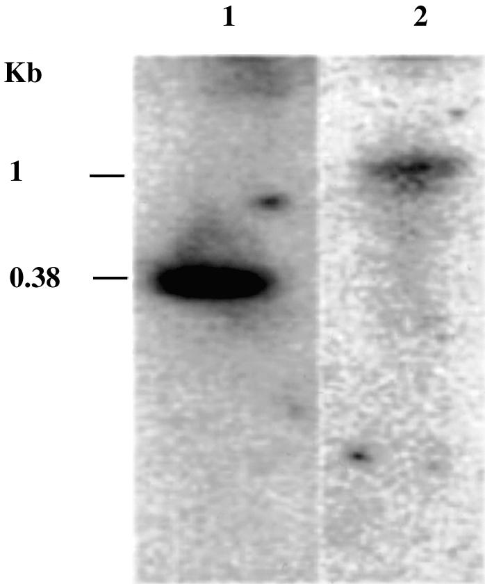
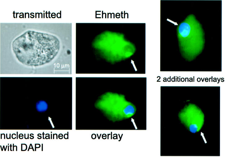
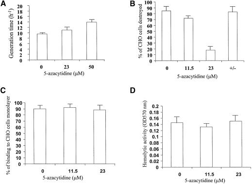
The DNA methylation status of the protozoan parasite Entamoeba histolytica was heretofore unknown. In the present study, we developed a new technique, based on the affinity of methylated DNA to 5-methylcytosine antibodies, to identify methylated DNA in this parasite. Ribosomal DNA and ribosomal DNA circles were isolated by this method and we confirmed the validity of our approach by sodium bisulfite sequencing. We also report the identification and the characterization of a gene, Ehmeth, encoding a DNA methyltransferase strongly homologous to the human DNA methyltransferase 2 (Dnmt2). Immunofluorescence microscopy using an antibody raised against a recombinant Ehmeth showed that Ehmeth is concentrated in the nuclei of trophozoites. The recombinant Ehmeth has a weak but significant methyltransferase activity when E.histolytica genomic DNA is used as substrate. 5-Azacytidine (5-AzaC), an inhibitor of DNA methyltransferase, was used to study in vivo the role of DNA methylation in E.histolytica. Genomic DNA of trophozoites grown with 5-AzaC (23 microM) was undermethylated and the ability of 5-AzaC-treated trophozoites to kill mammalian cells or to cause liver abscess in hamsters was strongly impaired.


Figures

Figure 1
Figure 1
Immunological detection of m5C in E.histolytica genomic DNA. (A) PCR-synthesized DNA (lane 1), PCR-synthesized DNA treated in vitro with M.SssI DNA methyltransferase (lane 2) and E.histolytica genomic DNA (lane 3) were spotted on nitrocellulose paper and incubated with antibodies to m5C. Ehmeth DNA was used as template for the production of PCR-synthesized DNA. The time of exposure for the upper line was 2 ms and for the lower line 12 ms. (B) The specificity of the reaction between m5C antibody and the genomic DNA of E.histolytica (lane 1) was demonstrated by competition experiments involving m5C (10–5 M) (lane 2). 5-AzaC is an inhibitor of 5-cytosine DNA methyltransferase currently used as a potent demethylating agent. Genomic DNA of trophozoites grown with 5-AzaC (23 µM) for 48 h (lane 3) and then cultivated for 2 weeks without 5-AzaC (lane 4) were also spotted on nitrocellulose paper and incubated with antibodies to m5C. The time of exposure was 12 ms. For both experiments (A and B), 0.5 µg of DNA was systematically spotted on the nitrocellulose paper.
Figure 2
Figure 2
Purification and analysis of E.histolytica methylated DNA regions. (A) E.histolytica genomic DNA was restricted with DpnII, ligated to R-Bgl-24 adaptors and amplified with primers R-Bgl-24 before (lane 2) and after (lane 3) m5C antibody affinity chromatography. Lane 1, 100 bp ladder (New England Biolabs). Note the difference of pattern before and after affinity chromatography. (B) Sequence analysis of rDNA region (accession no. X65163). The DpnII fragment isolated by m5C antibody affinity chromatography is underlined. The ORF for the senescence-related protein is represented in bold, and its putative amino acid sequence represented below. (C) Genomic sodium bisulfite analysis sequencing of the rDNA region that includes SRP (accession no. X65163) in trophozoites grown without 5-AzaC (upper chromatogram) or with 5-AzaC for 1 week (lower chromatogram). The methylated C residues are resistant to the bisulfite treatment (underlined). The unmethylated C residues are converted by the bisulfite treatment (designated by a star). The same distribution of methylated cytosine was found following the sequencing of four clones coming from two separate experiments with sodium bisulfite.
Figure 2
Figure 2
Purification and analysis of E.histolytica methylated DNA regions. (A) E.histolytica genomic DNA was restricted with DpnII, ligated to R-Bgl-24 adaptors and amplified with primers R-Bgl-24 before (lane 2) and after (lane 3) m5C antibody affinity chromatography. Lane 1, 100 bp ladder (New England Biolabs). Note the difference of pattern before and after affinity chromatography. (B) Sequence analysis of rDNA region (accession no. X65163). The DpnII fragment isolated by m5C antibody affinity chromatography is underlined. The ORF for the senescence-related protein is represented in bold, and its putative amino acid sequence represented below. (C) Genomic sodium bisulfite analysis sequencing of the rDNA region that includes SRP (accession no. X65163) in trophozoites grown without 5-AzaC (upper chromatogram) or with 5-AzaC for 1 week (lower chromatogram). The methylated C residues are resistant to the bisulfite treatment (underlined). The unmethylated C residues are converted by the bisulfite treatment (designated by a star). The same distribution of methylated cytosine was found following the sequencing of four clones coming from two separate experiments with sodium bisulfite.
Figure 3
Figure 3
Northern blot analysis of Ehmeth. Total RNA (10 µg) was size fractionated on 4% polyacrylamide denaturing gel containing 8 M urea under denaturing conditions and subsequently blotted electrophoretically onto a nylon membrane. Hybridization was carried out by a probe for the small subunit of E.histolytica ribosomal RNA (ssrRNA) (lane 1) or for the Ehmeth ORF (lane 2). The exposure time of X-ray film was 120 h for Ehmeth and 5 h for ssrRNA.
Figure 4
Figure 4
SDS–PAGE and western blot analysis of Ehmeth. (A) GelCode Blue staining of GST-Ehmeth (1 µg; lane 1) and His-Ehmeth (1 µg; lane 2) resolved under reducing conditions on a 10% SDS–polyacrylamide gel. (B) Western blot analysis under reducing conditions of trophozoites total lysate probed with the anti-Ehmeth antibody (1/500). Lane 1, GST-Ehmeth (10 ng); lane 2, His-Ehmeth (10 ng); lane 3, total lysate of trophozoites (25 µg); lane 4, trophozoites total lysate (25 µg) probed with rabbit pre-immune serum (1/500).
Figure 5
Figure 5
Cellular localization of Ehmeth in E.histolytica trophozoites. Ehmeth was detected by immunofluorescence microscopy using anti-Ehmeth antibody. Ehmeth distribution is shown in green using a primary anti-Ehmeth antibody and a secondary antibody conjugated with FITC. Nuclei (blue) were stained by DAPI. Computer-assisted image overlay analysis of the signal given by Ehmeth antibody and by DAPI, shows that Ehmeth is concentrated in the nuclei of trophozoites. Two additional examples showing a nuclear localization of Ehmeth are presented on the right.
Figure 6
Figure 6
Growth, virulence and adherence assays in trophozoites grown with 5-AzaC. (A) Dose–response analysis of 5-AzaC concentration on the growth of E.histolytica. Data represent the mean and standard deviation of three independent experiments. (B) Cytopathic activity measuring the in vitro ability of amoeba grown in the presence of 5-AzaC (23 µM) to destroy a monolayer of CHO cells. The column +/– shows the cytopathic activity of trophozoites grown for 2 days with 5-AzaC (23 µM) and then cultivated without 5-AzaC for 2 weeks. Data represent the mean and standard deviation of three independent experiments done in duplicate. (C) Adherence ability of trophozoites grown in the presence of 5-AzaC (23 µM) to a formaldehyde-fixed monolayer of CHO cells. Data represent the mean and standard deviation of three independent experiments done in duplicate. (D) Hemolytic assay measuring the ability of trophozoites grown in the presence of 5-AzaC (23 µM) to lyse human red blood cells. Data represent the mean and standard deviation of three independent experiments done in duplicate.

Similar articles

Cited by

References

    1. World Health Organization (1997) Amoebiasis. WHO Weekly Epidemiologic Record, 72, 97–100.
    1. Espinosa-Cantellano M. and Martinez-Palomo,A. (2000) Pathogenesis of intestinal amebiasis: from molecules to disease. Clin. Microbiol. Rev., 13, 318–331. - PMC - PubMed
    1. Stanley S.L. (2001) Pathophysiology of amoebiasis. Trends Parasitol., 17, 280–285. - PubMed
    1. Lewin B., Stanley,S.L.,Jr and Reed,S.L. (1998) The mystique of epigenetics. Cell, 93, 301–303. - PubMed
    1. Bestor T.H., Verdine,G.L., Santi,D.V., Norment,A., Garrett,C.E., Gilchrist,C.A., Petri,W.A., Singh,V.K., Moskovitz,J., Wilkinson,B.J. et al. (1994) DNA methyltransferases. Curr. Opin. Cell Biol., 6, 380–389. - PubMed

Publication types

MeSH terms