Regulation of synaptojanin 1 by cyclin-dependent kinase 5 at synapses
- PMID: 14704270
- PMCID: PMC327184
- DOI: 10.1073/pnas.0307813100
Regulation of synaptojanin 1 by cyclin-dependent kinase 5 at synapses
Abstract
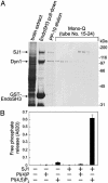
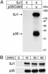
Synaptojanin 1 is a polyphosphoinositide phosphatase concentrated in presynaptic nerve terminals, where it dephosphorylates a pool of phosphatidylinositol 4,5-bisphosphate implicated in synaptic vesicle recycling. Like other proteins with a role in endocytosis, synaptojanin 1 undergoes constitutive phosphorylation in resting synapses and stimulation-dependent dephosphorylation by calcineurin. Here, we show that cyclin-dependent kinase 5 (Cdk5) phosphorylates synaptojanin 1 and regulates its function both in vitro and in intact synaptosomes. Cdk5 phosphorylation inhibited the inositol 5-phosphatase activity of synaptojanin 1, whereas dephosphorylation by calcineurin stimulated such activity. The activity of synaptojanin 1 was also stimulated by its interaction with endophilin 1, its major binding partner at the synapse. Notably, Cdk5 phosphorylated serine 1144, which is adjacent to the endophilin binding site. Mutation of serine 1144 to aspartic acid to mimic phosphorylation by Cdk5 inhibited the interaction of synaptojanin 1 with endophilin 1. These results suggest that Cdk5 and calcineurin may have an antagonistic role in the regulation of synaptojanin 1 recruitment and activity, and therefore in the regulation of phosphatidylinositol 4,5-bisphosphate turnover at synapses.
Figures


Comment in
-
Protein kinases talk to lipid phosphatases at the synapse.Proc Natl Acad Sci U S A. 2004 Feb 3;101(5):1112-3. doi: 10.1073/pnas.0308374101. Epub 2004 Jan 26. Proc Natl Acad Sci U S A. 2004. PMID: 14745021 Free PMC article. No abstract available.
Similar articles
-
Phosphorylation of Synaptojanin Differentially Regulates Endocytosis of Functionally Distinct Synaptic Vesicle Pools.J Neurosci. 2016 Aug 24;36(34):8882-94. doi: 10.1523/JNEUROSCI.1470-16.2016. J Neurosci. 2016. PMID: 27559170 Free PMC article.
-
Protein kinases talk to lipid phosphatases at the synapse.Proc Natl Acad Sci U S A. 2004 Feb 3;101(5):1112-3. doi: 10.1073/pnas.0308374101. Epub 2004 Jan 26. Proc Natl Acad Sci U S A. 2004. PMID: 14745021 Free PMC article. No abstract available.
-
Amphiphysin I is associated with coated endocytic intermediates and undergoes stimulation-dependent dephosphorylation in nerve terminals.J Biol Chem. 1997 Dec 5;272(49):30984-92. doi: 10.1074/jbc.272.49.30984. J Biol Chem. 1997. PMID: 9388246
-
Endophilin and synaptojanin hook up to promote synaptic vesicle endocytosis.Neuron. 2003 Nov 13;40(4):665-7. doi: 10.1016/s0896-6273(03)00726-8. Neuron. 2003. PMID: 14622570 Review.
-
Cdk5: a new player at synapses.Neurosignals. 2003 Sep-Oct;12(4-5):180-90. doi: 10.1159/000074619. Neurosignals. 2003. PMID: 14673204 Review.
Cited by
-
A threonine-based targeting signal in the human CD1d cytoplasmic tail controls its functional expression.J Immunol. 2010 May 1;184(9):4973-81. doi: 10.4049/jimmunol.0901448. Epub 2010 Apr 5. J Immunol. 2010. PMID: 20368272 Free PMC article.
-
Endophilin isoforms have distinct characteristics in interactions with N-type Ca2+ channels and dynamin I.Neurosci Bull. 2012 Oct;28(5):483-92. doi: 10.1007/s12264-012-1257-z. Epub 2012 Jul 13. Neurosci Bull. 2012. PMID: 22961472 Free PMC article.
-
Role of phosphoinositides at the neuronal synapse.Subcell Biochem. 2012;59:131-75. doi: 10.1007/978-94-007-3015-1_5. Subcell Biochem. 2012. PMID: 22374090 Free PMC article. Review.
-
Features of the Phosphatidylinositol Cycle and its Role in Signal Transduction.J Membr Biol. 2017 Aug;250(4):353-366. doi: 10.1007/s00232-016-9909-y. Epub 2016 Jun 8. J Membr Biol. 2017. PMID: 27278236 Review.
-
The phosphoinositide phosphatase Sjl2 is recruited to cortical actin patches in the control of vesicle formation and fission during endocytosis.Mol Cell Biol. 2005 Apr;25(8):2910-23. doi: 10.1128/MCB.25.8.2910-2923.2005. Mol Cell Biol. 2005. PMID: 15798181 Free PMC article.
References
-
- De Camilli, P., Emr, S. D., McPherson, P. S. & Novick, P. (1996) Science 271, 1533–1539. - PubMed
-
- Martin, T. F. J. (1998) Annu. Rev. Cell Dev. Biol. 14, 231–264. - PubMed
-
- Takenawa, T. & Itoh, T. (2001) Biochim. Biophys. Acta 1533, 190–206. - PubMed
-
- Yin, H. L. & Janmey, P. A. (2003) Annu. Rev. Physiol. 65, 761–789. - PubMed
Publication types
MeSH terms
Substances
Grants and funding
LinkOut - more resources
Full Text Sources
Other Literature Sources
Molecular Biology Databases

