. 2004 Jan 5;164(1):25-33.
doi: 10.1083/jcb.200306104. Epub 2003 Dec 29.

Stress-induced transcription of satellite III repeats

Affiliations

Stress-induced transcription of satellite III repeats

Caroline Jolly et al. J Cell Biol. .

Abstract

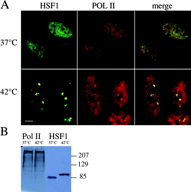
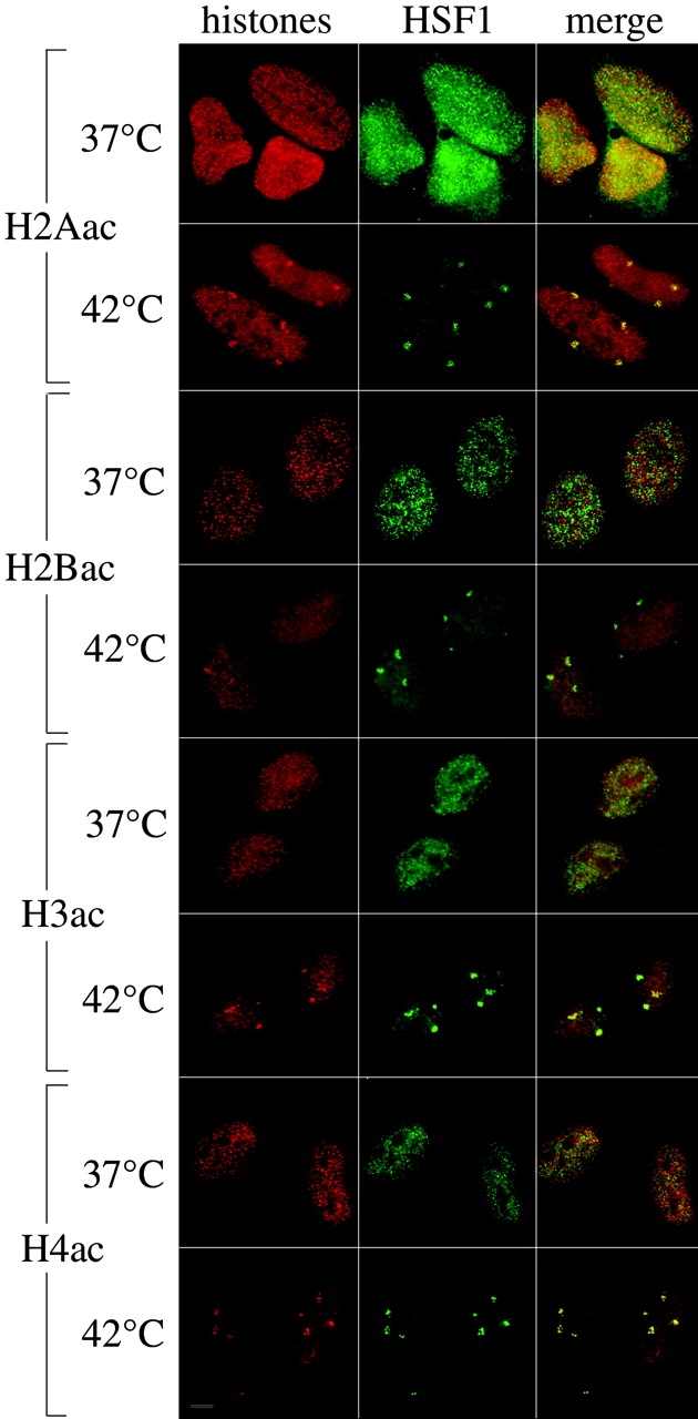
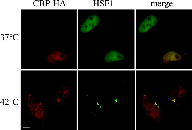
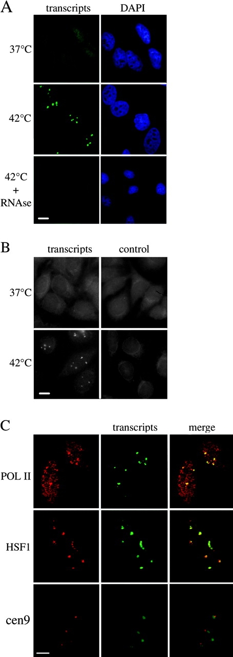
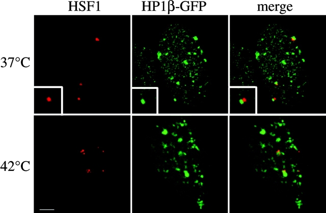
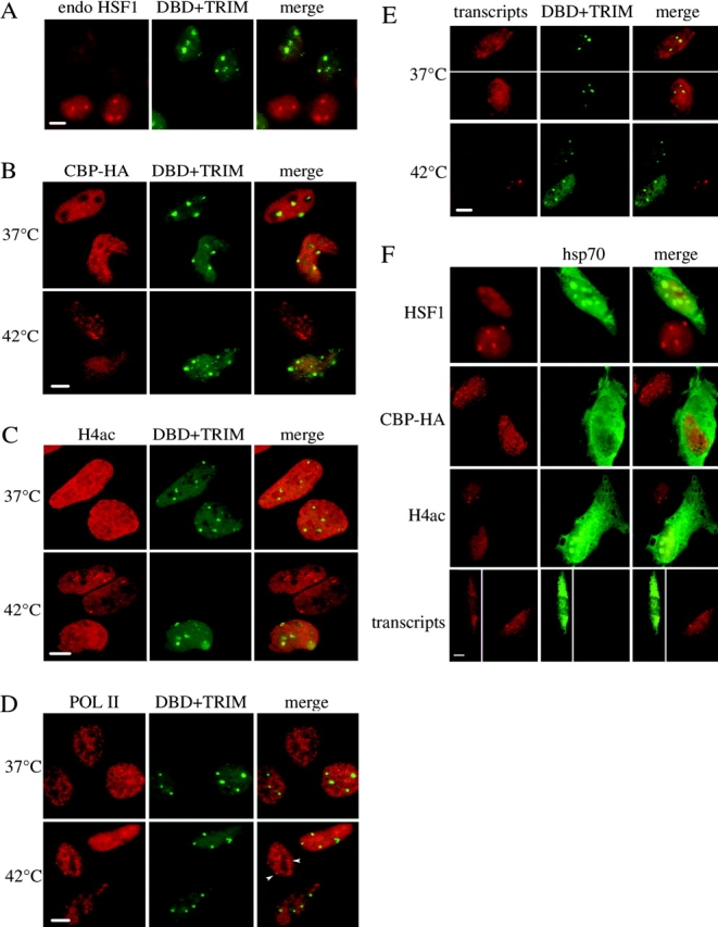
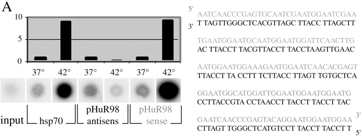
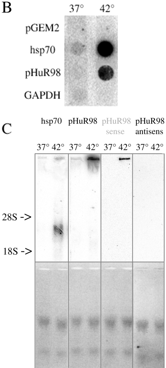
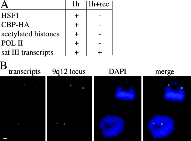
Exposure of mammalian cells to stress induces the activation of heat shock transcription factor 1 (HSF1) and the subsequent transcription of heat shock genes. Activation of the heat shock response also correlates with a rapid relocalization of HSF1 within a few nuclear structures termed nuclear stress granules. These stress-induced structures, which form primarily on the 9q12 region in humans through direct binding of HSF1 to satellite III repeats, do not colocalize with transcription sites of known hsp genes. In this paper, we show that nuclear stress granules correspond to RNA polymerase II transcription factories where satellite III repeats are transcribed into large and stable RNAs that remain associated with the 9q12 region, even throughout mitosis. This work not only reveals the existence of a new major heat-induced transcript in human cells that may play a role in chromatin structure, but also provides evidence for a transcriptional activity within a locus considered so far as heterochromatic and silent.


Figures

Figure 1.
Figure 1.
RNA polymerase II is concentrated within nuclear stress granules. (A) RNA polymerase II (red) was detected by immunofluorescence together with HSF1 (green) in non–heat-shocked and heat-shocked HeLa cells. At 37°C, HSF1 is diffusely distributed in the nucleoplasm and cytoplasm while RNA polymerase II displays a fine nuclear punctate staining. After 1 h at 42°C, HSF1 is massively recruited to nuclear stress granules, and RNA polymerase II is also found accumulated in these structures (yellow in the merged image), in addition to a remaining diffuse staining of the nucleus. Bar, 5 μm. (B) Total cell extracts prepared from non–heat-shocked (37°C) or heat-shocked (1 h at 42°C) cells were analyzed by Western blot. The blot was sequentially detected with 7C2 anti-RNA polymerase II antibody (left) and with rabbit anti-HSF1 antibody (right). No cross-reactivity of the antibodies is observed. The lowered HSF1 mobility observed in the 42°C sample is due to stress-induced posttranslational modifications of the protein (for review see Pirkkala et al., 2001).
Figure 2.
Figure 2.
Stress-induced HSF1 granules contain acetylated core histones. HSF1 (green) and acetylated forms of each core histone (red) were codetected by immunofluorescence in HeLa cells submitted or not to a 1-h heat shock at 42°C. At 37°C, HSF1 is diffusely distributed in the nucleus and cytoplasm, and acetylated histones exhibit a punctate distribution throughout the nucleus. After heat shock, acetylated forms of the four core histones are present to various extents within stress granules. Bar, 5 μm.
Figure 3.
Figure 3.
CBP is recruited to nuclear stress granules during heat shock. Transiently expressed CBP-HA (red) was codetected with HSF1 (green) in HeLa cells. In non–heat-shocked cells, CBP displays a fine punctate staining dispersed throughout the nucleoplasm. At 42°C, a fraction of the protein colocalizes with HSF1 in the granules. Bar, 5 μm.
Figure 4.
Figure 4.
Stress induces the transcription of chromosome 9 sat III repeats. (A) RNA FISH was performed on HeLa cells with clone pHuR98, which is specific for sat III repeats, as a probe. At 37°C, a faint nuclear and cytoplasmic staining is observed. In cells exposed to a 1-h heat shock at 42°C, three to four bright nuclear foci are detected in each nucleus, in addition to a persisting diffuse staining of the nucleus and cytoplasm. These foci are absent in heat-shocked cells treated with 100 μg/ml RNase A for 15 min before hybridization. (B) Comparison of the RNA FISH signals obtained with the pHuR98 probe and when the probe was omitted (control). Both at 37°C and 42°C, the intensity of the diffuse nuclear and cytoplasmic staining is comparable to the background level (control). (C) Codetection of pHuR98 transcripts by RNA FISH (green) with RNA polymerase II or HSF1 detected by immunofluorescence, or with chromosome 9 centromeres revealed by DNA FISH (red). Transcript foci coincide perfectly with HSF1 and RNA polymerase II within nuclear stress granules, and each transcript focus is found in the vicinity of a chromosome 9 centromere. Bars, 5 μm.
Figure 5.
Figure 5.
The heterochromatin protein HP1β is not concentrated in the 9q12 locus. HP1β-GFP construct (green) was transiently expressed in HeLa cells and codetected with either the DBD-TRIM mutant at 37°C or the endogenous HSF1 at 42°C (red). Projections of confocal series are shown. In both cases, nuclear stress granules are juxtaposed to, but never coincident with, HP1β-enriched foci (10 nuclei analyzed for each case). Insert shows a magnification of one stress granule. Bar, 5 μm.
Figure 6.
Figure 6.
Stress-induced sat III transcription is HSF1 dependent. The GFP-tagged DBF+TRIM mutant of HSF1 (green) was transiently transfected into HeLa cells and codetected with either (A) endogenous HSF1, (B) transiently expressed CBP-HA, (C) acetylated histone H4, (D) RNA polymerase II, or (E) pHuR98 transcripts (red). Overexpression of this mutant prevents the accumulation of endogenous HSF1, acetylated H4, CBP-HA, and RNA polymerase II into the granules, both at 37°C and at 42°C. Likewise, pHuR98 transcripts are not detected in the granules formed by the HSF1 mutant at 37°C and 42°C. Arrowheads in D point to granules of RNA pol II. (F) HeLa cells were transiently transfected with a plasmid coding for the HSP70 chaperone. HSP70 was then detected by immunofluorescence together with either the endogenous HSF1, transiently coexpressed CBP-HA, or acetylated histone H4 detected by immunofluorescence, or with pHuR98 transcripts revealed by RNA FISH. Only images of cells exposed for 1 h at 42°C are shown. In HSP70-overexpressing cells, the endogenous HSF1, CBP-HA, and acetylated histone H4 are not recruited to nuclear stress granules, and pHuR98 transcript foci are not present. The white lines in E and F indicate that the cells were taken from different fields. Bars, 10 μm.
Figure 7.
Figure 7.
Sat III transcripts are very large transcripts inducibly transcribed from one strand during stress. (A) Reverse transcription was performed on total RNA extracts prepared from HeLa cells submitted or not to a 1-h heat shock at 42°C, with either an hsp70 antisense, a pHuR98 antisense (black), or a pHuR98 sense (gray) primer. The reaction products were spotted onto nitrocellulose membrane and quantitated. The sequence of the double-stranded pHuR98 clone is shown on the right. A weak signal is observed for hsp70, pHuR98 antisense, and pHuR98 sense transcripts at 37°C. Heat shock induces a ninefold increase in the level of hsp70 transcripts. Likewise, a ninefold increase in the intensity of the signal corresponding to pHuR98 sense transcripts is observed at 42°C, while the signal corresponding to pHuR98 antisense transcripts decreases by two. (B) A run-on assay was performed with nuclei prepared from cells submitted or not to a 1-h heat shock at 42°C. The following probes were used: pGEM2 (control for nonspecific hybridization), pH2.3 (hsp70 gene), pHuR98 (sat III), and GAPDH (glyceraldehyde 3-phosphate dehydrogenase gene), which serves as a normalization control for transcription. Quantification of the spots by PhosphorImager® revealed that stress induces an 11.9-fold and a 13.4-fold induction in sat III and hsp70 transcription, respectively. (C) A Northern blot analysis was performed on total RNA extracts prepared from HeLa cells. Different probes were used: an hsp70 probe (Wu et al., 1985), the pHuR98 clone, an antisense pHuR98 oligonucleotide to reveal sense transcripts (gray), or a sense pHuR98 oligonucleotide to reveal antisense transcripts (black). Ethidium bromide staining of the gel before transfer confirms that equal amounts of RNA extracts were loaded in each lane. For hsp70 transcripts, a very weak signal is observed for the 37°C sample, while a signal at the expected size (2.3 kb) is observed in the 42°C sample (the positions of the 28S and 18S rRNAs are indicated). For pHuR98 transcripts, a faint signal is present with all three probes at 37°C, while in the 42°C sample, a very slowly migrating band is revealed, both with the pHuR98 probe and the antisense oligonucleotide (gray). No signal is observed with the sense oligonucleotide (black).
Figure 7.
Figure 7.
Sat III transcripts are very large transcripts inducibly transcribed from one strand during stress. (A) Reverse transcription was performed on total RNA extracts prepared from HeLa cells submitted or not to a 1-h heat shock at 42°C, with either an hsp70 antisense, a pHuR98 antisense (black), or a pHuR98 sense (gray) primer. The reaction products were spotted onto nitrocellulose membrane and quantitated. The sequence of the double-stranded pHuR98 clone is shown on the right. A weak signal is observed for hsp70, pHuR98 antisense, and pHuR98 sense transcripts at 37°C. Heat shock induces a ninefold increase in the level of hsp70 transcripts. Likewise, a ninefold increase in the intensity of the signal corresponding to pHuR98 sense transcripts is observed at 42°C, while the signal corresponding to pHuR98 antisense transcripts decreases by two. (B) A run-on assay was performed with nuclei prepared from cells submitted or not to a 1-h heat shock at 42°C. The following probes were used: pGEM2 (control for nonspecific hybridization), pH2.3 (hsp70 gene), pHuR98 (sat III), and GAPDH (glyceraldehyde 3-phosphate dehydrogenase gene), which serves as a normalization control for transcription. Quantification of the spots by PhosphorImager® revealed that stress induces an 11.9-fold and a 13.4-fold induction in sat III and hsp70 transcription, respectively. (C) A Northern blot analysis was performed on total RNA extracts prepared from HeLa cells. Different probes were used: an hsp70 probe (Wu et al., 1985), the pHuR98 clone, an antisense pHuR98 oligonucleotide to reveal sense transcripts (gray), or a sense pHuR98 oligonucleotide to reveal antisense transcripts (black). Ethidium bromide staining of the gel before transfer confirms that equal amounts of RNA extracts were loaded in each lane. For hsp70 transcripts, a very weak signal is observed for the 37°C sample, while a signal at the expected size (2.3 kb) is observed in the 42°C sample (the positions of the 28S and 18S rRNAs are indicated). For pHuR98 transcripts, a faint signal is present with all three probes at 37°C, while in the 42°C sample, a very slowly migrating band is revealed, both with the pHuR98 probe and the antisense oligonucleotide (gray). No signal is observed with the sense oligonucleotide (black).
Figure 8.
Figure 8.
Stress-induced sat III transcripts are stable transcripts that remain associated with the 9q12 locus. (A) Table showing the composition of nuclear stress granules after 1 h of heat shock at 42°C or for 3 h at 37°C after a 1-h heat shock at 42°C (200 nuclei analyzed in each case). While all components are detected in the granules after 1 h of heat shock (HSF1, overexpressed CBP-HA, acetylated histones, RNA polymerase II, sat III transcripts), only sat III transcripts are still detected in a large majority of cells after recovery. (B) Sat III transcripts (green) were detected by RNA FISH along with the 9q12 locus, revealed by DNA FISH with a probe specific for chromosome 9 classical satellites (D9Z1) in HeLa cells allowed to recover for 3 h at 37°C after a 1-h heat shock. Two mitotic cells with sat III transcripts (red) associated to the 9q12 locus (green) are shown. Only two spots are visible in each cell because the two others are not in the same focal plan. Bar, 5 μm.

Comment in

Similar articles

Cited by

References

    1. Avner, P., and E. Heard. 2001. X-chromosome inactivation: counting, choice and initiation. Nat. Rev. Genet. 2:59–67. - PubMed
    1. Bartlett, J.M., A.D. Watters, S.A. Ballantyne, J.J. Going, K.M. Grigor, and T.G. Cooke. 1998. Is chromosome 9 loss a marker of disease recurrence in transitional cell carcinoma of the urinary bladder? Br. J. Cancer. 77:2193–2198. - PMC - PubMed
    1. Besse, S., M. Vigneron, E. Pichard, and F. Puvion-Dutilleul. 1995. Synthesis and maturation of viral transcripts in herpes simplex virus type 1 infected HeLa cells: the role of interchromatin granules. Gene Expr. 4:143–161. - PMC - PubMed
    1. Buisson, M., F. Hans, I. Kusters, N. Duran, and A. Sergeant. 1999. The C-terminal region but not the Arg-X-Pro repeat of Epstein-Barr virus protein EB2 is required for its effect on RNA splicing and transport. J. Virol. 73:4090–4100. - PMC - PubMed
    1. Chiodi, I., M. Biggiogera, M. Denegri, M. Corioni, F. Weighardt, F. Cobianchi, S. Riva, and G. Biamonti. 2000. Structure and dynamics of hnRNP-labelled nuclear bodies induced by stress treatments. J. Cell Sci. 113:4043–4053. - PubMed

Publication types

MeSH terms