Review
. 2003:59:177-228.
doi: 10.1016/s0065-3527(03)59006-6.

5'- and 3'-noncoding regions in flavivirus RNA

Affiliations
Review

5'- and 3'-noncoding regions in flavivirus RNA

Lewis Markoff. Adv Virus Res. 2003.

Abstract

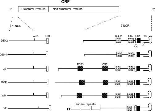
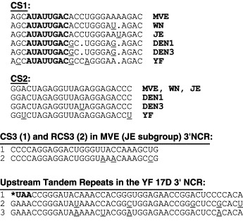
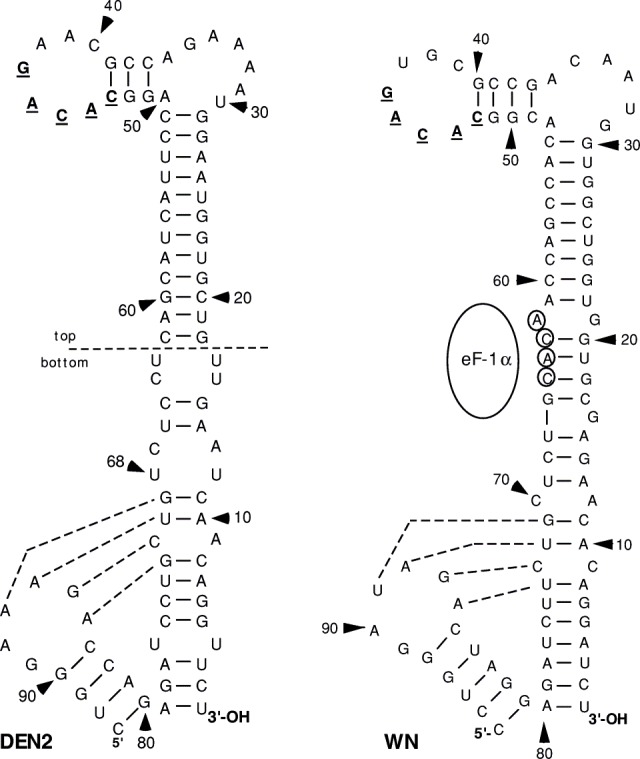
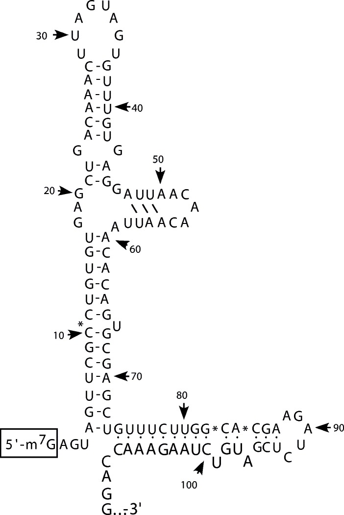
The flavivirus genome is a capped, positive-sense RNA approximately 10.5 kb in length. It contains a single long open reading frame (ORF), flanked by a 5´ noncoding regions (NCR), which is about 100 nucleotides in length, and a 3´ NCR ranging in size from about 400 to 800 nucleotides in length. The conserved structural and nucleotide sequence elements of these NCRs and their function in RNA replication and translation are the subjects of this chapter. The 5´ and 3´ NCRs play a role in the initiation of negative-strand synthesis on virus RNA released from entering virions, switching from negative-strand synthesis to synthesis of progeny plus strand RNA at late times after infection, and possibly in the initiation of translation and in the packaging of virus plus strand RNA into particles. The presence of conserved and nonconserved complementary nucleotide sequences near the 5´ and 3´ termini of flavivirus genomes suggests that ‘‘panhandle’’ or circular RNA structures are formed transiently by hydrogen bonding at some stage during RNA replication.


Figures

Fig 1
Fig 1
Nucleotide sequences of 5′ and 3′ termini of a selection of mosquito-borne flavivirus positive-strand (+) genome RNAs are shown aligned with the nucleotide sequences of 3′ and 5′ termini of the respective minus (−) strands, predicted to result when the positive strand is used as a template to form a replicative intermediate during virus replication. Dotted lines indicate the intervening nucleotides of the respective full-length genomes. Flavivirus conserved 5′- and 3′-terminal dinucleotides and their complementary nucleotides are boxed. The semiconserved pentanucleotide sequence ACACA is shown in bold in the 3′ termini of positive- and negative-strand RNAs, respectively, where present. Genome sequences are as follows: YF, yellow fever (Rice et al., 1985); WNV, West Nile (Wengler and Wengler, 1981); MVE, Murray Valley encephalitis (Genbank number NC00943); DEN3, dengue serotype 3 (Genbank number NC001475); KUN, Kunjin (Genbank numbers L24511 and L24512); and JE, Japanese encephalitis (Genbank number NC001437).
Fig 2
Fig 2
Some nucleotide sequence features of 5′ and 3′ NCRs of mosquito-borne flavivirus genomes are represented. Horizontal lines represent nucleotide sequences of DEN2, DEN4, JE, MVE, WN, and YF genome 5′ NCRs (aligned in vertical order on the left) and 3′ NCRs (aligned in vertical order on the right). Upward vertical ticks in the 5′ NCR indicate the location of the 5′-most start codon for translation of the single ORF in the flavivirus genome. Downward vertical ticks indicate the location(s) of translational stop codons at the 5′ ends of 3′ NCRs. The flavivirus-conserved stem and loop secondary structure predicted to form at the 3′ end of all genomes is indicated by a loop at the end of the linear sequence (SL). The relative locations of the conserved eight nucleotide cyclization sequences, 5′CS and CYC, respectively, at the 5′ and 3′ ends of the genome are indicated by open boxes with cross-hatching. The relative location of the conserved sequence, CS2, in all genomes is indicated by a stippled box, and the location of the CS2 tandem repeat sequence, RCS2, in all but the YF genome is indicated by a second stippled box upstream from CS2. Similarly, the relative locations of the conserved tandem repeat sequences, CS3 and RCS3, are indicated by black, cross-hatched boxes in genomes of viruses of the JE subgroup. The locations of three tandem repeat sequences in the upstream portion of the YF genome 3′ NCR are indicated by open rectangles.
Fig 3
Fig 3
Some nucleotide sequence features of 5′ and 3′ NCRs of TBE virus strain genomes are represented. The 5′ NCR of TBE virus is represented on the left as a horizontal line. The start codon for translation of a very short ORF in the 5′ NCR is represented by a single upward vertical tick, numbered one (1); the start codon for translation of the long ORF in TBE genomes is represented by an upward tick, numbered two (2). Stop codons for the short ORF in the 5′ NCR and for the long ORF at the 5′ end of the 3′ NCR are indicated by a downward vertical tick. Four of six different possible nucleotide sequence motifs for 3′ NCRs of TBE viruses are indicated on the right as horizontal lines (adapted from Wallner et al., 1995). Motif A, strains Neudorfl and 263; B, strain Ljubljana I; C, strain 132; and D, strain RK1424. The flavivirus-conserved 3′ stem and loop structure is indicated by a loop in the horizontal line and is labeled “SL.” The location of the restriction endonuclease site AgeI in TBE genomic DNA is indicated by an arrow defining the boundary between the variable region and the core element in the strain Neudorfl 3′ NCR. Black boxes indicate the relative locations of cyclization sequences C1 and C2 in the 5′ NCR and C1′ and C2′ in the 3′ NCR. Relative locations of TBE virus–conserved repeat sequences R1, R1′, R2, and R3 in the 3′ NCR are indicated by cross-hatched or stippled boxes. IR, conserved inverted repeat. PU⧸PY⧸PR, conserved homopurine, pyrimidine, and purine-rich segments in 3′ NCR RNA, respectively. Dotted lines indicate the locations of deletions in the genomes of some TBE strains relative to the motif found in strain Neudorfl RNA. The notation “AAAn” indicates the location of a poly(A)tract in the strains Neudorfl, 263, and 132 genomes. The location of a severely truncated poly(A)tract in the strain Llubljana 1 genome is specifically indicated by an arrow.
F<sc>ig</sc> 4
Fig 4
Nucleotide sequences of 3′ NCRs of dengue virus genomes representing each of the four serotypes are shown. Shaded areas indicate sequence homology. Dashes indicate gaps introduced into a sequence in order to maximize its alignment with the other sequences. Nucleotides that form the long stem and loop within the flavivirus-conserved 3′-SL (see text) are indicated by a horizontal line above sequence data, labeled 3′-LSH. The location of the flavivirus-conserved pentanucleotide sequence, 5′-CACAG-3′, within the 3′-SL, is indicated by a line below sequence data. The conserved sequences, CS1, the cyclization sequence, CS2, and RCS2 are indicated by horizontal lines above relevant sequence data. The conserved small stem and loop structures, TL1 and TL2 (Proutski et al., 1997b), are also so indicated. Nucleotides presumed to base pair in the formation of the long stem in the 3′-SL and in the formation of the short TL1 and TL2 stems are numbered.
F<sc>ig</sc> 5
Fig 5
Nucleotide sequences of some conserved motifs in 3′ NCRs of mosquito-borne flavivirus genomes. Deletions in CS1 with respect to the sequence for MVE (Hahn et al., 1987) are indicated by a period. Nucleotide substitutions with respect to the sequences of CS1 and CS2 in the MVE genome are indicated by underlining the substituted nucleotides. Nucleotides comprising the 3′ cyclization sequence (CYC) in CS1 are shown in bold print. The stop codon that comprises the first three nucleotides of the most upstream tandem repeat in the YF genome 3′ NCR (Rice et al., 1985) is also shown in bold and indicated by an asterisk. Nucleotide sequence data are from Genbank (Table I) or from the aforementioned references.
Fig 6
Fig 6
Nucleotide sequences of 3′-SLs in WN strain E101 and DEN2 strain NGC genomes (Zeng et al., 1998) are shown in the expected conformation and numbered from the 3′ end of each genome. The flavivirus-conserved pentanucleotide sequence 5′-CACAG-3′ is indicated in bold underlined type. The “top” and “bottom” portion of the DEN2 3′-SL as designated in a previous study with respect to the WN 3′-SL is shown by a labeled horizontal dashed line (Zeng et al., 1998). Essential nucleotides of the putative binding site for the translation elongation factor, eF-1α, to the WN 3′-SL are circled (Blackwell and Brinton, 1997). Nucleotides potentially involved in pseudoknot formation in both DEN2 and WN 3′-SLs are linked by dashed lines (Shi et al., 1996a).
F<sc>ig</sc> 7
Fig 7
Proposed secondary structure of the WN 5′ NCR (adapted from Brinton and Dispoto, 1988). Nucleotides are numbered from the 5′ terminus, excluding the cap, which is shown boxed. Nucleotides of the ORF, starting with nucleotide number 96, are shown in large, bold type (adapted from Brinton and Dispoto, 1988).
Fig 8
Fig 8
Chimeric DEN2⧸WN 3′ SL nucleotide sequences in DEN2 genomic DNA. The 3′ SL secondary structure is depicted as a line drawing. DEN2 nucleotide sequences are depicted as a thin line, and WN nucleotide sequences are depicted as a thick line. “Top” and “bottom” portions of the 3′ SL are indicated by horizontal dashed lines bisecting the long stem in the 3′ SL. The chimeric nucleotide sequences indicated were substituted for the wild-type nucleotide sequence of the 3′ SL in DEN2 infectious DNA, and the resulting mutant DNAs were used to derive RNAs that yielded DEN2 viruses with the indicated phenotypes after transfection of cultured monkey kidney cells (see text). Viable viruses all retained the DEN2-specific 11-bp segment comprising the top portion of the bottom section of the long stem in the 3′ SL. The DEN2mutF virus (asterisk) was notable for its host range-restricted phenotype in mosquito cells, where its replication was 100,000-fold reduced, compared to the wild-type DEN2 virus (Zeng et al., 1998).

Similar articles

Cited by

References

    1. Avni D., Shama S., Loreni F., Meyuhas O. Vertebrate mRNAs with a 5′-terminal pyrimidine tract are candidates for translational repression in quiescent cells: Characterization of the translational cis-regulatory element. Mol. Cell. Biol. 1994;14(6):3822–3833. - PMC - PubMed
    1. Behe M.J. An overabundance of long oligopurine tracts occurs in the genome of simple and complex eukaryotes. Nucleic Acids Res. 1995;23(4):689–695. - PMC - PubMed
    1. Bergamini G., Preiss T., Hentze M.W. Picornavirus IRESes and the poly(A) tail jointly promote cap-independent translation in a mammalian cell-free system. RNA. 2000;6(12):1781–1790. - PMC - PubMed
    1. Blackwell J.L., Brinton M.A. BHK cell proteins that bind to the 3′ stem-loop structure of the West Nile virus genome RNA. J. Virol. 1995;69(9):5650–5658. - PMC - PubMed
    1. Blackwell J.L., Brinton M.A. Translation elongation factor eF-1α interacts with the 3′ stem-loop region of West Nile Virus genomic RNA. J. Virol. 1997;71(9):6433–6444. - PMC - PubMed

LinkOut - more resources