

Essential requirement for Wnt signaling in proliferation of adult small intestine and colon revealed by adenoviral expression of Dickkopf-1
- PMID: 14695885
- PMCID: PMC314174
- DOI: 10.1073/pnas.2536800100
Essential requirement for Wnt signaling in proliferation of adult small intestine and colon revealed by adenoviral expression of Dickkopf-1
Abstract
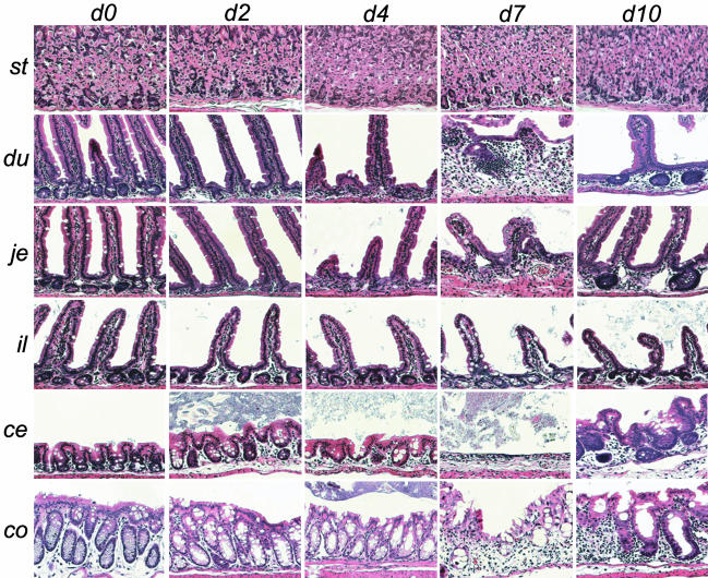
Whereas the adult gastrointestinal epithelium undergoes tremendous self-renewal through active proliferation in crypt stem cell compartments, the responsible growth factors regulating this continuous proliferation have not been defined. The exploration of physiologic functions of Wnt proteins in adult organisms has been hampered by functional redundancy and the necessity for conditional inactivation strategies. Dickkopf-1 (Dkk1) is a potent secreted Wnt antagonist that interacts with Wnt coreceptors of the LRP family. To address the contribution of Wnt signaling to gastrointestinal epithelial proliferation, adenoviral expression of Dkk1 was used to achieve stringent, conditional, and reversible Wnt inhibition in adult animals. Adenovirus Dkk1 (Ad Dkk1) treatment of adult mice repressed expression of the Wnt target genes CD44 and EphB2 within 2 days in both small intestine and colon, indicating an extremely broad role for Wnt signaling in the maintenance of adult gastrointestinal gene expression. In parallel, Ad Dkk1 markedly inhibited proliferation in small intestine and colon, accompanied by progressive architectural degeneration with the loss of crypts, villi, and glandular structure by 7 days. Whereas decreased Dkk1 expression at later time points (>10 days) was followed by crypt and villus regeneration, which was consistent with a reversible process, substantial mortality ensued from colitis and systemic infection. These results indicate the efficacy of systemic expression of secreted Wnt antagonists as a general strategy for conditional inactivation of Wnt signaling in adult organisms and illustrate a striking reliance on a single growth factor pathway for the maintenance of the architecture of the adult small intestine and colon.
Figures


Similar articles
-
Wnts as essential growth factors for the adult small intestine and colon.Cell Cycle. 2004 May;3(5):554-7. Epub 2004 May 15. Cell Cycle. 2004. PMID: 15044853
-
The Wnt antagonist Dkk1 regulates intestinal epithelial homeostasis and wound repair.Gastroenterology. 2011 Jul;141(1):259-68, 268.e1-8. doi: 10.1053/j.gastro.2011.03.043. Epub 2011 Mar 25. Gastroenterology. 2011. PMID: 21440550 Free PMC article.
-
Chimeric 5/35 adenovirus-mediated Dickkopf-1 overexpression suppressed tumorigenicity of CD44⁺ gastric cancer cells via attenuating Wnt signaling.J Gastroenterol. 2013 Jul;48(7):798-808. doi: 10.1007/s00535-012-0711-z. Epub 2012 Nov 28. J Gastroenterol. 2013. PMID: 23188090
-
Recombinant adenovirus as a methodology for exploration of physiologic functions of growth factor pathways.J Mol Med (Berl). 2008 Feb;86(2):161-9. doi: 10.1007/s00109-007-0261-7. Epub 2007 Sep 22. J Mol Med (Berl). 2008. PMID: 17891365 Review.
-
Dickkopf1 and the Spemann-Mangold head organizer.Int J Dev Biol. 2001;45(1):237-40. Int J Dev Biol. 2001. PMID: 11291852 Review.
Cited by
-
Rab8a vesicles regulate Wnt ligand delivery and Paneth cell maturation at the intestinal stem cell niche.Development. 2015 Jun 15;142(12):2147-62. doi: 10.1242/dev.121046. Epub 2015 May 26. Development. 2015. PMID: 26015543 Free PMC article.
-
Vangl2 promotes the formation of long cytonemes to enable distant Wnt/β-catenin signaling.Nat Commun. 2021 Apr 6;12(1):2058. doi: 10.1038/s41467-021-22393-9. Nat Commun. 2021. PMID: 33824332 Free PMC article.
-
Obesity under the moonlight of c-MYC.Front Cell Dev Biol. 2023 Dec 5;11:1293218. doi: 10.3389/fcell.2023.1293218. eCollection 2023. Front Cell Dev Biol. 2023. PMID: 38116204 Free PMC article. Review.
-
Krüppel-like Factor 5 Regulates Stemness, Lineage Specification, and Regeneration of Intestinal Epithelial Stem Cells.Cell Mol Gastroenterol Hepatol. 2020;9(4):587-609. doi: 10.1016/j.jcmgh.2019.11.009. Epub 2019 Nov 25. Cell Mol Gastroenterol Hepatol. 2020. PMID: 31778829 Free PMC article.
-
Wnt and FGF signals interact to coordinate growth with cell fate specification during limb development.Development. 2008 Oct;135(19):3247-57. doi: 10.1242/dev.023176. Development. 2008. PMID: 18776145 Free PMC article.
References
-
- Marshman, E., Booth, C. & Potten, C. S. (2002) BioEssays 24, 91-98. - PubMed
-
- Korinek, V., Barker, N., Moerer, P., van Donselaar, E., Huls, G., Peters, P. J. & Clevers, H. (1998) Nat. Genet. 19, 379-383. - PubMed
-
- van de Wetering, M., Sancho, E., Verweij, C., de Lau, W., Oving, I., Hurlstone, A., van der Horn, K., Batlle, E., Coudreuse, D., Haramis, A. P., et al. (2002) Cell 111, 241-250. - PubMed
Publication types
MeSH terms
Substances
Grants and funding
LinkOut - more resources
Full Text Sources
Other Literature Sources
Miscellaneous
