

Adult male rat hippocampus synthesizes estradiol from pregnenolone by cytochromes P45017alpha and P450 aromatase localized in neurons
- PMID: 14694190
- PMCID: PMC321772
- DOI: 10.1073/pnas.2630225100
Adult male rat hippocampus synthesizes estradiol from pregnenolone by cytochromes P45017alpha and P450 aromatase localized in neurons
Abstract
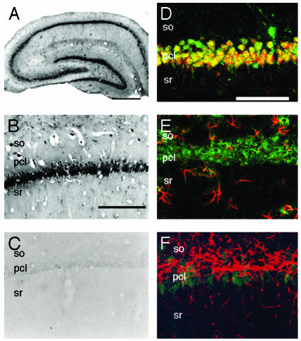
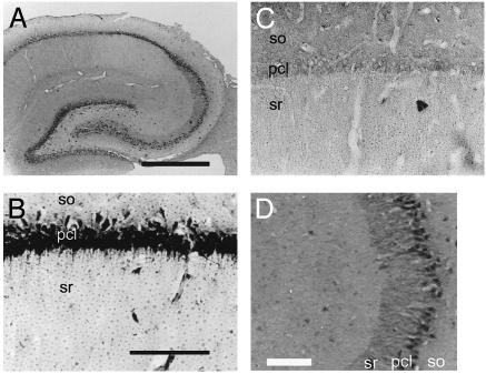
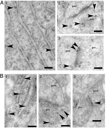
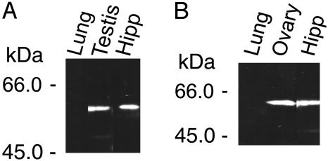
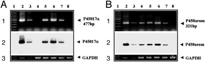
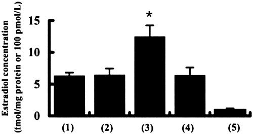
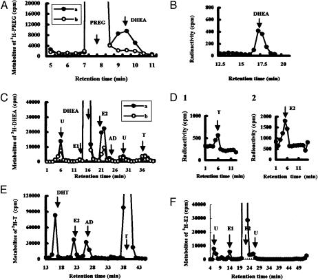
In adult mammalian brain, occurrence of the synthesis of estradiol from endogenous cholesterol has been doubted because of the inability to detect dehydroepiandrosterone synthase, P45017alpha. In adult male rat hippocampal formation, significant localization was demonstrated for both cytochromes P45017alpha and P450 aromatase, in pyramidal neurons in the CA1-CA3 regions, as well as in the granule cells in the dentate gyrus, by means of immunohistochemical staining of slices. Only a weak immunoreaction of these P450s was observed in astrocytes and oligodendrocytes. ImmunoGold electron microscopy revealed that P45017alpha and P450 aromatase were localized in pre- and postsynaptic compartments as well as in the endoplasmic reticulum in principal neurons. The expression of these cytochromes was further verified by using Western blot analysis and RT-PCR. Stimulation of hippocampal neurons with N-methyl-d-aspartate induced a significant net production of estradiol. Analysis of radioactive metabolites demonstrated the conversion from [(3)H]pregnenolone to [(3)H]estradiol through dehydroepiandrosterone and testosterone. This activity was abolished by the application of specific inhibitors of cytochrome P450s. Interestingly, estradiol was not significantly converted to other steroid metabolites. Taken together with our previous finding of a P450scc-containing neuronal system for pregnenolone synthesis, these results imply that 17beta-estradiol is synthesized by P45017alpha and P450 aromatase localized in hippocampal neurons from endogenous cholesterol. This synthesis may be regulated by a glutamate-mediated synaptic communication that evokes Ca(2+) signals.
Figures
Similar articles
-
Hippocampal cytochrome P450s synthesize brain neurosteroids which are paracrine neuromodulators of synaptic signal transduction.Biochim Biophys Acta. 2003 Feb 17;1619(3):301-16. doi: 10.1016/s0304-4165(02)00489-0. Biochim Biophys Acta. 2003. PMID: 12573490 Review.
-
Neurosteroid synthesis by cytochrome p450-containing systems localized in the rat brain hippocampal neurons: N-methyl-D-aspartate and calcium-dependent synthesis.Endocrinology. 2001 Aug;142(8):3578-89. doi: 10.1210/endo.142.8.8327. Endocrinology. 2001. PMID: 11459806
-
Retinoic acid stimulates 17beta-estradiol and testosterone synthesis in rat hippocampal slice cultures.Endocrinology. 2009 Sep;150(9):4260-9. doi: 10.1210/en.2008-1644. Epub 2009 Jun 4. Endocrinology. 2009. PMID: 19497980
-
Stimulation of estradiol biosynthesis by tributyltin in rat hippocampal slices.Endocr Res. 2014;39(4):168-72. doi: 10.3109/07435800.2013.875563. Epub 2014 Mar 28. Endocr Res. 2014. PMID: 24679120
-
Local neurosteroid production in the hippocampus: influence on synaptic plasticity of memory.Neuroendocrinology. 2006;84(4):255-63. doi: 10.1159/000097747. Epub 2006 Dec 1. Neuroendocrinology. 2006. PMID: 17142999 Review.
Cited by
-
Pharmacological administration of the isoflavone daidzein enhances cell proliferation and reduces high fat diet-induced apoptosis and gliosis in the rat hippocampus.PLoS One. 2013 May 31;8(5):e64750. doi: 10.1371/journal.pone.0064750. Print 2013. PLoS One. 2013. PMID: 23741384 Free PMC article.
-
The estrous cycle modulates hippocampal spine dynamics, dendritic processing, and spatial coding.bioRxiv [Preprint]. 2024 Aug 3:2024.08.02.606418. doi: 10.1101/2024.08.02.606418. bioRxiv. 2024. PMID: 39131375 Free PMC article. Preprint.
-
Locally Synthetized 17-β-Estradiol Reverses Amyloid-β-42-Induced Hippocampal Long-Term Potentiation Deficits.Int J Mol Sci. 2024 Jan 23;25(3):1377. doi: 10.3390/ijms25031377. Int J Mol Sci. 2024. PMID: 38338656 Free PMC article.
-
Xiao-Yao-San Formula Improves Cognitive Ability by Protecting the Hippocampal Neurons in Ovariectomized Rats.Evid Based Complement Alternat Med. 2020 Jun 19;2020:4156145. doi: 10.1155/2020/4156145. eCollection 2020. Evid Based Complement Alternat Med. 2020. PMID: 32655660 Free PMC article.
-
Estradiol reduction through aromatase inhibition impairs cocaine seeking in male rats.Front Behav Neurosci. 2024 Jan 16;17:1307606. doi: 10.3389/fnbeh.2023.1307606. eCollection 2023. Front Behav Neurosci. 2024. PMID: 38292056 Free PMC article.
References
-
- Foy, M. R. Xu, J. Xie, X. Brinton, R. D. Thompson, R. F. & Berger, T. W. (1999) J. Neurophisiol. 81, 925–929. - PubMed
-
- Azcoitia, I., Sierra, A., Veiga, S., Honda, S., Harada, N. & Garcia-Segura, L. M. (2001) J. Neurobiol. 47, 318–329. - PubMed
-
- Pozzo-Miller, L. D., Inoue, T. & Murphy, D. D. (1999) J. Neurophysiol. 81, 1404–1411. - PubMed
-
- Shibuya, K., Takata, N., Hojo, Y., Furukawa, A., Yasumatsu, N., Kimoto, T., Enami, T., Suzuki, K., Tanabe, N., Ishii, H., et al. (2002) Biochim. Biophys. Acta 25462, 1–16. - PubMed
Publication types
MeSH terms
Substances
Grants and funding
LinkOut - more resources
Full Text Sources
Molecular Biology Databases
Miscellaneous
