

Short-range functional interaction between connexin35 and neighboring chemical synapses
- PMID: 14681051
- PMCID: PMC1803252
- DOI: 10.1080/15419060390263254
Short-range functional interaction between connexin35 and neighboring chemical synapses
Abstract
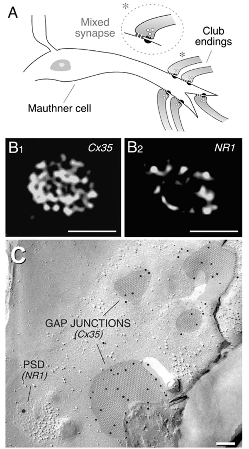
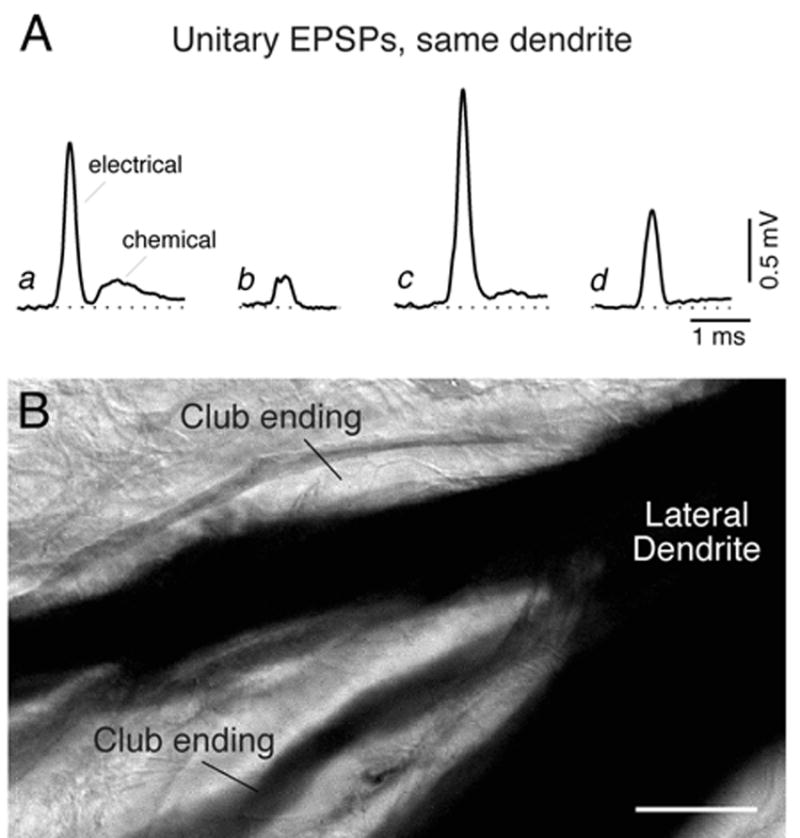
Auditory afferents terminating as mixed, electrical, and chemical, synapses on the goldfish Mauthner cells constitute an ideal experimental model to study the properties of gap junctions in the nervous system as well as to explore possible functional interactions with the other major form of interneuronal communication--chemically mediated synapses. By combining confocal microscopy and freeze-fracture replica immunogold labeling (FRIL), we found that gap junctions at these synapses contain connexin35 (Cx35), the fish ortholog of the neuron-specific human and mouse connexin36 (Cx36). Conductance of gap junction channels at these endings is known to be dynamically modulated by the activity of their co-localized chemically mediated glutamatergic synapses. By using simultaneous pre- and postsynaptic recordings at these single terminals, we demonstrate that such functional interaction takes place in the same ending, within a few micrometers. Accordingly, we also found evidence by confocal and FRIL double-immunogold labeling that the NR1 subunit of the NMDA glutamate receptor, proposed to be a key regulatory element, is present at postsynaptic densities closely associated with gap junction plaques containing Cx35. Given the widespread distribution of Cx35- and Cx36-mediated electrical synapses and glutamatergic synapses, our data suggest that the local functional interactions observed at these identifiable junctions may also apply to other electrical synapses, including those in mammalian brain.
Figures
Similar articles
-
Connexin35 mediates electrical transmission at mixed synapses on Mauthner cells.J Neurosci. 2003 Aug 20;23(20):7489-503. doi: 10.1523/JNEUROSCI.23-20-07489.2003. J Neurosci. 2003. PMID: 12930787 Free PMC article.
-
High-resolution proteomic mapping in the vertebrate central nervous system: close proximity of connexin35 to NMDA glutamate receptor clusters and co-localization of connexin36 with immunoreactivity for zonula occludens protein-1 (ZO-1).J Neurocytol. 2004 Jan;33(1):131-51. doi: 10.1023/B:NEUR.0000029653.34094.0b. J Neurocytol. 2004. PMID: 15173637 Free PMC article.
-
Heterotypic gap junctions at glutamatergic mixed synapses are abundant in goldfish brain.Neuroscience. 2015 Jan 29;285:166-93. doi: 10.1016/j.neuroscience.2014.10.057. Epub 2014 Nov 4. Neuroscience. 2015. PMID: 25451276 Free PMC article.
-
Dynamics of electrical transmission at club endings on the Mauthner cells.Brain Res Brain Res Rev. 2004 Dec;47(1-3):227-44. doi: 10.1016/j.brainresrev.2004.06.010. Brain Res Brain Res Rev. 2004. PMID: 15572174 Review.
-
Electrical synapses--gap junctions in the brain.Results Probl Cell Differ. 2006;43:99-128. doi: 10.1007/400_013. Results Probl Cell Differ. 2006. PMID: 17068969 Review.
Cited by
-
Abundance and ultrastructural diversity of neuronal gap junctions in the OFF and ON sublaminae of the inner plexiform layer of rat and mouse retina.Neuroscience. 2006 Nov 3;142(4):1093-117. doi: 10.1016/j.neuroscience.2006.08.020. Epub 2006 Sep 28. Neuroscience. 2006. PMID: 17010526 Free PMC article.
-
Voltage-dependent enhancement of electrical coupling by a subthreshold sodium current.J Neurosci. 2004 Apr 21;24(16):3999-4010. doi: 10.1523/JNEUROSCI.0077-04.2004. J Neurosci. 2004. PMID: 15102915 Free PMC article.
-
Two independent forms of activity-dependent potentiation regulate electrical transmission at mixed synapses on the Mauthner cell.Brain Res. 2012 Dec 3;1487:173-82. doi: 10.1016/j.brainres.2012.05.059. Epub 2012 Jul 4. Brain Res. 2012. PMID: 22771708 Free PMC article. Review.
-
Calcium-dependent binding of calmodulin to neuronal gap junction proteins.Biochem Biophys Res Commun. 2005 Oct 7;335(4):1191-8. doi: 10.1016/j.bbrc.2005.08.007. Biochem Biophys Res Commun. 2005. PMID: 16112650 Free PMC article.
-
Regulation of gap junction coupling through the neuronal connexin Cx35 by nitric oxide and cGMP.Cell Commun Adhes. 2006 Jan-Apr;13(1-2):41-54. doi: 10.1080/15419060600631474. Cell Commun Adhes. 2006. PMID: 16613779 Free PMC article.
References
-
- Furshpan EJ. Electrical transmission at an excitatory synapse in a vertebrate brain. Science. 1964;144:878–880. - PubMed
-
- Nakajima Y. Fine structure of the synaptic endings on the Mauthner cell of the goldfish. J Comp Neurol. 1974;156:375–402. - PubMed
-
- Wolszon L, Pereda A, Faber DS. A fast synaptic potential mediated by NMDA and non-NMDA receptors. J Neurophysiol. 1997;78:2693–2705. - PubMed
-
- Yang XD, Korn H, Faber DS. Long-term potentiation of electrotonic coupling at mixed synapses. Nature. 1990;348:542–545. - PubMed
Publication types
MeSH terms
Substances
Grants and funding
LinkOut - more resources
Full Text Sources
Miscellaneous