. 2004 Jan;24(1):420-7.
doi: 10.1128/MCB.24.1.420-427.2004.

Argonaute protein in the early divergent eukaryote Trypanosoma brucei: control of small interfering RNA accumulation and retroposon transcript abundance

Affiliations

Argonaute protein in the early divergent eukaryote Trypanosoma brucei: control of small interfering RNA accumulation and retroposon transcript abundance

Huafang Shi et al. Mol Cell Biol. 2004 Jan.

Abstract

Members of the Argonaute protein family have been linked through a combination of genetic and biochemical studies to RNA interference (RNAi) and related phenomena. Here, we describe the characterization of the first Argonaute protein (AGO1) in Trypanosoma brucei, the earliest divergent eukaryote where RNAi has been described so far. AGO1 is predominantly cytoplasmic and is found in a ribonucleoprotein particle with small interfering RNAs (siRNAs), and this particle is present in a soluble form, as well as associated with polyribosomes. A genetic knockout of AGO1 leads to a loss of RNAi, and concomitantly, endogenous retroposon-derived siRNAs as well as siRNAs derived from transgenic double-stranded RNA are reduced to almost undetectable levels. Furthermore, AGO1 deficiency leads to an increase in retroposon transcript abundance via mechanisms operating at the transcriptional level and at the RNA stability level. Our results suggest that AGO1 function is required for production and/or stabilization of siRNAs and provide the first evidence for an Argonaute protein being involved in the regulation of retroposon transcript levels.


Figures
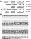
FIG. 1.
FIG. 1.
Argonaute family members. (A) Domain structure of selected members of the Argonaute family. Abbreviations: At AGO1, A. thaliana Argonaute 1 (accession number U91995); Nc QDE-2, Neurospora crassa QDE-2 (accession number AF217760); Ce RDE-1, C. elegans RDE-1 (accession number AF180730); Dm AGO2, D. melanogaster Argonaute 2 (accession number AE003530); Tb AGO1, T. brucei Argonaute 1. The drawing is not to scale. (B) Protein sequence of T. brucei Argonaute 1. The domains schematically indicated in panel A are highlighted.
FIG. 2.
FIG. 2.
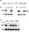
The AGO1 gene is essential for RNAi. (A) Schematic representation of the complementation cassette, which contains in a 5′-to-3′ direction 330 nt of AGO1 5′-flanking sequences including processing signals for 5′-end formation of the AGO1 mRNA (AGO1 5′UTR), the AGO1 open reading frame of 2,712 bp (AGO1), the procyclic acidic repetetive protein intergenic region of 990 bp (PARP), the neomycin resistance gene (NEO), and finally 780 bp of AGO 3′-flanking sequences (AGO1 3′UTR). (B) Northern blot analysis. RNA isolated from wild-type cells (wt; lane 1), AGO1-knockout cells (ago1−/−; lane 2), and ago1−/− cells complemented with the AGO1 gene (ago1c; lane 3) was probed for AGO1 mRNA (upper panel). α-Tubulin mRNA served as a control for RNA recovery and loading (lower panel). (C) Western blot. Total cell extracts from wild-type cells (wt; lane 1), AGO1-knockout cells (ago1−/−; lane 2), and ago1−/− cells complemented with the AGO1 gene (ago1c; lane 3) were analyzed by Western blotting for the level of AGO1 protein by using anti-AGO1 polyclonal antibodies. An immunologically cross-reacting protein was used as a loading control (indicated by an asterisk). (D) Assay for RNAi. Wild-type cells (wt; lanes 1 and 2), AGO1-knockout cells (ago1−/−; lanes 3 and 4), and ago1−/− cells complemented with the AGO1 gene (ago1c; lanes 5 and 6) were challenged with poly(dI-dC) (lanes 1, 3, and 5) or α-tubulin dsRNA (lanes 2, 4, and 6), and the level of α-tubulin mRNA was monitored by Northern blotting (upper panel). Paraflagellar rod (PFR) mRNA served as a control for RNA recovery and loading (lower panel).
FIG. 3.
FIG. 3.
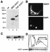
Cellular localization of AGO1. (A) Western blot with anti-BB2 antibodies of extracts from wild-type (lane 1), BB2-tagged AGO1 (lane 2), and TAP-tagged AGO1 (lane 3) cells. (B) Cells expressing TAP-tagged AGO1 were processed for indirect immunofluorescence as described previously (26) by staining with a rabbit anti-protein A antibody (subpanel AGO1). DNA was stained with 4′,6′-diamidino-2-phenylindole (subpanel DAPI). The small dots represent the kinetoplast DNA. (C) Western blot of TAP-tagged AGO1 with a rabbit anti-protein A polyclonal antibody of equivalent amounts of total extract (lane 1), postnuclear pellet (lane 2), supernatant (lane 3), S100 fraction (lane 4), and ribosomal pellet (lane 5). (D) Sucrose density gradient analysis of a cytoplasmic extract from T. brucei cells expressing TAP-tagged AGO1 (9). The upper panel shows the absorbance profile at 254 nm, and the positions of the 80S monosome and polyribosomes are indicated. The lower panel shows a Western blot analysis of the sucrose density gradient fractions using a rabbit anti-protein A polyclonal antibody.
FIG. 4.
FIG. 4.
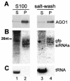
AGO1 is associated with siRNAs. S100 and ribosome salt-washed fractions from cells expressing BB2-tagged AGO1 and GFP dsRNA were subjected to immunoprecipitations with anti-BB2 antibodies, and supernatant (S) and pellet fractions (P) were processed for Western blot analysis for AGO1 (A) and for Northern blot analysis for GFP siRNAs (B), as well as for initiator methionyl tRNA to control for immunoprecipitation specificity (C). A DNA size marker of 26 nt is indicated in panel B.
FIG. 5.
FIG. 5.
AGO1 cofractionates with GFP siRNAs. The salt-extracted material from the ribosome pellet of cells expressing BB2-tagged AGO1 and GFP dsRNA was loaded onto a Superdex 200 column, and selected fractions were subjected to Western blot analysis with anti-BB2 antibodies (A) and Northern blotted for the presence of GFP siRNAs (B). The elution positions of carbonic anhydrase (30 kDa), bovine serum albumin (66 kDa), and thyroglobulin (669 kDa) are indicated. A DNA size marker of 26 nt is indicated in panel B.
FIG. 6.
FIG. 6.
siRNA accumulation is dependent on AGO1. (A) The level of Ingi siRNAs was probed by Northern blotting in wild-type cells (wt; lane 1) and four independently cloned cell lines deficient in the AGO1 protein (ago1−/−; lanes 2 to 5). RNA size markers in nucleotides are indicated on the right. The hybridization to initiator methionyl tRNA (tRNA) served as a loading control. (B) Abundance of Ingi siRNAs in wild-type (lane 1), AGO1-deficient (lane 2), BB2-AGO1-complemented (lane 3), and TAP-AGO1-complemented (lane 4) cells. The level of 5S rRNA was used as a loading control. (C) Northern blot analysis for GFP siRNAs of RNA isolated from wild-type cells expressing GFP dsRNA (lane 1) and four independent clonal ago1−/− cell lines expressing GFP dsRNA (lanes 2 to 5). RNA size markers in nucleotides are indicated on the left. The hybridization to initiator methionyl tRNA (tRNA) served as a loading control. (D) Detection of GFP dsRNA in the cell lines described in panel C, with β-tubulin mRNA (β-tub) serving as a loading control.
FIG. 7.
FIG. 7.
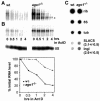
AGO1 affects the accumulation of Ingi and SLACS transcripts. (A) Northern blot of Ingi transcripts in wild-type (wt) and ago1−/− cells over a period of 4 h following the addition of actinomycin D. The asterisk indicates a transcript which appeared to be more long lived in ago1−/− cells than in wild-type cells. (B) Northern blot of SLACS transcripts in wild-type and ago1−/− cells over a period of 4 h following the addition of actinomycin D (upper panel). Hybridization to 5S RNA served as a loading control (middle panel). SLACS transcripts were quantitated relative to 5S RNA (lower panel). (C) Newly synthesized RNA from wild-type and ago1−/− cells was hybridized to the following DNAs spotted onto a nitrocellulose filter: large rRNAs (rib), 5S rRNA (5S), α-tubulin (tub), SLACS, and Ingi. The numbers next to the SLACS and Ingi hybridizations indicate the fold increase in ago1−/− cells relative to wild-type cells. The experiment was repeated three times, and the standard deviation is indicated.

Similar articles

Cited by

References

    1. Aksoy, S., T. M. Lalor, J. Martin, L. H. Van der Ploeg, and F. F. Richards. 1987. Multiple copies of a retroposon interrupt spliced leader RNA genes in the African trypanosome, Trypanosoma gambiense. EMBO J. 6:3819-3826. - PMC - PubMed
    1. Bastin, P., Z. Bagherzadeh, K. R. Matthews, and K. Gull. 1996. A novel epitope tag system to study protein targeting and organelle biogenesis in Trypanosoma brucei. Mol. Biochem. Parasitol. 77:235-239. - PubMed
    1. Bohmert, K., I. Camus, C. Bellini, D. Bouchez, M. Caboche, and C. Benning. 1998. AGO1 defines a novel locus of Arabidopsis controlling leaf development. EMBO J. 17:170-180. - PMC - PubMed
    1. Carmell, M. A., Z. Xuan, M. Q. Zhang, and G. J. Hannon. 2002. The Argonaute family: tentacles that reach into RNAi, developmental control, stem cell maintenance, and tumorigenesis. Genes Dev. 16:2733-2742. - PubMed
    1. Catalanotto, C., G. Azzalin, G. Macino, and C. Cogoni. 2002. Involvement of small RNAs and role of the qde genes in the gene silencing pathway in Neurospora. Genes Dev. 16:790-795. - PMC - PubMed

Publication types

LinkOut - more resources