. 2003 Dec 12:1:2.
doi: 10.1186/1741-7007-1-2.

TbAGO1, an argonaute protein required for RNA interference, is involved in mitosis and chromosome segregation in Trypanosoma brucei

Affiliations

TbAGO1, an argonaute protein required for RNA interference, is involved in mitosis and chromosome segregation in Trypanosoma brucei

Mickaël Durand-Dubief et al. BMC Biol. .

Abstract

Background: RNA silencing processes are widespread in almost all eukaryotic organisms. They have various functions including genome protection, and the control of gene expression, development and heterochromatin formation. RNA interference (RNAi) is the post-transcriptional destruction of RNA, which is mediated by a ribonucleoprotein complex that contains, among several components, RNA helicases and Argonaute proteins. RNAi is functional in trypanosomes, protozoan parasites that separated very early from the main eukaryotic lineage and exhibit several intriguing features in terms of the control of gene expression. In this report, we investigated the functions of RNAi in Trypanosoma brucei.

Results: By searching through genome databases, novel Argonaute-like proteins were identified in several protozoa that belong to the kinetoplastid order, a group of organisms that diverged early from the main eukaryotic lineage. T. brucei possesses two Argonaute-like genes termed TbAGO1 and TbPWI1. Dual transient transfection assays suggest that TbAGO1, but not TbPWI1, is involved in RNAi. The entire coding region of TbAGO1 was deleted by double gene knockout. TbAGO1-/- cells turned out to be completely resistant to RNAi generated either by transfected double-stranded RNA or by expression of an inverted repeat. TbAGO1-/- cells were viable but showed a dramatically reduced growth rate. This was probably due to defects in mitosis and abnormal chromosome segregation as revealed by in situ analysis. The RNAi and growth phenotypes were complemented by the inducible expression of a GFP::TbAGO1 fusion protein that revealed the cytoplasmic location of the protein.

Conclusions: The requirement of TbAGO1 for RNAi in trypanosomes demonstrates the evolutionary ancient involvement of Argonaute proteins in RNAi silencing processes. RNAi-deficient TbAGO1-/- cells showed numerous defects in chromosome segregation and mitotic spindle assembly. We propose a working hypothesis in which RNAi would be involved in heterochromatin formation at the centromere and therefore in chromosome segregation.


Figures
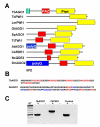
Figure 1
Figure 1
Argonaute-like proteins in protists. A. Schematic representation of Argonaute-like proteins. TbAGO1, Trypanosoma brucei AGO1 (accession number AY433802); TbPWI1, Trypanosoma brucei PWI1 (AY433803); LmPWI1, Leishmania major AGO1 (AL446005); GlAGO1, Giardia intestinalis AGO1 (AY142143); SpAGO1 Schizosaccharomyces pombe AGO1 (CAA19275); TtTWI1 Tetrahymena thermophila TWI1 (AB084111); AtAGO1, Arabidopsis thaliana AGO1 (U91995), CeRDE1, Caenorhabditis elegans RDE1 (AF180730); NcQDE2, Neurospora crassa QDE2 (AF217760); DmAGO2, Drosophila melanogaster AGO2 (NM168626). The red and yellow boxes indicate PAZ and Piwi domains respectively. The TbAGO1 RGG box is shown in green, the DmAGO2 poly-Q box is shown in blue and the N-terminal extension of AtAGO1 is annotated as a poly-Q box but also containing RGG motives and is shown in blue and green. B. Amino-terminal extensions of TbAGO1 and AtAGO1 contain RGG boxes. The RGG motifs are shown in red and the poly-Q box in blue. C. TbAGO1 and TbPWI1 RNA are expressed in the culture-adapted procyclic stage of T. brucei. Total RNA extracted from wild-type trypanosomes was incubated with (+) or without (-) reverse transcriptase and PCR-amplified using specific primers for the indicated genes.
Figure 2
Figure 2
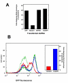
TbAGO1 is required for RNAi in transient transfection assays. A. Wild-type trypanosomes were first electroporated with dsRNA corresponding to the indicated genes, returned to culture for 10 h and then transfected with PFRA dsRNA. Samples were fixed 15 h later and cells exhibiting PFRA silencing were identified by immunofluorescence using an anti-PFRA specific monoclonal antibody. The experiment was repeated six times, yielding similar results. B. FACS analysis of trypanosomes transformed with plasmid pGFPTbAGO1430 grown in the absence (non-induced; thin green line) or in the presence of tetracycline (induced; thick lines). Induced cells were not transfected (thick green line) or transfected with TbAGO1 dsRNA (thick red line) or with TbGRP1 dsRNA as negative control (thick blue line). GFP::TbAGO1 protein levels were monitored by FACS analysis. Only TbAGO1 dsRNA was able to reduce GFP::TbAGO1 fluorescent protein levels (notice shift of thick red line compared to blue or green thick lines). Cells were transfected with the PFRA dsRNA 10 h later and PFRA silencing was monitored as above (inset). This experiment was carried out twice and yielded similar results.
Figure 3
Figure 3
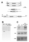
Generation of TbAGO1-/- (KO) and TbAGO1-/- +GFP::TbAGO1Ti cell lines. A. Schematic representation of the endogenous TbAGO1 locus (top), and of the constructs used for double TbAGO1 knock-out (middle) and for expression of GFP::TbAGO1 fusion protein (bottom). Large and small boxes represent protein coding sequences and TbAGO1 flanking regions used for the knockout construct. The tetracycline-inducible EP promoter of the pGFP::TbAGO1430 vector is indicated by a star. B. Southern blot analysis showing the absence of TbAGO1 from the TbAGO1-/- (KO) cell line. Genomic DNA was purified from the indicated cell lines, digested with Sca I and Xba I, and fragments were separated and transferred on a nylon membrane for hybridisation with a TbAGO1 probe. C. Northern blot analysis. Total RNA (10 μg) from the indicated cell lines was run on a gel and transferred onto a nylon membrane for hybridisation with a TbAGO1 probe (top panel) or with an α-tubulin probe (central panel). The bottom panel shows ethidium bromide staining of the membrane after transfer.
Figure 4
Figure 4
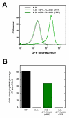
TbAGO1-/- cells are resistant to RNAi. A. FACS profile of TbAGO1-/- (KO), or of TbAGO1-/- +GFP::TbAGO1Ti cells grown in the absence (-TET, non-induced, thin green lines) or in the presence of tetracycline (+TET, induced, thick green lines). B. The indicated cell lines were transfected with PFRA dsRNA and returned to culture for 15 h before fixation and monitoring of the RNAi phenotype by immunofluorescence.
Figure 5
Figure 5
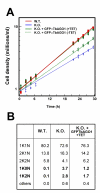
TbAGO1-/- cells show delayed growth rate and abnormal cell types. A. Growth curve of wild type (W.T., red squares), TbAGO1-/- (K.O., blue crosses), or of TbAGO1-/- +GFP::TbAGO1Ti cells grown in the absence (-TET, non-induced, open diamonds and dotted green line) or in the presence of tetracycline (+TET, induced, closed diamonds and full green line). B. Analysis of cell types within populations of wild-type (W.T.), TbAGO1-/- (K.O.), or of TbAGO1-/- +GFP::TbAGO1Ti cells grown in the presence of tetracycline (K.O.+GFP::TbAGO1+TET). 1K1N, cells with one kinetoplast and one nucleus; 2K1N, cells with two kinetoplasts and one nucleus; 2K2N, cells with two kinetoplasts and two nuclei; 1K0N, cells with one kinetoplast but without nucleus (zoids) and 1K2N, cells with one kinetoplast and two nuclei. More than 1 000 cells were counted per experiment.
Figure 6
Figure 6
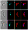
Spindle formation is frequently aberrant in TbAGO1-/- cells. Trypanosomes were stained with the anti-tubulin KMX-1 monoclonal antibody and counterstained with DAPI to show nuclear and mitochondrial DNA. Mitotic cells from wild type (WT, top panels) or TbAGO1-/- (KO, central and bottom panels). From left to right, phase contrast image, DAPI staining (blue), tubulin staining (red) and merged DAPI and tubulin staining. The typical elongated mitotic spindle found in wild-type trypanosomes is frequently poorly developed (central panels) or completely bent on one side of the nucleus (bottom panels) in TbAGO1-/- cells.
Figure 7
Figure 7
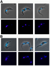
Nucleolus segregation is not equal in TbAGO1-/- cells. Immunofluorescence of trypanosomes stained with the anti-nucleolus marker L1C6 monoclonal antibody and counterstained with DAPI to show nuclear and mitochondrial DNA. A. Wild-type trypanosomes, B. TbAGO1-/- trypanosomes. In both cases, the top series shows a phase contrast image superimposed onto the DAPI image (blue), and the bottom series shows the DAPI image (blue) merged with nucleolus staining (red). Left panels, cells at the anaphase stage of nuclear mitosis. Middle and right panels, cells at the telophase stage of nuclear mitosis. In wild-type cells (A), both nuclei inherit similar amounts of nucleolar material, whereas clear differences are visible between the two nuclei in a large number of binucleated cells from the TbAGO1-/- mutant (B).
Figure 8
Figure 8
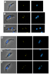
Chromosome segregation is modified in TbAGO1-/- cells. Wild-type (A-C) and TbAGO1-/- (D-F) cells were fixed and processed for FISH using a probe for the 5S tandem repeats, localised on chromosome I and stained with DAPI. Left, phase-contrast image merged to DAPI (blue), middle, chromosome I signal (yellow) and right, merged DAPI-FISH signals. A. G1 cell; B-F. Mitotic cells. Defects in chromosome segregation are frequently observed in mutant cells.
Figure 9
Figure 9
GFP::TbAGO1 is localised in the cytoplasm. Live cells from cell line TbAGO1-/- +GFP::TbAGO1Ti were observed by phase contrast (top panels) or by direct GFP fluorescence (bottom panels, images were not digitally enhanced). The position in the cell cycle can be evaluated by the number and position of flagellar pockets (indicated by yellow stars) and the distance separating them. Arrows indicate the position of the nucleolus (dark spot within the nucleus). A. Cell with one kinetoplast and one nucleus. B, C. Cells with two kinetoplasts and one mitotic nucleus. D. Post-mitotic cell with two kinetoplasts and two nuclei.

Similar articles

Cited by

References

    1. van der Krol AR, Mur LA, Beld M, Mol JN, Stuitje AR. Flavonoid genes in petunia: addition of a limited number of gene copies may lead to a suppression of gene expression. Plant Cell. 1990;2:291–299. doi: 10.1105/tpc.2.4.291. - DOI - PMC - PubMed
    1. Cogoni C, Irelan JT, Schumacher M, Schmidhauser TJ, Selker EU, Macino G. Transgene silencing of the al-1 gene in vegetative cells of Neurospora is mediated by a cytoplasmic effector and does not depend on DNA-DNA interactions or DNA methylation. EMBO J. 1996;15:3153–3163. - PMC - PubMed
    1. Ruiz F, Vayssie L, Klotz C, Sperling L, Madeddu L. Homology-dependent gene silencing in Paramecium. Mol Biol Cell. 1998;9:931–943. - PMC - PubMed
    1. Fire A, Xu S, Montgomery MK, Kostas SA, Driver SE, Mello CC. Potent and specific genetic interference by double-stranded RNA in Caenorhabditis elegans. Nature. 1998;391:806–811. doi: 10.1038/35888. - DOI - PubMed
    1. Elbashir SM, Lendeckel W, Tuschl T. RNA interference is mediated by 21- and 22-nucleotide RNAs. Genes Dev. 2001;15:188–200. doi: 10.1101/gad.862301. - DOI - PMC - PubMed

Publication types

MeSH terms

Substances

LinkOut - more resources