. 2004 Feb;15(2):575-87.
doi: 10.1091/mbc.e03-06-0401. Epub 2003 Dec 2.

The zinc transporter ZnT3 interacts with AP-3 and it is preferentially targeted to a distinct synaptic vesicle subpopulation

Affiliations

The zinc transporter ZnT3 interacts with AP-3 and it is preferentially targeted to a distinct synaptic vesicle subpopulation

Gloria Salazar et al. Mol Biol Cell. 2004 Feb.

Abstract

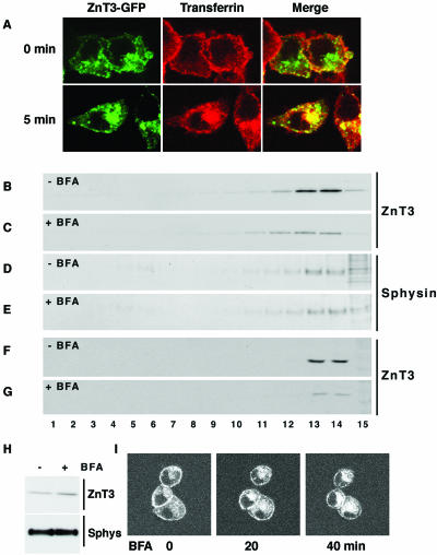
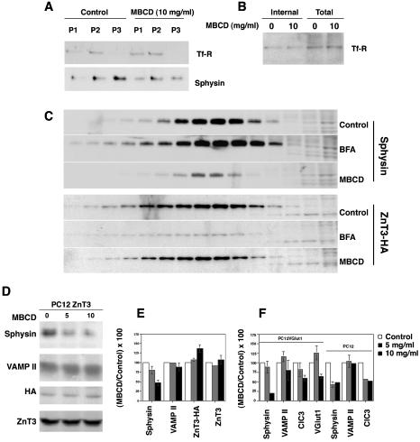
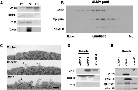
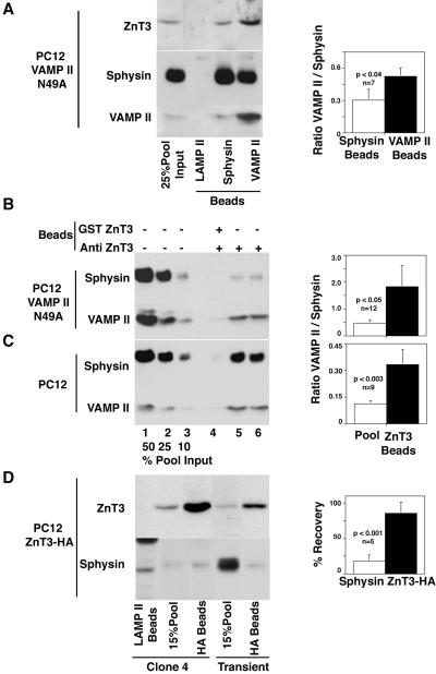
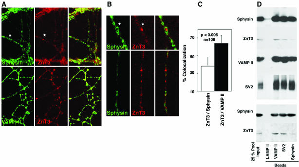
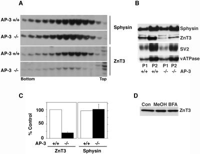
Synaptic vesicles (SV) are generated by two different mechanisms, one AP-2 dependent and one AP-3 dependent. It has been uncertain, however, whether these mechanisms generate SV that differ in molecular composition. We explored this hypothesis by analyzing the targeting of ZnT3 and synaptophysin both to PC12 synaptic-like microvesicles (SLMV) as well as SV isolated from wild-type and AP-3-deficient mocha brains. ZnT3 cytosolic tail interacted selectively with AP-3 in cell-free assays. Accordingly, pharmacological disruption of either AP-2- or AP-3-dependent SLMV biogenesis preferentially reduced synaptophysin or ZnT3 targeting, respectively; suggesting that these antigens were concentrated in different vesicles. As predicted, immuno-isolated SLMV revealed that ZnT3 and synaptophysin were enriched in different vesicle populations. Likewise, morphological and biochemical analyses in hippocampal neurons indicated that these two antigens were also present in distinct but overlapping domains. ZnT3 SV content was reduced in AP-3-deficient neurons, but synaptophysin was not altered in the AP-3 null background. Our evidence indicates that neuroendocrine cells assemble molecularly heterogeneous SV and suggests that this diversity could contribute to the functional variety of synapses.


Figures

Figure 1.
Figure 1.
AP-3 interacts with the carboxy-terminal tail of ZnT3. (A) Glutathione beads were coated without (lanes 2, 3, 6, 7, 10, and 11) or with equal amounts of GST fusion proteins encompassing the carboxy-terminal cytosolic domain of synaptophysin (sphysin, lanes 4-5), the ZnT3 amino-terminal (lanes 8-9), and carboxy-terminal tails (lanes 12-13). Bead-bound fusion proteins were incubated in the absence (even lanes) or presence of rat brain cytosol (odd lanes). After washing, protein complexes bound to beads were analyzed by immunoblot with anti-delta antibodies (top) and the GST fusion proteins by Coomassie staining (bottom). Lane 1 represents 1/50 of the brain cytosol input. Only the carboxy-terminal tail of ZnT3 bound AP-3 from rat brain cytosol (n = 3). (B) Tagged neuronal AP-3 subunits are assembled in AP-3 complexes by HEK293 cells. HEK293 cells were transiently transfected with empty vector (lanes 1 and 4), myc-His6-tagged-μ3b (lanes 2 and 5), or myc-His6-tagged-β3b (lanes 3 and 6). Detergent cell extracts were immunoprecipitated using preimmune or σ3 antibodies, and the immunocomplexes were resolved in SDS-PAGE gels. Tagged subunits were detected in the immunoprecipitated material by using anti-myc 9E10 antibodies, indicating their incorporation into AP-3 complexes. (C) Tagged subunits incorporation into in AP-3 complexes was analyzed by sucrose gradient sedimentation (Dell'Angelica et al., 1997) and compared with HEK293 endogenous AP-3 complexes. Immunoblots with β3 or myc epitope antibodies were used to identify the complexes. No differences in the sedimentation between endogenous AP-3 and AP-3 containing tagged subunits were detected. Thyroglobulin (Thyr, 669 kDa), albumin (BSA, 66 kDa), and carbonic anhydrase (Anh, 29 kDa) were used as molecular weight markers. (D) Neuronal AP-3 interacts with the carboxy-terminal tail of ZnT3. Myc-His6-tagged neuronal AP-3 reconstituted in HEK293 cells was bound to nonantibody-coated (lanes 2, 5, and 8) or 9E10 antibody-coated protein G beads (lanes 3, 4, 6, 7, 9, and 10). Lane 1 corresponds to input. Beads were incubated with equal amounts of the GST fusion proteins encoding the synaptophysin and ZnT3 cytosolic tails. After washing, complexes were resolved by SDS-PAGE and analyzed by immunoblot. Bound AP-3 and GST fusion proteins were detected with antibodies against the hinge domain of β3B and GST, respectively. Only the carboxy-terminal end of ZnT3 is retained by the neuronal AP-3 complex (n = 3).
Figure 2.
Figure 2.
ZnT3 targeting from endosomes is perturbed by brefeldin A. (A) ZnT3 is localized in early endosomes. To label cell surface, PC12 cells carrying ZnT3-GFP were incubated at 4°C with Alexa568-transferrin (50 μg/ml) and washed to remove unbound ligand. Cell were kept either at 4°C or transferred to 37°C to resume internalization. Assays were stopped at 0°C. Perinuclear ZnT3 is extensively decorated by internalized transferrin, indicating its early endosome nature. (B-G) Brefeldin A preferentially hinders ZnT3-HA targeting to SLMV. ZnT3 clone 4 PC12 cells (B-E) or untransfected PC12 cells (F and G) were incubated in the absence (B, D, and F) or presence of brefeldin A (10 μg/ml; C, E, and G) for 2 h. Cells were homogenized and equal protein amounts of SLMV-enriched S1 supernatants were resolved in 10-45% sucrose velocity gradients (Lichtenstein et al., 1998). Fractions were analyzed by immunoblot with antibodies against synaptophysin (sphysin) and ZnT3. Although SLMV synaptophysin content was minimally affected by brefeldin A, ZnT3 was robustly reduced. Brefeldin A reduces the targeting of endogenous ZnT3 to SLMV (compare F and G). (H) ZnT3 present in SLMV is redistributed to P1 membranes were ZnT3 content increases after drug treatment. (I) Endosomal ZnT3-GFP perinuclear distribution is modified by brefeldin A. PC12 cells transfected with ZnT3-GFP were incubated at 37°C in the presence of brefeldin A (10 μg/ml) and imaged by time-lapse confocal microscopy. Time 0 represents a frame taken before the addition of the drug. Brefeldin A induced a redistribution of ZnT3-GFP. Shown a representative section from a Z-time series (n = 3).
Figure 3.
Figure 3.
Differential effects of plasma membrane cholesterol depletion upon synaptic vesicle protein targeting to SLMV. (A) PC12 cells were incubated in the absence or presence of MBCD (10 mg/ml) for 45 min at 37°C to deplete plasma membrane cholesterol. Incubations were stopped at 4°C and cell homogenates fractionated as described in MATERIALS AND METHODS. Equal protein amounts from pellets P1-P3 were resolved on SDS-PAGE gels and the distribution of TfR and synaptophysin (sphysin) was determined by immunoblot. P3 pellets were substantially enriched in SV markers, yet devoid of detectable transferrin receptor. Synaptophysin content on SLMV (P3) was substantially reduced after MBCD. (B) Cholesterol depletion does not modify the intracellular pool of transferrin receptor. PC12 cells were biotinylated at 4°C with the disulfide cleavable biotinylation agent NHS-Biotin. Cells were warmed to 37°C, in either the absence or presence of MBCD for 45 min. Reactions were stopped and the cell surface biotin was removed with glutathione at 4°C. Cell extracts were precipitated with streptavidin-agarose beads and the presence of transferrin receptor was assessed by immunoblot. No differences in the intracellular pool of transferrin receptor were detected, therefore excluding a generalized endocytic trafficking defect induced by cholesterol depletion. (C) Cholesterol depletion selectively reduces synaptophysin targeting to SLMV. PC12 cells transfected with ZnT3-HA were treated in the absence or presence MBCD (10 mg/ml) or BFA (10 μg/ml). Reactions were stopped in ice and equal protein amounts of cell homogenate were resolved by differential and glycerol gradient centrifugation. SLMV content of synaptophysin and ZnT3 were determined by immunoblot. In contrast to ZnT3, synaptophysin targeting is affected by cholesterol depletion but not by BFA (n = 3). (D) ZnT3 targeting to SLMV is not affected by cholesterol depletion in P3 membranes. Untransfected PC12 or cells transfected with ZnT3-HA were treated or not with MBCD. Equal amounts of P3 protein were analyzed by immunoblot. Although the synaptophysin content on P3 vesicles was substantially reduced after MBCD treatment, both endogenous (ZnT3) and HA-tagged ZnT3 (ZnT3-HA) did not exhibit decreased content on SLMV after cholesterol depletion. (E) Results expressed as percentages of the control value obtained in the D). (F) Cholesterol depletion affects the SLMV targeting of different SV proteins. PC12 cells or a clone bearing Vglut1 were treated with increasing concentrations MBCD. P3 SLMV were isolated and their content of synaptophysin (sphysin), VAMP II, ClC3, and Vglut1 was determined by immunoblot on equal amounts of P3 protein. Results are expressed as percentages of the control value. SLMV protein contents in the absence of drug were arbitrarily set to 100%. PC12Vglut1 (n = 2), PC12 (n = 7), and PC12 ZnT3-HA (n = 3). Absent error bars correspond to errors below the graphing threshold.
Figure 4.
Figure 4.
Biochemical and ultrastructural characterization of the ZnT3 and synaptophysin-containing SLMV. (A) PC12 clone 4 cell expressing ZnT3-HA were differentially fractionated to generate P1, P2, and SLMV-enriched S2 fractions. Equal protein amounts of these fractions were resolved in SDS-PAGE and tested by immunoblot with antibodies against ZnT3, KDELr, TrfR, and TGN38. S2 fraction does not contain appreciable contamination with exocytic or endocytic membranes. (B) SLMV-enriched S2 fractions from PC12 clone 4 cells were size fractionated in 5-25% glycerol gradients and probed with antibodies against synaptophysin, VAMP II, and ZnT3. All SV proteins comigrated on the same region of the gradient. (C) Magnetic beads coated with anti-LAMP II antibodies (control), anti-synaptophysin, and anti-HA tag (ZnT3) were incubated with glycerol gradient-purified vesicles (SMLV pool, B). Beads were extensively washed and processed for transmission electron microscopy. Asterisks mark isolated vesicles bound over the horizon of the magnetic bead. Size distribution analysis of these vesicles was identical for ZnT3 and synaptophysin isolated SLMV. (D) and E) SLMV were isolated with control beads coated with an antibody against LAMP II or with antibodies against VAMP II or rabaptin 5 (rabapt5). Beads bound membranes were resolved by SDS-PAGE and analyzed by immunoblot against the antigens described in the figure. VAMP II concentrated SLMV as assessed by ZnT3, VAMP II, and synaptophysin immunoreactivity but not other membranes. Rabaptin 5-coated beads did not enriched SLMV markers. Double asterisks denote IgG chains.
Figure 5.
Figure 5.
ZnT3 and synaptophysin are enriched in different SLMV populations. (A) Glycerol gradient isolated SLMV derived from PC12 VAMP II N49A cells were immuno-magnetically isolated with beads coated with control (LAMP II), synaptophysin, or VAMP II antibodies. After washing, bead-bound immuno-complexes were resolved in SDS-PAGE gels and analyzed by immunoblot with antibodies against VAMP II and synaptophysin. Isolations performed with VAMP II antibodies consistently brought down vesicles enriched in VAMP II over synaptophysin compared with vesicles isolated with synaptophysin antibodies. Control beads did not bind SLMV. First lane from the left represents 25% of the vesicle input. Quantifications of seven assays performed in three independent experiments are shown to the right. Glycerol gradient-isolated SLMV derived from PC12 VAMP II N49A cells (B) or untransfected PC12 cells (C) were immuno-magnetically isolated with beads coated with affinity-purified anti-ZnT3 amino-terminal antibodies. Controls were performed by competing vesicle binding with an excess of the ZnT3 amino-terminal GST fusion protein. Immunocomplexes were resolved by SDS-PAGE and analyzed by immunoblot. Depicted from left to right are decreasing concentrations of the SLMV input (percentage of the total), control reaction competed with the amino-terminal tail of ZnT3, and two immuno-magnetic anti-ZnT3 isolations. ZnT3 isolated vesicles are enriched in VAMP II over synaptophysin at least threefold compared with the SLMV pool. Right, quantifications of 12 and 9 assays performed in four and three independent experiments, respectively. (D) Glycerol gradient isolated SLMV derived from PC12 ZnT3-HA clone 4 cells or transiently transfected cells were immuno-magnetically isolated with beads coated with control (LAMP II, first lane from the left) or anti-HA antibodies. Although nearly all ZnT3-HA-containing vesicles were isolated with anti-HA antibodies, the immuno-magnetic beads only retrieved a 17% of all the vesicle-bound synaptophysin protein present in the assay. Low levels of synaptophysin were also detected in SLMV isolated with anti-HA antibodies, by using vesicles purified from transiently transfected cells. Quantifications of five assays performed in three independent experiments with SLMV derived from clone 4 are shown at the right. Values represent the percentage of recovery of the total amount of vesicle-bound ZnT3 and synaptophysin added to the assay.
Figure 6.
Figure 6.
Localization of ZnT3 and SV antigens in primary cultures of hippocampal neurons. Primary cultures of hippocampal neurons (14 DIV) were double stained with antibodies against ZnT3, synaptophysin, or VAMP II antibodies. Images of the processes were acquired by confocal microscopy. (B) Magnification of the area marked by an asterisk in A, and a process from a field obtained in an independent experiment. Asterisk show regions positive for ZnT3, but with reduced levels of synaptophysin immunoreactivity. The extent of colocalization was determined in single optical sections of isolated processes (14 DIV). (C) Quantification of the extent of colocalization between ZnT3 and the other SV antigens by using MetaMorph. (D) Purified brain SV were immuno-magnetically isolated with beads coated with control (LAMP II), synaptophysin, SV2, or VAMP II antibodies. Beads were washed and immuno-complexes were resolved in SDS-PAGE gels and analyzed by immunoblot with antibodies against ZnT3, VAMP II, SV2, and synaptophysin. Isolations performed with VAMP II and SV2 antibodies consistently brought down vesicles containing ZnT3. In contrast, isolations performed with synaptophysin antibodies retrieved low levels of vesicle-associated ZnT3. Control beads did not bind SV. First lane from the left represents 25% of the vesicle input. Depicted are two representative experiments of four performed at least in duplicate. Two independent SV preparations were used (top and bottom).
Figure 7.
Figure 7.
ZnT3 and synaptophysin targeting to synaptic vesicles from AP-3-deficient mouse. High-speed supernatants obtained from wild-type and mocha brains were sedimented in 5-25% glycerol velocity gradients to resolve SV. SV were identified by immunoblot with antibodies against synaptophysin (sphysin) and ZnT3. (A) In contrast to synaptophysin SV, ZnT3 content is decreased in AP-3 deficiencies. (B) Immunoblot analysis of synaptophysin, ZnT3, SV2, and the p116 subunit of the vacuolar ATPase in P1 and P2 membranes of wild-type and mocha brain membranes. ZnT3 is the only SV antigen whose levels are affected by the mocha allele. (C) Quantitation of ZnT3 and synaptophysin content in the synaptic vesicle peak (fractions 7-9) in four independent experiments. A similar decrease in ZnT3 content was observed by quantifying the transporter in P1 or P2 membranes (n = 5). (D) Brefeldin A-induced block in ZnT3 targeting to SLMV does not decrease the total cellular levels of ZnT3. PC12 ZnT3-HA clone 4 cells were incubated with BFA as described in Figures 1 and 2. The total cellular levels of ZnT3 were determined by immunoblot. ZnT3 cell content remained constant in the presence of BFA (n = 2).
Figure 8.
Figure 8.
Hippocampal neuron secretory compartments are heterogeneous in composition. (A) Hippocampal neurons were zinc-loaded and double stained with zinquin and LysoSensor DND-189 and the excess of dyes washed away. Neurons were time-lapse imaged and challenged with 10 nM α-latrotoxin at 37°C. LysoSensor DND-189 fluorescence is translocated to the plasma membrane and then decreases, whereas the zinquin signal is progressively reduced over time. The effect of the toxin was monitored in the transmitted image channel through the bouton engorgement. Depicted is one experiment of three performed in duplicate. (B) Hippocampal neurons (14 DIV) were double-labeled as described. Cell were washed and simultaneously imaged in vivo by two-photon microscopy (zinquin) and confocal microscopy (LysoSensor DND-189). Asterisks (top) mark region magnified in the bottom. Boutons and interbouton processes were stained with both dyes; however, zinquin-positive areas were not stained to the same extent with Lysosensor. Arrowheads depict an individual process were boutons positive for zinquin show a wide range of LysoSensor staining. Note the zinquin-positive boutons that do not stain with LysosSensor. Arrows mark LysoSensor-positive puncta that do not stain for zinquin. Similar results were obtained in five independent experiments performed at least in duplicate.

Similar articles

Cited by

References

    1. Atwood, H.L., and Karunanithi, S. (2002). Diversification of synaptic strength: presynaptic elements. Nat. Rev. Neurosci. 3, 497-516. - PubMed
    1. Baumert, M., Maycox, P.R., Navone, F., De Camilli, P., and Jahn, R. (1989). Synaptobrevin: an integral membrane protein of 18, 000 daltons present in small synaptic vesicles of rat brain. EMBO J. 8, 379-384. - PMC - PubMed
    1. Bellocchio, E.E., Reimer, R.J., Fremeau, R.T., Jr., and Edwards, R.H. (2000). Uptake of glutamate into synaptic vesicles by an inorganic phosphate transporter. Science 289, 957-960. - PubMed
    1. Bennett, M.K., Calakos, N., Kreiner, T., and Scheller, R.H. (1992). Synaptic vesicle membrane proteins interact to form a multimeric complex. J. Cell Biol. 116, 761-775. - PMC - PubMed
    1. Blagoveshchenskaya, A.D., Hewitt, E.W., and Cutler, D.F. (1999). A complex web of signal-dependent trafficking underlies the triorganellar distribution of P-selectin in neuroendocrine PC12 cells. J. Cell Biol. 145, 1419-1433. - PMC - PubMed

Publication types

MeSH terms

LinkOut - more resources