Gene expression in the normal adult human kidney assessed by complementary DNA microarray
- PMID: 14657249
- PMCID: PMC329285
- DOI: 10.1091/mbc.e03-06-0432
Gene expression in the normal adult human kidney assessed by complementary DNA microarray
Abstract
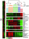
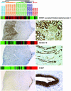
The kidney is a highly specialized organ with a complex, stereotyped architecture and a great diversity of functions and cell types. Because the microscopic organization of the nephron, the functional unit of the kidney, has a consistent relationship to the macroscopic anatomy of the kidney, knowledge of the characteristic patterns of gene expression in different compartments of the kidney could provide insight into the functions and functional organization of the normal nephron. We studied gene expression in dissected renal lobes of five adult human kidneys using cDNA microarrays representing approximately 30,000 different human genes. Total RNA was isolated from sections of the inner and outer cortex, inner and outer medulla, papillary tips, and renal pelvis and from glomeruli isolated by sieving. The results revealed unique and highly distinctive patterns of gene expression for glomeruli, cortex, medulla, papillary tips, and pelvic samples. Immunohistochemical staining using selected antisera confirmed differential expression of several cognate proteins and provided histological localization of expression within the nephron. The distinctive patterns of gene expression in discrete portions of the kidney may serve as a resource for further understanding of renal physiology and the molecular and cellular organization of the nephron.
Figures


Similar articles
-
Effect of methionine sulfoximine on glutathione and amino acid levels in the nephron.Am J Physiol. 1976 Nov;231(5 Pt. 1):1536-40. doi: 10.1152/ajplegacy.1976.231.5.1536. Am J Physiol. 1976. PMID: 998800
-
Transport in isolated cells from defined nephron segments.Methods Enzymol. 1990;191:380-409. doi: 10.1016/0076-6879(90)91025-2. Methods Enzymol. 1990. PMID: 2074768 No abstract available.
-
The structural organization of the kidney of the desert rodent Psammomys obesus.Anat Embryol (Berl). 1975 Dec 23;148(2):121-43. doi: 10.1007/BF00315265. Anat Embryol (Berl). 1975. PMID: 1211658
-
Na K ATPase in the rat nephron related to sodium transport; results with quantitative histochemistry. Part II.Curr Probl Clin Biochem. 1971;3:320-44. Curr Probl Clin Biochem. 1971. PMID: 4280963 Review. No abstract available.
-
Ammonia transport in the mammalian kidney.Am J Physiol. 1985 Apr;248(4 Pt 2):F459-71. doi: 10.1152/ajprenal.1985.248.4.F459. Am J Physiol. 1985. PMID: 3885755 Review.
Cited by
-
Profiling native pulmonary basement membrane stiffness using atomic force microscopy.Nat Protoc. 2024 May;19(5):1498-1528. doi: 10.1038/s41596-024-00955-7. Epub 2024 Mar 1. Nat Protoc. 2024. PMID: 38429517 Review.
-
Isolation of Rat Glomeruli and Propagation of Mesangial Cells to Study the Kidney in Health and Disease.Methods Mol Biol. 2023;2664:31-39. doi: 10.1007/978-1-0716-3179-9_3. Methods Mol Biol. 2023. PMID: 37423980
-
Systems biology and machine learning approaches identify drug targets in diabetic nephropathy.Sci Rep. 2021 Dec 6;11(1):23452. doi: 10.1038/s41598-021-02282-3. Sci Rep. 2021. PMID: 34873190 Free PMC article.
-
Identification of a foetal epigenetic compartment in adult human kidney.Epigenetics. 2022 Mar;17(3):335-355. doi: 10.1080/15592294.2021.1900027. Epub 2021 Mar 30. Epigenetics. 2022. PMID: 33783321 Free PMC article.
-
Basement membrane stiffness determines metastases formation.Nat Mater. 2021 Jun;20(6):892-903. doi: 10.1038/s41563-020-00894-0. Epub 2021 Jan 25. Nat Mater. 2021. PMID: 33495631
References
-
- Akaiwa, M., Yae, Y., Sugimoto, R., Suzuki, S.O., Iwaki, T., Izuhara, K., and Hamasaki, N. (1999). Hakata antigen, a new member of the ficolin/opsonin p35 family, is a novel human lectin secreted into bronchus/alveolus and bile. J. Histochem. Cytochem. 47, 777-786. - PubMed
-
- Alizadeh, A.A. et al. (2000). Distinct types of diffuse large B-cell lymphoma identified by gene expression profiling. Nature 403, 503-511. - PubMed
-
- Amara, N., Palapattu, G.S., Schrage, M., Gu, Z., Thomas, G.V., Dorey, F., Said, J., and Reiter, R.E. (2001). Prostate stem cell antigen is overexpressed in human transitional cell carcinoma. Cancer Res. 61, 4660-4665. - PubMed
-
- Arystarkhova, E., Wetzel, R.K., Asinovski, N.K., and Sweadner, K.J. (1999). The gamma subunit modulates Na(+) and K(+) affinity of the renal Na, K-ATPase. J. Biol. Chem. 274, 33183-33185. - PubMed
-
- Bonilha, V.L., and Rodriguez-Boulan, E. (2001). Polarity and developmental regulation of two PDZ proteins in the retinal pigment epithelium. Invest. Ophthalmol. Vis. Sci. 42, 3274-3282. - PubMed
Publication types
MeSH terms
Grants and funding
LinkOut - more resources
Full Text Sources
Other Literature Sources
Miscellaneous

