. 2003 Nov;12(5):1251-60.
doi: 10.1016/s1097-2765(03)00427-1.

Direct binding of the PDZ domain of Dishevelled to a conserved internal sequence in the C-terminal region of Frizzled

Affiliations

Direct binding of the PDZ domain of Dishevelled to a conserved internal sequence in the C-terminal region of Frizzled

Hing-C Wong et al. Mol Cell. 2003 Nov.

Abstract

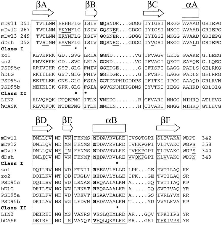
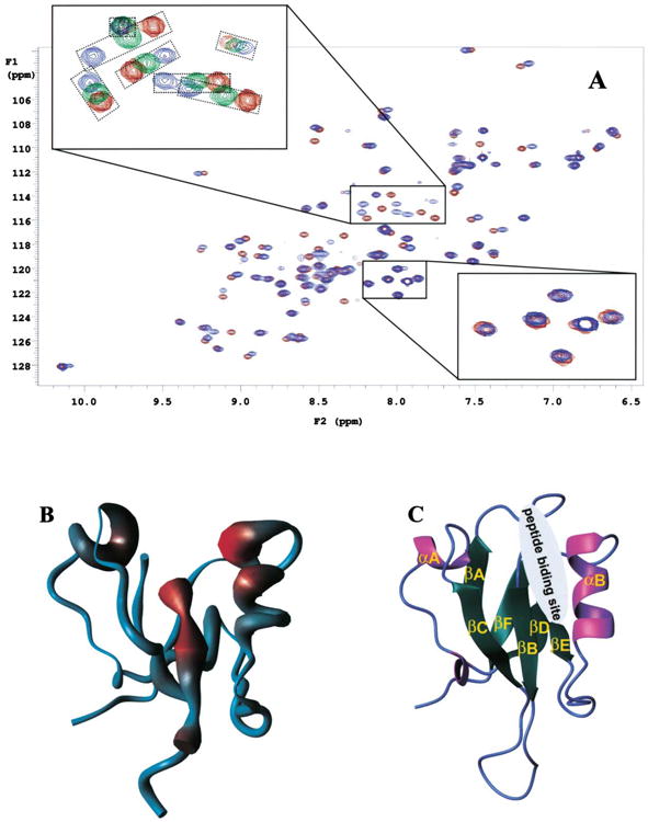
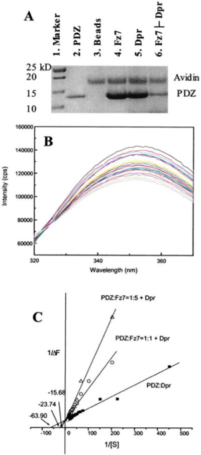
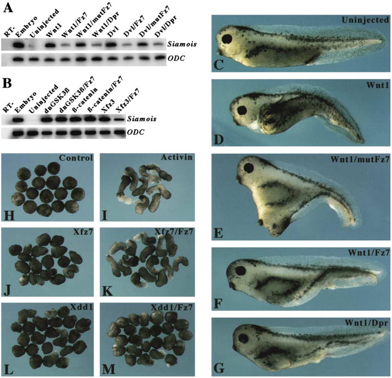
The cytoplasmic protein Dishevelled (Dvl) and the associated membrane-bound receptor Frizzled (Fz) are essential in canonical and noncanonical Wnt signaling pathways. However, the molecular mechanisms underlying this signaling are not well understood. By using NMR spectroscopy, we determined that an internal sequence of Fz binds to the conventional peptide binding site in the PDZ domain of Dvl; this type of site typically binds to C-terminal binding motifs. The C-terminal region of the Dvl inhibitor Dapper (Dpr) and Frodo bound to the same site. In Xenopus, Dvl binding peptides of Fz and Dpr/Frodo inhibited canonical Wnt signaling and blocked Wnt-induced secondary axis formation in a dose-dependent manner, but did not block noncanonical Wnt signaling mediated by the DEP domain. Together, our results identify a missing molecular connection within the Wnt pathway. Differences in the binding affinity of the Dvl PDZ domain and its binding partners may be important in regulating signal transduction by Dvl.


Figures

Figure 1
Figure 1. Structure-Based Alignment of the Amino Acid Sequences of the PDZ Domains of Dvl Homologs and Other Proteins
Secondary structural elements are indicated above the sequences. Residues at the GH positions are in boldface type. The asterisk denotes the binding pocket for the ligand's C terminus. Sequence differences among the PDZ domains are indicated by underlining.
Figure 2
Figure 2. Interaction between the mDvl1 PDZ Domain and Fz7
(A) 15N-HSQC spectra of free Fz7 peptide and Fz7 peptide bound to the PDZ domain of mDvl1. The red contour lines represent spectra of the free form of the PDZ domain when no Fz7 peptide (GKTLQSWRRFYH) was present, the green lines (upper inset) represent spectra of partially bound forms of the PDZ domain when 0.86 mM Fz7 peptide was present, and the blue lines represent spectra of the fully bound forms of the PDZ domain when 10 mM Fz7 peptide was present. The concentration of the PDZ domain was 1.1 mM. The two insets show the enlarged regions where the chemical-shift perturbations were small (lower inset) and large (upper inset). In the upper inset, the signals from the same residue but in different spectra were placed in smaller boxes. (B) The figure shows the worm representation of the backbone structure of the mDvl1 PDZ domain. The thickness of the worm is proportional to the weighted sum (in Hz) of the 1H and 15N shifts upon binding by the Fz7 peptide (see A), and the increasing chemical-shift perturbation is shown (blue, low; red, high). (C) Ribbon diagram of the PDZ domain structure. The binding site of the Fz7 peptide identified from the chemical-shift perturbation studies is indicated.
Figure 3
Figure 3. Fz7 and Dpr/Frodo Peptides and Their Binding to the PDZ Domain In Vitro
(A) Biotinylated Fz7 or Dpr/Frodo peptide was coupled to the UltraLink Immobilized Monomeric Avidin, and the avidin-coupled peptides were then incubated with purified PDZ domain. After extensive washing, peptide-interacting proteins were resolved and visualized by SDS-PAGE. Besides those bound to the PDZ domain, some proteins that became detached from the avidin-immobilized beads were also observed. Lane 1, marker. Lane 2, PDZ domain. Lane 3, The PDZ domain and the avidin-agarose beads. Lane 4, The PDZ domain and beads coupled to the Fz7 peptide. Lane 5, the PDZ domain and beads coupled to the Dpr/Frodo peptide. Lane 6: the PDZ domain bound to the Fz7 peptide-coupled beads after elution by the Dpr/Frodo peptide. The experimental condition was similar to that used for the mixture depicted in lane 4, except that before SDS-PAGE, the beads were washed with a buffer that contained Dpr/Frodo peptide. (B) Fluorescence spectra (labeled with different colors) of the PDZ domain in the presence of various concentrations of Dpr/Frodo peptides. Intrinsic tryptophan fluorescence of the PDZ domain was quenched by bound peptide. (C) Reciprocal plot of fluorescence data were fitted to a competitive inhibition model in which the Fz7 peptide disrupts the interaction between the Dpr/Frodo peptide and the PDZ domain. Extracted from the fitting, the binding affinity of Dpr/Frodo peptide and the PDZ domain was 16.1 ± 0.7 μM, and the affinity of the Fz7 peptide and the PDZ domain, extracted from the two apparent KI values at two concentrations (5.9 μM and 12.8 μM, respectively), was 9.5 ± 7.9 μM.
Figure 4
Figure 4. Effects of PDZ Binding Peptides on Canonical Wnt Signaling in Xenopus Embryos
(A) The Fz7 and Dpr/Frodo peptides inhibited the canonical Wnt pathway. RT-PCR was conducted to analyze the expression of the Xenopus Wnt target gene Siamois in ectodermal explants. Synthetic mRNA corresponding to Wnt1 (1 pg) and Dvl (500 ng) were injected alone or were coinjected with PDZ binding peptides (100 ng; corresponding concentration after injection, approximately 200 μM). Injections were administered to the animal-pole region at the 8 cell stage, and ectodermal explants were cultured until they reached the early gastrula stage, at which time they underwent RT-PCR analysis. (B) The Fz7 peptide had no effect on Siamois expression induced by dominant-negative GSK3β (dnGSK3β) or by β-catenin, but it blocked Siamois expression induced by Xfz3. RT-, whole-embryo sample analyzed in the absence of reverse transcription. ODC (ornithine decarboxylase) was used as a loading control. (C–G) Blockage of secondary axis formation by PDZ binding peptides. Ventro-vegetal injections of Wnt1 mRNA (1 pg) and PDZ binding peptides (100 ng; corresponding concentration after injection, approximately 200 μM) were done at the 4 cell stage. Embryos that received injections were cultured until they reached the larval stage; at that point they were analyzed to determine whether any secondary axis was present. (C) A control embryo that received no injection. (D) An embryo that received an injection of Wnt1 mRNA developed a complete secondary axis. (E) An embryo that received coinjections of the mutated Fz7 peptide, mutFz7, and Wnt1 mRNA developed a secondary axis. (F and G) An embryo that received coinjections of Wnt1 mRNA and either Fz7 (F) or Dpr/Frodo (G) peptide developed a partial secondary axis. (H–M) The Fz7 peptide differentially rescued the effect of Xfz7 and Xdd1 on elongation of Xenopus explants. The animal-pole region of embryos at the 4 cell stage received injections of 500 ng synthetic mRNA corresponding to Xfz7 or Xdd1 that were given alone or together with the Fz7 peptide. Ectodermal explants dissected at the early blastula stage were treated with activin and cultured until they reached the neurula stage. (H) Untreated explants from control embryos did not show elongation. (I) Activin-treated explants from control embryos exhibited extensive elongation. (J) Xfz7 blocked explant elongation induced by activin. (K) The coinjected Fz7 peptide rescued explant elongation. (L) Xdd1 blocked explant elongation to a similar degree as did Xfz7. (M) The coinjected Fz7 peptide did not rescue explant elongation.

Similar articles

Cited by

References

    1. Axelrod JD. Unipolar membrane association of Dishevelled mediates Frizzled planar cell polarity signaling. Genes Dev. 2001;15:1182–1187. - PMC - PubMed
    1. Axelrod JD, Miller JR, Shulman JM, Moon RT, Perrimon N. Differential recruitment of Dishevelled provides signaling specificity in the planar cell polarity and Wingless signaling pathways. Genes Dev. 1998;12:2610–2622. - PMC - PubMed
    1. Bezprozvanny I, Maximov A. Classification of PDZ domains. FEBS Lett. 2001;509:457–462. - PubMed
    1. Boutros M, Mlodzik M. Dishevelled: at the crossroads of divergent intracellular signaling pathways. Mech Dev. 1999;83:27–37. - PubMed
    1. Boutros M, Paricio N, Strutt DI, Mlodzik M. Dishevelled activates JNK and discriminates between JNK pathways in planar polarity and wingless signaling. Cell. 1998;94:109–118. - PubMed

Publication types

MeSH terms

Substances

Associated data