Essential roles for angiotensin receptor AT1a in bleomycin-induced apoptosis and lung fibrosis in mice
- PMID: 14633624
- PMCID: PMC1892377
- DOI: 10.1016/S0002-9440(10)63607-3
Essential roles for angiotensin receptor AT1a in bleomycin-induced apoptosis and lung fibrosis in mice
Abstract
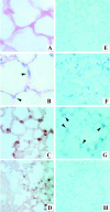
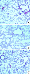
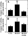
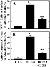
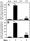
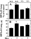
Apoptosis of alveolar epithelial cells (AECs) has been implicated as a key event in the pathogenesis of lung fibrosis. Recent studies demonstrated a role for the synthesis and binding of angiotensin II to receptor AT1 in the induction of AEC apoptosis by bleomycin (BLEO) and other proapoptotic stimuli. On this basis we hypothesized that BLEO-induced apoptosis and lung fibrosis in mice would be inhibited by the AT1 antagonist losartan (LOS) or by targeted deletion of the AT1 gene. Lung fibrosis was induced by intratracheal administration of BLEO (1 U/kg) to wild-type C57BL/6J mice. Co-administration of LOS abrogated BLEO-induced increases in total lung caspase 3 activity detected 6 hours after in vivo administration and reduced by 57% BLEO-induced caspase 3 activity in blood-depleted lung explants exposed to BLEO ex vivo (both P < 0.05). Co-administration of LOS in vivo reduced DNA fragmentation and immunoreactive caspase 3 (active form) in AECs, measured at 14 days after intratracheal BLEO, by 66% and 74%, respectively (both P < 0.05). LOS also inhibited the accumulation of lung hydroxyproline by 45%. The same three measures of apoptosis and lung fibrosis were reduced by 89%, 85%, and 75%, respectively (all P < 0.01), in mice with a targeted disruption of the AT1a receptor gene (C57BL/6J-Agtr1a(tm1Unc)). These data indicate an essential role for angiotensin receptor AT1a in the pathogenesis of BLEO-induced lung fibrosis in mice and suggest that AT1 receptor signaling is required for BLEO-induced apoptosis of AECs in mice as it is in rat and human AECs.
Figures

Similar articles
-
Bleomycin-induced apoptosis of alveolar epithelial cells requires angiotensin synthesis de novo.Am J Physiol Lung Cell Mol Physiol. 2003 Mar;284(3):L501-7. doi: 10.1152/ajplung.00273.2002. Epub 2002 Nov 15. Am J Physiol Lung Cell Mol Physiol. 2003. PMID: 12573988
-
Attenuation of bleomycin-induced pulmonary fibrosis by intratracheal administration of antisense oligonucleotides against angiotensinogen mRNA.Curr Pharm Des. 2007;13(12):1257-68. doi: 10.2174/138161207780618867. Curr Pharm Des. 2007. PMID: 17504234
-
Abrogation of bleomycin-induced epithelial apoptosis and lung fibrosis by captopril or by a caspase inhibitor.Am J Physiol Lung Cell Mol Physiol. 2000 Jul;279(1):L143-51. doi: 10.1152/ajplung.2000.279.1.L143. Am J Physiol Lung Cell Mol Physiol. 2000. PMID: 10893213
-
Sphingolipids in pulmonary fibrosis.Adv Biol Regul. 2015 Jan;57:55-63. doi: 10.1016/j.jbior.2014.09.008. Epub 2014 Oct 13. Adv Biol Regul. 2015. PMID: 25446881 Free PMC article. Review.
-
Breakdown of Epithelial Barrier Integrity and Overdrive Activation of Alveolar Epithelial Cells in the Pathogenesis of Acute Respiratory Distress Syndrome and Lung Fibrosis.Biomed Res Int. 2015;2015:573210. doi: 10.1155/2015/573210. Epub 2015 Oct 7. Biomed Res Int. 2015. PMID: 26523279 Free PMC article. Review.
Cited by
-
Hepatocyte growth factor inhibits apoptosis by the profibrotic factor angiotensin II via extracellular signal-regulated kinase 1/2 in endothelial cells and tissue explants.Mol Biol Cell. 2010 Dec;21(23):4240-50. doi: 10.1091/mbc.E10-04-0341. Epub 2010 Oct 6. Mol Biol Cell. 2010. PMID: 20926686 Free PMC article.
-
Bulbus Fritillariae Cirrhosae as a Respiratory Medicine: Is There a Potential Drug in the Treatment of COVID-19?Front Pharmacol. 2022 Jan 20;12:784335. doi: 10.3389/fphar.2021.784335. eCollection 2021. Front Pharmacol. 2022. PMID: 35126123 Free PMC article. Review.
-
Chitinase 3-like 1 suppresses injury and promotes fibroproliferative responses in Mammalian lung fibrosis.Sci Transl Med. 2014 Jun 11;6(240):240ra76. doi: 10.1126/scitranslmed.3007096. Sci Transl Med. 2014. PMID: 24920662 Free PMC article.
-
Angiotensin II: tapping the cell cycle machinery to kill endothelial cells.Am J Physiol Lung Cell Mol Physiol. 2012 Oct 1;303(7):L575-6. doi: 10.1152/ajplung.00260.2012. Epub 2012 Aug 10. Am J Physiol Lung Cell Mol Physiol. 2012. PMID: 22886501 Free PMC article. No abstract available.
-
Treatment of idiopathic pulmonary fibrosis with losartan: a pilot project.Lung. 2012 Oct;190(5):523-7. doi: 10.1007/s00408-012-9410-z. Epub 2012 Jul 19. Lung. 2012. PMID: 22810758 Free PMC article. Clinical Trial.
References
-
- Selman M, King T, Pardo A: Idiopathic pulmonary fibrosis: prevailing and evolving hypotheses about its pathogenesis and implications for therapy. Ann Intern Med 2001, 134:136-151 - PubMed
-
- Haschek WM, Witschi HP: Pulmonary fibrosis: a possible mechanism. Toxicol Appl Pharmacol 1979, 51:475-487 - PubMed
-
- Hagimoto N, Kuwano K, Nomoto Y, Kunitake R, Hara N: Apoptosis and expression of FAS/FAS ligand mRNA in bleomycin-induced pulmonary fibrosis in mice. Am J Respir Cell Mol Biol 1997, 16:91-101 - PubMed
Publication types
MeSH terms
Substances
Grants and funding
LinkOut - more resources
Full Text Sources
Other Literature Sources
Medical
Molecular Biology Databases
Research Materials

