. 2004 Feb;15(2):637-48.
doi: 10.1091/mbc.e03-02-0097. Epub 2003 Nov 14.

Distinct roles for the AAA ATPases NSF and p97 in the secretory pathway

Affiliations

Distinct roles for the AAA ATPases NSF and p97 in the secretory pathway

Seema Dalal et al. Mol Biol Cell. 2004 Feb.

Abstract

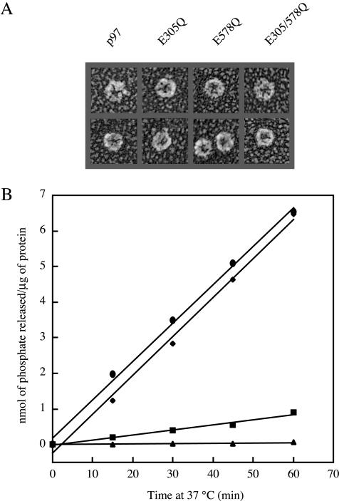
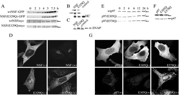
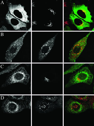
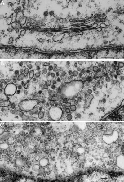
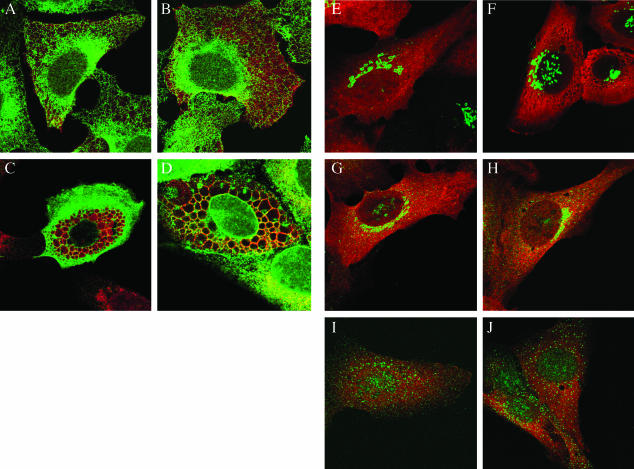
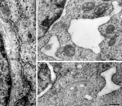
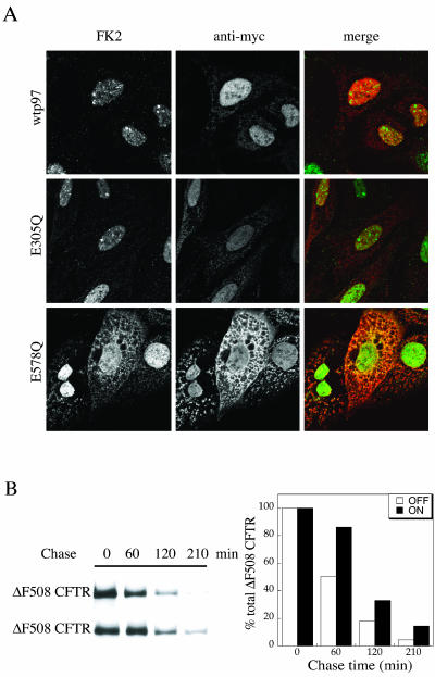
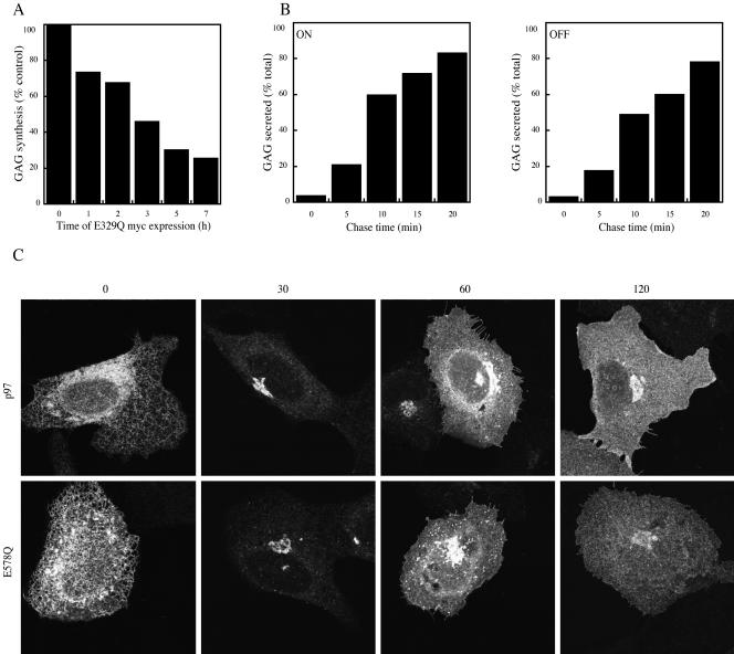
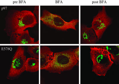
NSF and p97 are related AAA proteins implicated in membrane trafficking and organelle biogenesis. p97 is also involved in pathways that lead to ubiquitin-dependent proteolysis, including ER-associated degradation (ERAD). In this study, we have used dominant interfering ATP-hydrolysis deficient mutants (NSF(E329Q) and p97(E578Q)) to compare the function of these AAA proteins in the secretory pathway of mammalian cells. Expressing NSF(E329Q) promotes disassembly of Golgi stacks into dispersed vesicular structures. It also rapidly inhibits glycosaminoglycan sulfation, reflecting disruption of intra-Golgi transport. In contrast, expressing p97(E578Q) does not affect Golgi structure or function; glycosaminoglycans are normally sulfated and secreted, as is the VSV-G ts045 protein. Instead, expression of p97(E578Q) causes ubiquitinated proteins to accumulate on ER membranes and slows degradation of the ERAD substrate cystic-fibrosis transmembrane-conductance regulator. In addition, expression of p97(E578Q) eventually causes the ER to swell. More specific assessment of effects of p97(E578Q) on organelle assembly shows that the Golgi apparatus disperses and reassembles normally after treatment with brefeldin A and during mitosis. These findings demonstrate that ATP-hydrolysis-dependent activities of NSF and p97 in the cell are not equivalent and suggest that only NSF is directly involved in regulating membrane fusion.


Figures

Figure 1.
Figure 1.
The D2 domain of p97 is its dominant ATPase domain. (A) Deep-etch electron microscopy of bacterially expressed and purified p97 demonstrates that Walker B DExx box mutations in D1(p97(E305Q)), D2 (p97(E578Q)), or both (p97(E305/578Q)) do not alter its normal hexameric cylindrical structure. Molecules are 13-15 nm in diameter. (B) Normal ATPase activity of wtp97 (♦) and p97(E305Q) (•) versus diminished ATPase activity in p97(E578Q) (▪) and p97(E305/578Q) (▴) indicates that its D2 domain is responsible for the majority of the enzyme's ATPase activity. Data shown are an average of two independent measurements and are representative of four independent trials.
Figure 2.
Figure 2.
Properties of U2OS cell lines expressing NSF and p97 variants. (A) Time courses of induction of NSF-GFP versus NSFmyc in human osteosarcoma U2OS cells expressing wild-type and mutant proteins after addition of tetracycline. GFP-tagged proteins were detected with an anti-GFP antibody, and myc-tagged and endogenous NSF were seen using a monoclonal anti-NSF antibody. Tagged proteins are indicated with an asterisk, and endogenous protein is marked with an arrow. (B) Immunoprecipitation from extracts of cells expressing NSF-GFP or NSF(E329Q)-GFP with anti-GFP antibody retrieves endogenous NSF. Samples immunoprecipitated with GFP antibodies were blotted with a monoclonal antibody recognizing NSF. Control represents a GFP expressing cell line. The endogenous protein is marked with an arrow. HC is the IgG heavy chain. (C) α-SNAP immunoprecipitates with NSF(E329Q)myc in the presence of Mg2+ATP and does not immunoprecipitate with NSFmyc. Control represents untransfected U2OS cells. Anti-myc antibody was used for immunoprecipitation, and samples were blotted with antibody recognizing α-SNAP. α-SNAP migrates just behind IgG light chain, seen along the bottom of the blot. (D) Intracellular distribution of NSF and NSF(E329Q) before (-) and after (+) saponin permeabilization shows retention of the mutant on intracellular membranes. Each box is 83 × 83 μm. (E) Time courses of induction of wtp97, p97(E305Q), and p97(E578Q)myc after addition of tetracycline to stably transfected cell lines. Tagged proteins are indicated with an asterisk, and endogenous protein is marked with an arrow. (F) Top panel: Immunoprecipitation with anti-myc antibody shows that endogenous p97 coassembles with induced wtp97, p97(E305Q), and p97(E578Q)myc in vivo. Precipitated proteins are stained with Ponceau Red and the identity of the bands was confirmed by immunoblotting with p97 antibodies (unpublished observations). Tagged proteins are indicated with an asterisk, and endogenous protein is marked with an arrow. Immunoblots with anti-p97 antibody of anti-myc antibody depleted p97(E578Q) lysates confirmed that a majority of the endogenous p97 coassembles with the p97(E578Q) (our unpublished observations). Bottom panel: The same immunoprecipitates blotted with an antibody against p47. (G) Intracellular distribution of wtp97, p97(E305Q), and p97(E578Q)myc before (-) and after (+) permeabilization with saponin. All images were collected at the same laser power on a confocal microscope. Each box is 83 × 83 μm.
Figure 3.
Figure 3.
NSF(E329Q) disrupts the Golgi ribbon but does not affect the ER. Immunofluorescence shows (A) cytoplasmic localization of NSF-GFP (green) and normal-looking Golgi ribbons visualized with anti-giantin antibody (red), (B) punctate distribution of NSF(E329Q)-GFP (green) and disrupted Golgi membranes (antigiantin, red). In cells expressing (C) NSF myc and (D) NSF(E329Q)myc the endoplasmic reticulum (anti-PDI, green) and Golgi apparatus (anti-giantin, red) are differentially affected. These images were collected on a confocal microscope. Each box is 83 × 83 μm.
Figure 4.
Figure 4.
Ultrastructure of NSF(E329Q)-expressing cells. Thin-section EM showing normal Golgi cisternae in NSF-GFP-expressing cells (A). Similar cisternae are absent from cells expressing NSF(E329Q)-GFP. Apparent Golgi remnants (B) and pools of vesicles (C) appear adjacent to the nucleus. Scale bars, 0.2 μm.
Figure 5.
Figure 5.
p97(E578Q) perturbs the ER but does not affect Golgi morphology. (A) p97myc (anti-myc, red) and (B) p97(E305Q)myc (anti-myc, red) do not affect normal ER morphology as observed using the ER marker anti-PDI (green). (C) p97(E578Q)myc (antimyc, green) leads to swelling of the ER (anti-PDI, red) in ∼37% of cells after a 12-h induction. (D) Staining of p97(E578Q)myc with an ER membrane marker (anti-calnexin, green) and lumenal marker (anti-PDI, red) confirms that vacuoles are ER derived. (E, G, and I) p97myc (anti-myc, red) and (F, H, and J) p97(E578Q)myc (anti-myc, red) do not perturb (E and F) giantin (anti-giantin, green), (G and H) COPI (anti-β-COP, green), and (I and J) COPII (anti-Sec23, green). These images were collected on a confocal microscope. Each box is 83 × 83 μm.
Figure 6.
Figure 6.
Dilated ER and nuclear envelope in p97(E578Q) expressing cells. Traditional thin section EM of (A) a p97(E578Q)myc expressing cell in which the ER looks relatively normal, (B) a p97(E578Q)myc expressing cell containing swollen ribosome-studded ER. (C) Thin-section EM of freeze substituted p97(E578Q)myc-expressing cell showing swollen ER and nuclear envelope together with apparently normal Golgi. Scale bars, 0.2 μm.
Figure 7.
Figure 7.
p97(E578Q) causes accumulation of ubiquitinated proteins on ER membranes and a delay in ΔF508 CFTR degradation. (A) After saponin permeabilization, retained ubiquitin-conjugates (FK2, green), and p97myc, p97(E305Q)myc, or p97(E578Q)myc (anti-myc, red) were visualized by immunofluorescence. These images were collected on a confocal microscope. Each box is 73 × 73 μm. (B) Pulse chase analysis of ΔF508 CFTR turnover in cells expressing (ON) or not (OFF) p97(E578Q). Immunoprecipitated ΔF508 CFTR is shown in the autoradiographs on the left (OFF, top; ON, bottom), and quantitated by densitometry on the right. The data presented are representative of four independent trials.
Figure 8.
Figure 8.
NSF(E329Q) disrupts Golgi function, whereas p97(E578Q) does not. (A) Synthesis of 35S-sulfated GAGs as a function of the time of NSF(E329Q)myc induction. Data for the different time points were normalized relative to 100% for the 0 time point. (B) Secretion of 35S-sulfated GAGs relative to the total 35S-sulfated GAGs synthesized for different chase times in p97(E578Q)myc cells that were induced for 12 h (ON) or were not induced (OFF). (C) VSV-G ts045-GFP distribution in p97myc cells (top panel) and p97(E578Q)myc cells induced for 12 h (bottom panel) at 0, 30, 60, and 120 min after shifting the cells from the nonpermissive temperature (38.5°C) to the permissive temperature (32°C). These images were obtained on a confocal microscope. Each box is 92 × 92 μm.
Figure 9.
Figure 9.
p97(E578Q) does not affect Golgi distribution mitosis. p97(E578Q)myc cells at different stages of mitosis stained with anti-giantin (green) and anti-α-tubulin (red). (A) metaphase, (B) anaphase, (C) telophase I, (D-F) telophase II. Images were obtained on a confocal microscope. (A-C) Each box is 40 × 40 μm. (D-F) Each box is 83 × 83 μm.
Figure 10.
Figure 10.
Disruption and reassembly of Golgi ribbon in response to BFA are unaffected by p97(E578Q). p97myc cells (top panel) and p97(E578Q)myc cells (bottom panel) are shown from left to right before BFA addition, after BFA treatment for 40 min, and after recovery from BFA for 120 min, respectively. Giantin is in green (anti-giantin) and p97(E578Q)myc is in red (anti-myc). Images were obtained on a confocal microscope. Each box is 83 × 83 μm.

Similar articles

Cited by

References

    1. Abrami, L., Fivaz, M., Glauser, P., Parton, R., and van der Goot, F. (1998). A pore-forming toxin interacts with a GPI-anchored protein and causes vacuolation of the endoplasmic reticulum. J. Cell Biol. 140, 525-540. - PMC - PubMed
    1. Acharya, U., Jacobs, R., Peters, J., Watson, N., Farquhar, M., and Malhotra, V. (1995). The formation of Golgi stacks from vesiculated Golgi membranes requires two distinct fusion events. Cell 82, 895-904. - PubMed
    1. Alvarez, C., and Sztul, E. (1999). Brefeldin A (BFA) disrupts the organization of the microtubule and actin cytoskeletons. Eur. J. Cell Biol. 78, 1-14. - PubMed
    1. Bays, N., and Hampton, R. (2002). Cdc48-Ufd1-Npl 4, stuck in the middle with Ub. Curr. Biol. 12, R366-R371. - PubMed
    1. Coppolino, M.G., Kong, C., Mohtashami, M., Schreiber, A.D., Brumell, J.H., Finlay, B.B., Grinstein, S., Trimble, W.S. (2001). Requirement for N-ethylmaleimide-sensitive factor activity at different stages of bacterial invasion and phagocytosis. J. Biol. Chem. 276, 4772-4780. - PubMed

Publication types

MeSH terms

LinkOut - more resources