. 2003 Dec;23(23):8563-75.
doi: 10.1128/MCB.23.23.8563-8575.2003.

Acetylation of androgen receptor enhances coactivator binding and promotes prostate cancer cell growth

Affiliations

Acetylation of androgen receptor enhances coactivator binding and promotes prostate cancer cell growth

Maofu Fu et al. Mol Cell Biol. 2003 Dec.

Abstract

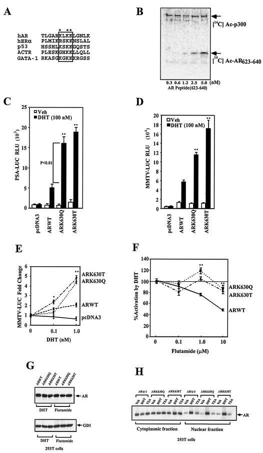
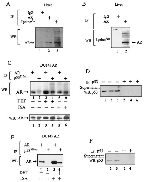
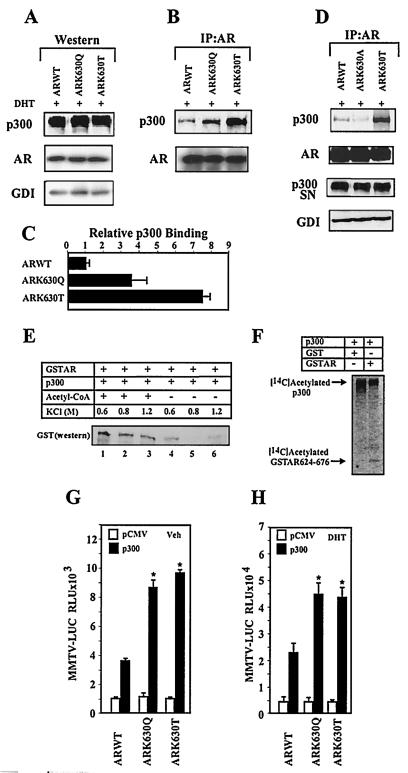
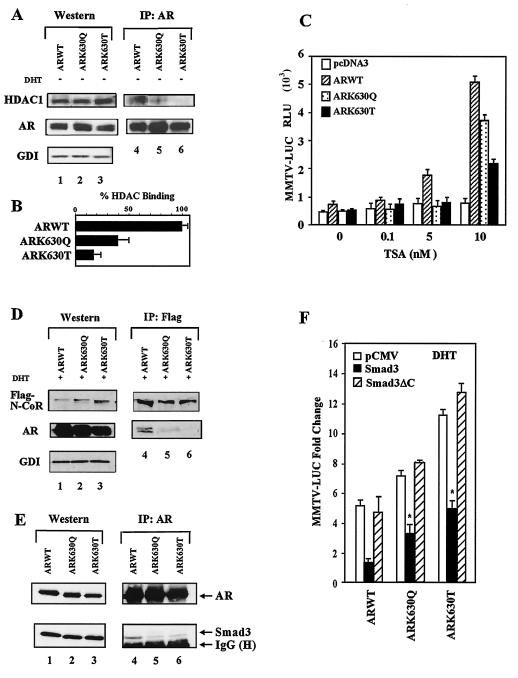
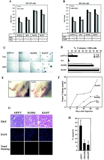
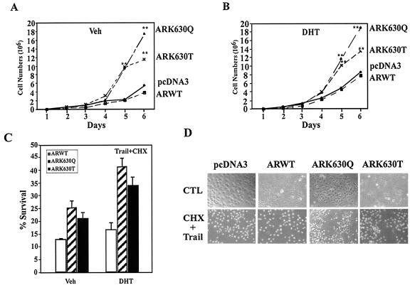
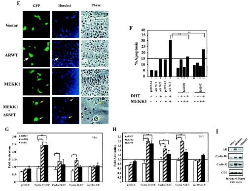
Modification by acetylation occurs at epsilon-amino lysine residues of histones and transcription factors. Unlike phosphorylation, a direct link between transcription factor acetylation and cellular growth or apoptosis has not been established. We show that the nuclear androgen receptor (AR), a DNA-binding transcriptional regulator, is acetylated in vivo. The acetylation of the AR is induced by ligand dihydrotestosterone and by histone deacetylase (HDAC) inhibitors in living cells. Direct AR acetylation augmented p300 binding in vitro. Constructs mimicking neutral polar substitution acetylation (AR(K630Q), AR(K630T)) enhanced p300 binding and reduced N-CoR/HDAC/Smad3 corepressor binding, whereas charged residue substitution (AR(K630R)) reduced p300 binding and enhanced corepressor binding. The AR acetylation mimics promoted cell survival and growth of prostate cancer cells in soft agar and in nude mice and augmented transcription of a subset of growth control target gene promoters. Thus, transcription factor acetylation regulates coactivator/corepressor complex binding, altering expression of specific growth control genes to promote aberrant cellular growth in vivo.


Figures

FIG.1.
FIG.1.
AR acetylation site regulates ligand sensitivity and specificity. (A) The AR acetylation motif indicating lysine 630 (*), with homology shown to the acetylation motif of human p53 and ACTR proteins. (B) In vitro histone acetyltransferase (HAT) assays used wild-type AR peptides incubated with baculovirus-purified p300 and [14C]acetyl coenzyme A. (C) Activity of the PSA-Luc and (D) MMTV-Luc reporters was assessed in DU145 cells with the wild-type and mutant ARs and is shown as relative luciferase activity (mean ± standard error of the mean for n > 6 separate transfections). (E) The expression plasmids encoding wild-type AR or AR mutants were transfected and examined for DHT dose responsiveness in DU145 cells with the MMTV-Luc reporter. Cells were treated with either vehicle or DHT (24 h). (F) The wild-type AR and AR acetylation site mutants were assessed for responsiveness to flutamide with induction shown compared to equal amounts of the empty expression vector cassette (pSG5). The data are means ± standard error of the mean for n > 9. (G) The wild-type AR and AR acetylation site mutants were transfected into 293T cells, treated with 100 nM DHT or 10 nM flutamide for 24 h. The cell lysates were then subjected to Western blotting with the antibodies to the AR or GDI as the protein-loading control. (H) The wild-type AR and AR acetylation site mutants were transfected into 293T cells, treated either with vehicle, DHT (100 nM), or TSA (100 nM) for 24 h. Western blotting of the nuclear and cytoplasmic fractions is shown.
FIG. 2.
FIG. 2.
AR acetylation in vivo. (A) The cell lysate was extracted from wild-type male liver and subjected to immunoprecipitation (IP) with either normal rabbit IgG, anti-AR, or anti-acetyl-lysine antibody. The immunoprecipitate was separated by SDS-PAGE and blotted with anti-AR antibody (Upstate Biotech). (B) The mouse liver lysate was also immunoprecipitated with normal rabbit IgG or anti-AR antibody and blotted with anti-acetyl-lysine antibody. (C, E) The DU145ARwt stable cell lines were treated with vehicle (ethanol) or DHT (100 nM) for 12 h and then treated with TSA (30 nM) for 6 h. Immunoprecipitation was done with anti-AR or anti-acetyl-lysine peptide (p53320) as indicated. Prior to immunoprecipitation with anti-acetyl-p53320, cell lysates were first cleared with an anti-p53 antibody (anti-p53[1-393]; sc-4246; Santa Cruz Biotech). The immunoprecipitate was resolved by SDS-PAGE and blotted with anti-AR antibody. (D, F) Western blot (WB) of cell lysates precleared with anti-p53 antibody (sc-4246).
FIG. 3.
FIG. 3.
Direct AR acetylation enhances p300 binding. (A, B, and D) Extracts from cells transfected with the AR expression vectors were subjected to Western blot analysis (A) or AR immunoprecipitation Western blotting (B and D) to detect p300 and the AR. The AR immunoprecipitation supernatant (SN) was immunoblotted for p300 to show the proportion of p300 not bound to the AR. (C) The relative binding of the AR mutants to p300 were shown as mean ± standard error of the mean (n = 3). (E) Acetylation of the AR enhances p300 binding in vitro. GST pull-down was performed with p300 and either acetylated AR (lanes 1 to 3) or unacetylated AR (lanes 4 to 6). Western blotting of the GST-AR pull-down product is shown. (F) p300 in vitro HAT assay. Baculovirus-expressed p300 protein (100 ng) was incubated with either GST or GST-AR624-676 in the presence of [14C]acetyl coenzyme A, resolved by SDS-PAGE, and exposed to a phosphoimaging screen for 24 h. The autoradiograms of the 14C-acetylated p300 and AR fusion protein are indicated. (G and H) Increased p300-mediated trans-activation of AR acetylation mutants. The MMTV-Luc reporter was assessed with the wild-type AR and AR acetylation site mutants, with p300 or equal amounts of the empty expression vector cassettes (pCMV), and treated with vehicle or DHT (10−7 M) for 24 h. The data shown were normalized to the DHT-induced activity for the AR construct (mean ± standard error of the mean, n > 9 separate transfections).
FIG. 4.
FIG. 4.
Reduced N-CoR and HDAC1 binding of AR acetylation site mutants. (A) 293T cells transfected with the ARs were subjected to either direct Western blotting or AR immunoprecipitation and Western blotting for HDAC1 and AR. GDI served as the loading control. (B) The binding of HDAC1 to the AR is shown as percent binding for multiple separate transfections. (C) DU145 cells were cotransfected with the MMTV-Luc reporter and the expression vector for the wild-type AR, mutant AR, or empty expression vector cassette (pcDNA3) and treated with TSA for 24 h. (D) 293T cells were transfected with the ARs and Flag-N-CoR, treated with DHT (10−7M), and the cellular extracts were subjected to Western blotting or immunoprecipitation (anti-M2-Flag antibody for N-CoR) and subsequent AR Western blotting. (E) AR immunoprecipitation of cells transfected with AR acetylation site mutants and Smad3 with sequential Western blotting for Smad3. (F) DU145 Cells were transfected with the MMTV-Luc reporter and expression vectors for AR, Smad3, or Smad3ΔC. The DHT-induced change in MMTV-Luc activity is shown as the mean ± standard error of the mean.
FIG.5.
FIG.5.
AR acetylation mutants convey contact-independent growth. (A and B) MTT assay of DU145 stable cell lines expressing either pcDNA3, wild-type AR, or AR acetylation site mutants. Equal numbers of cells were seeded into 96-well plates, treated with either DHT, TSA, or SAHA for 24 h, and the MTT assay was conducted, measuring absorbance at 570 nm. (C and D) DU145 cells stably expressing wild-type AR or AR acetylation site mutants were seeded in soft agar. Phase contrast image of the colonies from a representative experiment is shown (magnification, ×100). Colony numbers and size (percentage of colonies with >100 cells) are shown at day 14. (E and F) Nude mice were implanted with 106 cells of stable lines expressing either wild-type AR or AR acetylation site mutants. The mean volume of DU145 tumors grown in nude mice were shown at each time point. (G and H) Apoptosis in implanted tumors was assessed by TUNEL staining for wild-type AR, ARK630Q, and ARK630T (n = 4).
FIG. 6.
FIG. 6.
AR acetylation site governs cellular proliferation and MEKK1-dependent apoptosis. (A and B) Enhanced cellular proliferation rate of AR acetylation site mutants. A total of 2 × 10⁴ DU145 cells stably transfected with the expression vector for either wild-type AR, AR mutants, or control vector pcDNA3 were seeded and treated either with vehicle (A) or DHT (10−7M) (B). The representative results of three independent experiments are shown. (C) AR acetylation site prostate cancer cell lines are resistant to TRAIL-induced apoptosis. Cell survival rates of DU145 stable cell lines exposed to TRAIL (10 ng/ml) and cycloheximide (CHX; 10 μg/ml) are shown compared with untreated cells (100%). (D) Phase contrast of cell lines is shown. (E, F). AR acetylation site mutants evade MEKK-1-mediated apoptosis. DU145 cells were transfected with MEKK1, AR, and pCMV-GFP as indicated and treated with either vehicle or DHT (10 nM) for 24 h. The morphology of the transfected DU145 cells is shown in phase contrast. (E) White arrows indicate GFP-positive cells, and yellow arrows indicate GFP-positive cells showing chromatin condensation. (F) The graph represents independent experiments in which 200 green fluorescent cells were counted and scored for cytoplasmic blebbing and chromatin condensation; *, P < 0.01 for the effect of MEKK1 on liganded AR-induced apoptosis. (G and H) AR acetylation site mutants alter regulation of cell cycle control genes. Reporter assays showing regulation of cell cycle control promoters by wild-type AR or AR acetylation site mutants in DU145 cells treated with vehicle (G) or DHT (H). (I) Western blot analysis of DU145 stable cell lines stably expressing wild-type AR and AR acetylation mutants. Cells were starved for 24 h and harvested 6 h after treatment (10% charcoal-stripped fetal bovine serum plus 100 nM DHT). GDI served as a protein-loading control.
FIG. 6.
FIG. 6.
AR acetylation site governs cellular proliferation and MEKK1-dependent apoptosis. (A and B) Enhanced cellular proliferation rate of AR acetylation site mutants. A total of 2 × 10⁴ DU145 cells stably transfected with the expression vector for either wild-type AR, AR mutants, or control vector pcDNA3 were seeded and treated either with vehicle (A) or DHT (10−7M) (B). The representative results of three independent experiments are shown. (C) AR acetylation site prostate cancer cell lines are resistant to TRAIL-induced apoptosis. Cell survival rates of DU145 stable cell lines exposed to TRAIL (10 ng/ml) and cycloheximide (CHX; 10 μg/ml) are shown compared with untreated cells (100%). (D) Phase contrast of cell lines is shown. (E, F). AR acetylation site mutants evade MEKK-1-mediated apoptosis. DU145 cells were transfected with MEKK1, AR, and pCMV-GFP as indicated and treated with either vehicle or DHT (10 nM) for 24 h. The morphology of the transfected DU145 cells is shown in phase contrast. (E) White arrows indicate GFP-positive cells, and yellow arrows indicate GFP-positive cells showing chromatin condensation. (F) The graph represents independent experiments in which 200 green fluorescent cells were counted and scored for cytoplasmic blebbing and chromatin condensation; *, P < 0.01 for the effect of MEKK1 on liganded AR-induced apoptosis. (G and H) AR acetylation site mutants alter regulation of cell cycle control genes. Reporter assays showing regulation of cell cycle control promoters by wild-type AR or AR acetylation site mutants in DU145 cells treated with vehicle (G) or DHT (H). (I) Western blot analysis of DU145 stable cell lines stably expressing wild-type AR and AR acetylation mutants. Cells were starved for 24 h and harvested 6 h after treatment (10% charcoal-stripped fetal bovine serum plus 100 nM DHT). GDI served as a protein-loading control.

Similar articles

Cited by

References

    1. Abate-Shen, C., and M. M. Shen. 2000. Molecular genetics of prostate cancer. Genes Dev. 14:2410-2434. - PubMed
    1. Abreu-Martin, M. T., A. Chari, A. A. Palladino, N. A. Craft, and C. L. Sawyers. 1999. Mitogen-activated protein kinase kinase kinase 1 activates androgen receptor-dependent transcription and apoptosis in prostate cancer. Mol. Cell. Biol. 19:5143-5154. - PMC - PubMed
    1. Alland, L., R. Muhle, H. J. Hou, J. Potes, L. Chin, N. Schreiber-Agus, and R. A. DePinho. 1997. Role for N-CoR and histone deacetylase in Sin3-mediated transcriptional repression. Nature 387:49-55. - PubMed
    1. Chen, D., H. Ma, H. Hong, S. S. Koh, S. M. Huang, B. T. Schurter, D. W. Aswad, and M. R. Stallcup. 1999. Regulation of transcription by a protein methyltransferase. Science 284:2174-2177. - PubMed
    1. Chen, J. D., and R. M. Evans. 1995. A transcriptional co-repressor that interacts with nuclear hormone receptors. Nature 377:454-457. - PubMed

Publication types

MeSH terms