. 2003 Dec;77(23):12753-63.
doi: 10.1128/jvi.77.23.12753-12763.2003.

COX-2 induction during murine gammaherpesvirus 68 infection leads to enhancement of viral gene expression

Affiliations

COX-2 induction during murine gammaherpesvirus 68 infection leads to enhancement of viral gene expression

Tonia L Symensma et al. J Virol. 2003 Dec.

Abstract

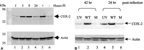
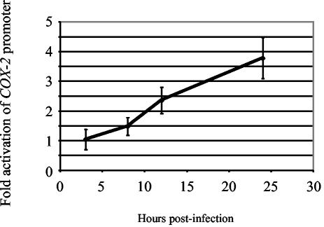
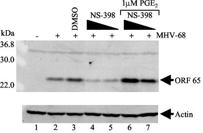
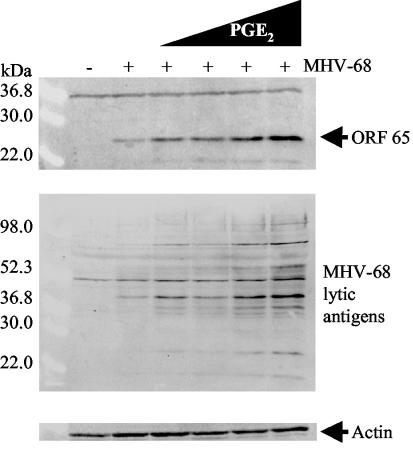
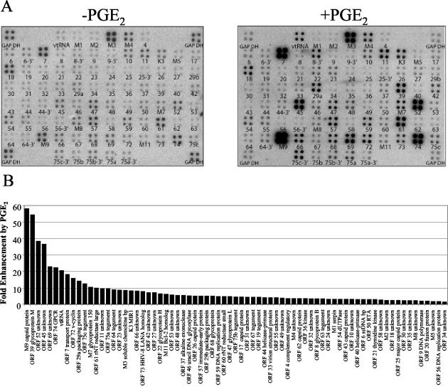
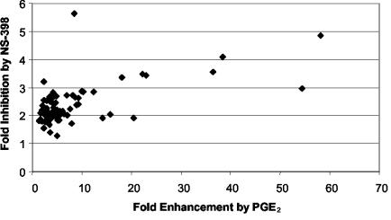
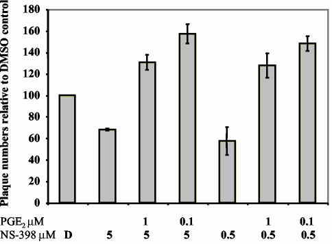
The murine gammaherpesvirus 68 (MHV-68 or gammaHV-68) model provides many advantages for studying virus-host interactions involved in gammaherpesvirus replication, including the role of cellular responses to infection. We examined the effects of cellular cyclooxygenase-2 (COX-2) and its by-product prostaglandin E(2) (PGE(2)) on MHV-68 gene expression and protein production following de novo infection of cultured cells. Western blot analyses revealed an induction of COX-2 protein in MHV-68-infected cells but not in cells infected with UV-irradiated MHV-68. Luciferase reporter assays demonstrated activation of the COX-2 promoter during MHV-68 replication. Two nonsteroidal anti-inflammatory drugs, a COX-2-specific inhibitor (NS-398) and a COX-1-COX-2 inhibitor (indomethacin), substantially reduced MHV-68 protein production in infected cells. Inhibition of viral protein expression and virion production by NS-398 was reversed in the presence of exogenous PGE(2). Global gene expression analysis using an MHV-68 DNA array showed that PGE(2) increased production of multiple viral gene products, and NS-398 inhibited production of many of the same genes. These studies suggest that COX-2 activity and PGE(2) production may play significant roles during MHV-68 de novo infection.


Figures

FIG. 1.
FIG. 1.
MHV-68 de novo infection increases COX-2 protein levels and requires viral transcription. (A) COX-2 protein expression was induced during MHV-68 replication. NIH 3T3 cells in low-serum conditions were uninfected (lane 6) or infected with EGFP-MHV-68 (lanes 1 to 4). As a positive control, uninfected NIH 3T3 cells were treated with a serum shock (lane 5). Cell extracts were harvested at 1, 5, 9, or 24 h postinfection (PI) and analyzed by Western blotting using a COX-2 polyclonal antibody (top panel). Each lane contained 15 μg of total protein as determined by Bradford assays. The membrane was reprobed with antiactin providing a loading control (bottom panel). (B) COX-2 protein expression was not induced by UV-irradiated MHV-68 compared to wt MHV-68. NIH 3T3 cells in low-serum conditions were infected with wt MHV-68 (lanes 2 and 5), infected with an equal volume of UV-irradiated wt MHV-68 (lanes 1 and 4), or uninfected (lanes 3 and 6). Cell extracts were harvested 24 and 42 h postinfection for Western blot analysis using a COX-2 polyclonal antibody. Each lane contains 15 μg of total protein as determined by Bradford assays. The membrane was reprobed with antiactin, providing a loading control (bottom panel).
FIG. 2.
FIG. 2.
MHV-68 replication activates the COX-2 promoter. NIH 3T3 cells were transfected with a COX-2 promoter-luciferase construct (74) and a pRL-SV40 control plasmid. Transfected cells were infected with wt MHV-68 or mock infected. Cell extracts were harvested at 3, 8, 12, and 24 h postinfection, and luciferase activities were measured and normalized to control samples. Each data point represents four separate experiments performed in duplicate.
FIG. 3.
FIG. 3.
NS-398 and indomethacin suppress MHV-68 protein expression. NIH 3T3 cells were untreated (lanes 1 and 2) or pretreated with DMSO (lane 3) or 2 (lane 4), 10 (lane 5), or 50 (lane 6) μM NS-398. Similarly, NIH 3T3 cells were pretreated with ethanol (EtOH) only (lane 7) or 0.2 (lane 8), 1 (lane 9), or 5 (lane 10) μM indomethacin. Subsequently, cells were infected with wt MHV-68 (lanes 2 to 10) and maintained in the appropriate diluent-drug concentrations. Cell extracts were harvested 8 h postinfection for Western blot analyses using anti-MHV-68 serum (top panel) or an antibody to MHV-68 ORF 26 (center panel). Control cells were uninfected (lane 1) or uninfected and treated with NS-398 (lane 11) or indomethacin (lane 12). As a loading control, the blot was probed with antiactin (bottom panel). Each lane contains 30 μg of total protein as determined by Bradford assays.
FIG. 4.
FIG. 4.
NS-398 suppression of MHV-68 viral protein expression is relieved in the presence of exogenous PGE2. NIH 3T3 cells were untreated (lanes 1 and 2) or pretreated with DMSO (lane 3) or 5 (lanes 4 and 6) or 0.5 (lanes 5 and 7) μM NS-398. Cells were infected with wt MHV-68 (lanes 2 to 7) and maintained in the appropriate DMSO-NS-398 concentrations. Subsequently, infected cells treated with NS-398 were also treated with 1 μM PGE2 (lanes 6 and 7), and cell extracts were harvested at 12 h postinfection for Western blot analysis using an antibody to a late lytic gene product, MHV-68 M9-ORF 65 (upper panel). Control cells were uninfected (lane 1) or infected with no treatment (lane 2). As a loading control, the blot was probed with antiactin (lower panel). Each lane contains 15 μg of total protein as determined by Bradford assays.
FIG. 5.
FIG. 5.
PGE2 exhibits dose-dependent enhancement of MHV-68 viral protein expression. NIH 3T3 cells were uninfected (lane 1) or infected for 1 h with wt MHV-68 (lanes 2 to 6). Immediately following inoculation, cells were treated with increasing concentrations of PGE2: 0.05 (lane 3), 0.25 (lane 4), 1.25 (lane 5), and 6.25 (lane 6) μM. Control cells were infected and untreated (lane 2). Cell extracts were harvested 12 h postinfection, and protein expression was assayed by Western blot analysis using an antibody to MHV-68 M9-ORF 65 (top panel). The blot was stripped and reprobed with anti-MHV-68 serum (center panel). As a loading control, the blot was probed with antiactin (bottom panel). Each lane contains 30 μg of total protein as determined by Bradford assays.
FIG. 6.
FIG. 6.
Upregulation of MHV-68 gene expression by PGE2. (A) BHK-21 cells were untreated (left panel) or pretreated with 1 μM PGE2 (right panel) before infection with wt MHV-68. Cells were maintained in medium containing 1 μM PGE2 during the infection and postinfection incubation (right panel). Total RNA was harvested 8 h postinfection, and labeled cDNA probe was generated for hybridization to MHV-68 DNA arrays. The ORFs are printed below each array element. (B) A STORM phosphorimager and ImageQuant system were used to quantitate the signal from the array elements corresponding to 73 known and predicted MHV-68 ORFs. GAPDH-normalized values from the array with PGE2 (A, right panel) were divided by the corresponding GAPDH-normalized values from the untreated array (A, left panel) to derive the fold induction of gene expression relative to the untreated level for each array element. These values and their corresponding MHV-68 ORFs are ordered in the bar graph based on increasing fold induction of gene expression relative to the untreated level. Statistical significance of differences in expression was assessed by paired t test.
FIG. 7.
FIG. 7.
Downregulation of MHV-68 gene expression by NS-398. (A) BHK-21 cells were untreated (left panel) or pretreated with 5 μM NS-398 (right panel) before infection with wt MHV-68. Cells were maintained in medium containing 5 μM NS-398 during the infection and postinfection incubations (right panel). Total RNA was harvested 8 h postinfection, and labeled cDNA probe was generated for hybridization to MHV-68 DNA arrays. The ORFs are printed below each array element. (B) A STORM phosphorimager and ImageQuant system were used to quantitate the signal from the array elements corresponding to 73 known and predicted MHV-68 ORFs. GAPDH-normalized values from the array with NS-398 (A, right panel) were divided by the corresponding GAPDH-normalized values from the untreated array (A, left panel) to derive the fold inhibition of gene expression relative to the untreated level for each array element. These values and their corresponding MHV-68 ORFs are ordered in the bar graph based on increasing fold inhibition of gene expression relative to the untreated level. Statistical significance of differences in expression was assessed by paired t test.
FIG. 8.
FIG. 8.
Relationship between MHV-68 gene induction in response to PGE2 and suppression in response to NS-398. Change in expression of each ORF was quantified as described above (Fig. 6 and 7), and magnitude of gene induction in response to PGE2 (horizontal axis) was compared with magnitude of gene suppression in response to NS-398 (vertical axis). Each point represents 1 of 73 assayed MHV-68 ORFs, and the best-fit line was produced by linear regression. The strength of relationship between a gene's induction by PGE2 and its suppression by NS-398 was quantified by Pearson correlation coefficient (r).
FIG. 9.
FIG. 9.
Inhibition of MHV-68 virion production by NS-398 is reversed by PGE2. NIH 3T3 cells were pretreated with DMSO (D) or NS-398 (0.5 or 5 μM). Subsequently, cells were infected with wt MHV-68 and maintained in the appropriate DMSO-NS-398 concentrations. Immediately after infection, NS-398-treated cells were also treated with PGE2 (0.1 or 1 μM). Supernatants were harvested at 12 h postinfection, and viral titer was quantitated using plaque assays on BHK-21 monolayers.

Similar articles

Cited by

References

    1. Ahn, J. W., K. L. Powell, P. Kellam, and D. G. Alber. 2002. Gammaherpesvirus lytic gene expression as characterized by DNA array. J. Virol. 76:6244-6256. - PMC - PubMed
    1. Alcaraz, M. J., A. Habib, M. Lebret, C. Creminon, S. Levy-Toledano, and J. Maclouf. 2000. Enhanced expression of haem oxygenase-1 by nitric oxide and antiinflammatory drugs in NIH 3T3 fibroblasts. Br. J. Pharmacol. 130:57-64. - PMC - PubMed
    1. Barnes, A., H. Dyson, N. P. Sunil-Chandra, P. Collins, and A. A. Nash. 1999. 2′-Deoxy-5-ethyl-beta-4′-thiouridine inhibits replication of murine gammaherpesvirus and delays the onset of virus latency. Antivir. Chem. Chemother. 10:321-326. - PubMed
    1. Bratcher, D. F., C. J. Harrison, N. Bourne, L. R. Stanberry, and D. I. Bernstein. 1993. Effect of indomethacin on ultraviolet radiation-induced recurrent herpes simplex virus disease in guinea-pigs. J. Gen. Virol. 74:1951-1954. - PubMed
    1. Brock, T. G., R. W. McNish, and M. Peters-Golden. 1999. Arachidonic acid is preferentially metabolized by cyclooxygenase-2 to prostacyclin and prostaglandin E2. J. Biol. Chem. 274:11660-11666. - PubMed

Publication types

MeSH terms