. 2003 Dec;77(23):12679-91.
doi: 10.1128/jvi.77.23.12679-12691.2003.

Strand-specific RNA synthesis defects in a poliovirus with a mutation in protein 3A

Affiliations

Strand-specific RNA synthesis defects in a poliovirus with a mutation in protein 3A

Natalya L Teterina et al. J Virol. 2003 Dec.

Abstract

Substitution of a methionine residue at position 79 in poliovirus protein 3A with valine or threonine caused defective viral RNA synthesis, manifested as delayed onset and reduced yield of viral RNA, in HeLa cells transfected with a luciferase-containing replicon. Viruses containing these same mutations produced small or minute plaques that generated revertants upon further passage, with either wild-type 3A sequences or additional nearby compensating mutations. Translation and polyprotein processing were not affected by the mutations, and 3AB proteins containing the altered amino acids at position 79 showed no detectable loss of membrane-binding activity. Analysis of individual steps of viral RNA synthesis in HeLa cell extracts that support translation and replication of viral RNA showed that VPg uridylylation and negative-strand RNA synthesis occurred normally from mutant viral RNA; however, positive-strand RNA synthesis was specifically reduced. The data suggest that a function of viral protein 3A is required for positive-strand RNA synthesis but not for production of negative strands.


Figures
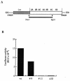
FIG. 1.
FIG. 1.
Mutant PV replicon RNAs with defects in RNA synthesis. (A) PV replicon RNAs with structural genes replaced by a luciferase reporter gene are diagrammed. PV coding sequences are indicated in the white box, and the sequence coding for luciferase is shaded. XhoI-BglII fragments produced by PCR under conditions favoring random mutations are shown by a black line. The RNAs contain two nonviral guanylate residues at the 5′ end, generated by use of the T7 promoter. Luciferase is released from the PV polyprotein by 2A protease cleavage. (B) Luciferase expression was measured in HeLa cell extracts transfected with RNA transcripts produced from individual clones of mutated pLuc-PV. Luciferase activity (in relative light units [RLU]) is shown in extracts from 1.4 × 10⁴ cells at 7 h after transfection.
FIG. 2.
FIG. 2.
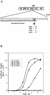
Mutation in the sequence coding for PV protein 3A causing defects in RNA replication. (A) The amino acid sequence of the C-terminal part of wild-type PV 3A protein is shown with the residue numbers in 3A indicated above it. The cleavage site between 3A and 3B is indicated after amino acid 87. The hydrophobic domain of protein 3A is underlined. Residue methionine 79 is circled. The codon for methionine and the changes introduced by site-directed mutagenesis are shown. (B) Time course of RNA replication measured by luciferase accumulation following RNA transfection of HeLa cells at 37°C.
FIG. 3.
FIG. 3.
Plaque morphology of the PV-3A-79 mutants. HeLa cell monolayers were transfected with serial dilutions of wild-type or mutant RNA transcripts and overlaid with minimal essential medium containing 0.4% agarose 1 h after transfection. Plates were incubated at 37°C and stained with crystal violet 48 h after transfection. The plate transfected with the indicated amount of each RNA is shown.
FIG. 4.
FIG. 4.
In vitro translation of replicon RNAs. T7 RNA transcripts (20 and 10 nM) from wild-type pRLuc-31 (3A-wt) (lanes 2 and 3), pLuc-3A-M79V (lanes 4 and 5), pLuc-3A-M79V-H86Y (lanes 6 and 7), pLuc-3A-H86Y (lanes 8 and 9), pLuc-3A-M79T (lanes 10 and 11), and pLuc-3A-M79L (lane 12; 20 nM) were used to program translation in HeLa S10 extracts supplemented with ribosomal salt wash in the presence of [35S]methionine. Lane 1 shows the products of translation programmed with 10 nM PV1 virion RNA. Reaction products were subjected to electrophoresis on a 12.5% polyacrylamide-SDS gel. The asterisk indicates Luc-related protein bands.
FIG. 5.
FIG. 5.
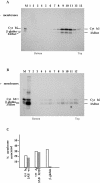
Sucrose density gradient analysis of 3AB proteins. (A and B) Translation reactions were programmed with FLAG epitope-tagged wild-type 3AB and cytochrome b5 in the absence (A) or presence (B) of canine pancreas microsomal membranes. Products were sedimented through discontinuous sucrose gradients, and fractions were collected and immunoprecipitated with anti-FLAG antibody before analysis on SDS-PAGE gels. A mixture of individually translated FLAG epitope-tagged wild-type 3AB, cytochrome b5, and β-globin proteins served asmarkers for the gel (lane M); lane T in panel B represents a sample of the translation reaction not subjected to gradient sedimentation. (C) Radioactivity in each fraction of the sucrose gradients containing proteins from translation reactions programmed with cytochrome b5 mRNA and either wild-type 3AB, 3AB-M79T, or β-globin mRNAs, in the presence of canine pancreas microsomal membranes, was quantitated by phosphorimager. The percentage of total radioactivity cosedimenting with membranes (fractions 2 to 6) was calculated for each protein and plotted.
FIG. 6.
FIG. 6.
Accumulation of PV replicon-specific RNAs in transfected cells. (A) HeLa cell monolayers were transfected with RNA transcripts in the presence of DEAE-dextran. Cells were harvested at the indicated times after transfection; total cytoplasmic RNAs isolated from approximately 104 cells were bound to a nylon membrane and hybridized to a 32P-labeled riboprobe complementary to nt 220 to 460 of PV RNA. Mutant replicon 3D-K276L bearing a mutation in 3D was used as negative control. The vRNA standards show increasing amounts of purified PV RNA (0.1 to 3 ng) hybridized in parallel. (B) Quantitative presentation of data obtained by analysis with a PhosphorImager and ImageQuant software (Molecular Dynamics).
FIG. 7.
FIG. 7.
Effect of substitutions at residue M79 in PV protein 3A on VPg uridylylation and negative-strand RNA synthesis. Translation-replication extracts were programmed with full-length RNA transcripts as indicated. (A) Synthesis and processing of viral proteins were measured in reactions carried out in the presence of [35S]methionine, and labeled viral proteins were analyzed as described in the legend to Fig. 4. (B and C) VPg uridylylation (B) and negative-strand RNA synthesis (C) were measured in reactions containing preinitiation RNA replication complexes isolated from translation reactions containing 2 mM GuHCl and the indicated RNAs after sedimentation and resuspension in the absence of guanidine (lanes 1, 3, 4, 5, and 6). GuHCl was added to the samples in lane 2. Negative-strand RNA synthesis and VPg uridylylation assays were performed as described in Materials and Methods; the data were analyzed with a PhosphorImager and are expressed as a percentage of the product observed with wild-type PV RNA transcript. The major uridylylated VPg band in panel B is thought to be diuridylylated, but it has not been rigorously characterized.
FIG. 8.
FIG. 8.
Replication of RNA transcripts with an authentic PV 5′ end. (A) Preinitiation RNA replication complexes were isolated from translation-replication reactions programmed with RNA transcripts produced from constructs that did not (lane 1) or did (lane 2) contain the cis-active hammerhead ribozyme attached to the 5′ end of the poliovirus genome sequence. Preinitiation RNA replication complexes were incubated for 60 min at 37°C in reaction mixtures containing [α-32P]CTP; RNA was phenol extracted and analyzed on nondenaturing 1% agarose gels. (B) RNA transcripts produced from constructs containing the cis-active hammerhead ribozyme (5′Rz-PV-3Awt [lanes 1 and 2], 5′Rz-PV-3A-M79V [lane 3], and 5′Rz-PV-3A-M79T [lane 4[) or vRNA (lane 5) were used to program translation-replication reactions and form preinitiation RNA replication complexes. The complexes were isolated and incubated as above in the presence (lane 2) or absence (lanes 1, 3, 4, and 5) of 2 mM GuHCl, and labeled RNAs were analyzed in denaturing gels, as for Fig. 7. The replication activity of each substrate as the percentage of synthesis observed with wild-type 5′Rz-PV RNA transcript (lane 1) is shown under each lane and was measured using a PhosphorImager.

Similar articles

Cited by

References

    1. Aldabe, R., and L. Carrasco. 1995. Induction of membrane proliferation by poliovirus proteins 2C and 2BC. Biochem. Biophys. Res. Commun. 206:64-76. - PubMed
    1. Andino, R., G. E. Rieckhof, P. L. Achacoso, and D. Baltimore. 1993. Poliovirus RNA synthesis utilizes an RNP complex formed around the 5′-end of viral RNA. EMBO J. 12:3587-3598. - PMC - PubMed
    1. Ansardi, D. C., R. Pal-Ghosh, D. Porter, and C. D. Morrow. 1995. Encapsidation and serial passage of a poliovirus replicon which expresses an inactive 2A proteinase. J. Virol. 69:1359-1366. - PMC - PubMed
    1. Banerjee, R., A. Echeverri, and A. Dasgupta. 1997. Poliovirus-encoded 2C polypeptide specifically binds to the 3′-terminal sequences of viral negative-strand RNA. J. Virol. 71:9570-9578. - PMC - PubMed
    1. Banerjee, R., W. Tsai, W. Kim, and A. Dasgupta. 2001. Interaction of poliovirus-encoded 2C/2BC polypeptides with the 3′ terminus negative-strand cloverleaf requires an intact stem-loop b. Virology 280:41-51. - PubMed

LinkOut - more resources