. 2003 Nov;23(22):8282-94.
doi: 10.1128/MCB.23.22.8282-8294.2003.

Poly(ADP-ribose) polymerase 1 and Ste20-like kinase hKFC act as transcriptional repressors for gamma-2 herpesvirus lytic replication

Affiliations

Poly(ADP-ribose) polymerase 1 and Ste20-like kinase hKFC act as transcriptional repressors for gamma-2 herpesvirus lytic replication

Yousang Gwack et al. Mol Cell Biol. 2003 Nov.

Abstract

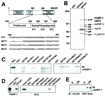
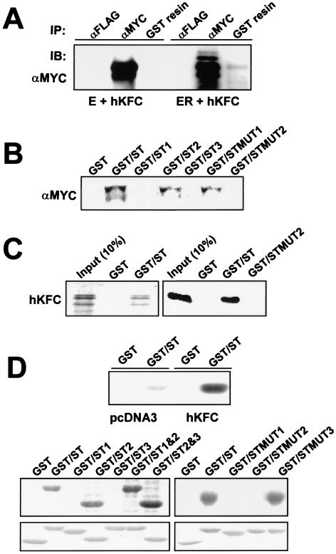
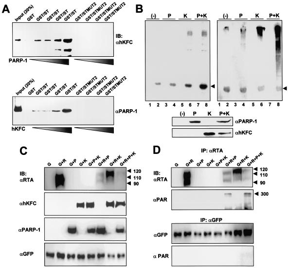
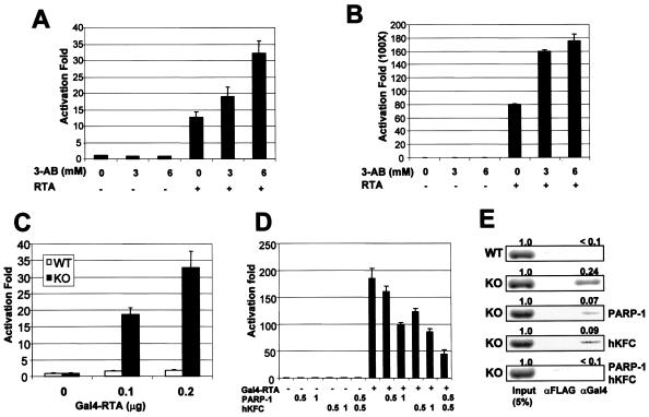
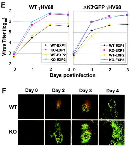
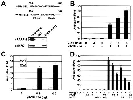
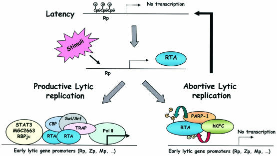
The replication and transcription activator (RTA) of gamma-2 herpesvirus is sufficient to drive the entire virus lytic cycle. Hence, the control of RTA activity should play an important role in the maintenance of viral latency. Here, we demonstrate that cellular poly(ADP-ribose) polymerase 1 (PARP-1) and Ste20-like kinase hKFC interact with the serine/threonine-rich region of gamma-2 herpesvirus RTA and that these interactions efficiently transfer poly(ADP-ribose) and phosphate units to RTA. Consequently, these modifications strongly repressed RTA-mediated transcriptional activation by inhibiting its recruitment onto the promoters of virus lytic genes. Conversely, the genetic ablation of PARP-1 and hKFC interaction or the knockout of the PARP-1 gene and activity considerably enhanced gamma-2 herpesvirus lytic replication. Thus, this is the first demonstration that cellular PARP-1 and hKFC act as molecular sensors to regulate RTA activity and thereby, herpesvirus latency.


Figures

FIG. 1.
FIG. 1.
Purification and identification of KSHV RTA binding proteins. (A) Schematic diagram of the proline-rich and ST-rich regions of RTA and its mutants. Amino acid numbers indicate the proline-rich and ST-rich regions of RTA. LZ, leucine zipper region; TAD, transcriptional activation domain. Red letters indicate the mutations described in the text. (B) Identification of RTA binding proteins. Glutathione-Sepharose beads containing 5 μg of GST, GST-Pro, or GST-ST fusion protein were mixed with lysates of 35S-labeled BJAB-B cells. RTA binding proteins were resolved by SDS-PAGE and autoradiographed with a PhosphorImager. Lines indicate the proteins that specifically interacted with GST-ST. The proteins identified by mass spectrometry are indicated at the right side of the figure. (C) Specific interaction of the RTA ST-rich region with PARP-1. BJAB cell lysates were used for pull-down assay with GST, GST-ST, or GST-ST mutant fusion proteins followed by immunoblotting (IB) with an anti-PARP-1 (αPARP-1) antibody. Whole-cell lysates (WCL) were included as a control. (D) Direct interaction between PARP-1 and RTA. [35S]methionine-labeled PARP-1 and RTA proteins from in vitro translation were mixed with GST or GST-ST protein and GST orGST-PARP-1 subdomain fusion protein, respectively. After extensive washing, 35S-labeled polypeptides associated with GST fusion proteins were separated by SDS-PAGE followed by autoradiography. The first lane indicates the 10% input amount of PARP-1 and RTA proteins used for the binding assay. (E) Interaction of PARP-1 and RTA in 293T cells. At 48 h posttransfection, with either EB-GST (E) or EB-GST-RTA vector (ER), 293T cell lysates were used for precipitation (IP) with an anti-FLAG (αFLAG) antibody or glutathione-Sepharose affinity column (GST resin). The polypeptides associated with anti-Flag and GST complex were separated by SDS-PAGE followed by immunoblotting with an anti-PARP-1 antibody. (F) Poly(ADP-ribosy)lation of RTA by PARP-1. GST, GST-ST, or GST-ST mutant fusion protein was incubated with 1 μg of recombinant PARP-1 and [32P]NAD+. 32P-labeled GST, GST-ST, or GST-ST mutant fusion protein was separated by SDS-PAGE followed by autoradiography. (G and H) After mixing with recombinant PARP-1, the glutathione beads containing GST, GST-ST, or GST-ST mutant fusion protein were extensively washed, subjected to a ribosylation reaction with NAD+, separated by SDS-PAGE, and reacted with an anti-PAR (αPAR) antibody (G) or an anti-PARP-1 antibody (H). A similar amount of each GST fusion protein was used in this assay (right bottom panel). αGST, anti-GST.
FIG. 1.
FIG. 1.
Purification and identification of KSHV RTA binding proteins. (A) Schematic diagram of the proline-rich and ST-rich regions of RTA and its mutants. Amino acid numbers indicate the proline-rich and ST-rich regions of RTA. LZ, leucine zipper region; TAD, transcriptional activation domain. Red letters indicate the mutations described in the text. (B) Identification of RTA binding proteins. Glutathione-Sepharose beads containing 5 μg of GST, GST-Pro, or GST-ST fusion protein were mixed with lysates of 35S-labeled BJAB-B cells. RTA binding proteins were resolved by SDS-PAGE and autoradiographed with a PhosphorImager. Lines indicate the proteins that specifically interacted with GST-ST. The proteins identified by mass spectrometry are indicated at the right side of the figure. (C) Specific interaction of the RTA ST-rich region with PARP-1. BJAB cell lysates were used for pull-down assay with GST, GST-ST, or GST-ST mutant fusion proteins followed by immunoblotting (IB) with an anti-PARP-1 (αPARP-1) antibody. Whole-cell lysates (WCL) were included as a control. (D) Direct interaction between PARP-1 and RTA. [35S]methionine-labeled PARP-1 and RTA proteins from in vitro translation were mixed with GST or GST-ST protein and GST orGST-PARP-1 subdomain fusion protein, respectively. After extensive washing, 35S-labeled polypeptides associated with GST fusion proteins were separated by SDS-PAGE followed by autoradiography. The first lane indicates the 10% input amount of PARP-1 and RTA proteins used for the binding assay. (E) Interaction of PARP-1 and RTA in 293T cells. At 48 h posttransfection, with either EB-GST (E) or EB-GST-RTA vector (ER), 293T cell lysates were used for precipitation (IP) with an anti-FLAG (αFLAG) antibody or glutathione-Sepharose affinity column (GST resin). The polypeptides associated with anti-Flag and GST complex were separated by SDS-PAGE followed by immunoblotting with an anti-PARP-1 antibody. (F) Poly(ADP-ribosy)lation of RTA by PARP-1. GST, GST-ST, or GST-ST mutant fusion protein was incubated with 1 μg of recombinant PARP-1 and [32P]NAD+. 32P-labeled GST, GST-ST, or GST-ST mutant fusion protein was separated by SDS-PAGE followed by autoradiography. (G and H) After mixing with recombinant PARP-1, the glutathione beads containing GST, GST-ST, or GST-ST mutant fusion protein were extensively washed, subjected to a ribosylation reaction with NAD+, separated by SDS-PAGE, and reacted with an anti-PAR (αPAR) antibody (G) or an anti-PARP-1 antibody (H). A similar amount of each GST fusion protein was used in this assay (right bottom panel). αGST, anti-GST.
FIG. 2.
FIG. 2.
hKFC interacts with and phosphorylates RTA. (A) Interaction of RTA with hKFC in 293T cells. At 48 h posttransfection, with either EB-GST (E) and the myc-tagged hKFC expression vector or EB-GST-RTA vector (ER) with the myc-tagged hKFC expression vector, 293T cell lysates were used for precipitation (IP) with anti-FLAG antibody (αFLAG), anti-myc antibody (αMYC), or glutathione-Sepharose affinity column (GST resin). The polypeptides associated with anti-Flag, anti-myc, or GST complex were separated by SDS-PAGE followed by immunoblotting (IB) with an anti-myc antibody. By comparison with hKFC in the anti-myc complex, approximately 5% of hKFC was detected in the GST-RTA complex. (B) Mutation at the basic amino acids of the RTA ST-rich region abolishes the interaction with hKFC. Lysates of 293T cells transfected with the myc-tagged hKFC expression vector were mixed with GST, GST-ST, and GST-ST mutant proteins. Polypeptides associated with GST complex were separated by SDS-PAGE followed by an anti-myc antibody. (C) Direct interaction between RTA and hKFC. (Left panel) [35S]methionine-labeled hKFC protein from in vitro translation was mixed with GST or GST-ST fusion protein. After extensive washing, 35S-labeled hKFC associated with GST or GST-ST fusion protein was separated by SDS-PAGE followed by autoradiography. (Right panel) Purified hKFC protein was mixed with GST, GST-ST, or GST-STMUT2 fusion protein. After extensive washings, hKFC associated with GST fusion protein was separated by SDS-PAGE followed by immunoblotting with an anti-hKFC antibody. The first lane indicates the 10% input amount of hKFC protein used for both binding assays. (D) Phosphorylation of the RTA ST-rich region by hKFC. Lysates of 293T cells transfected with pcDNA3 vector and the myc-tagged hKFC expression vector were mixed with GST or GST-ST fusion protein (top). Lysates of 293T cells transfected with the myc-tagged hKFC expression vector were mixed with GST, GST-ST, or GST-ST mutantfusion protein as indicated above the gels (bottom). After several washings, GST complex was subjected to an in vitro kinase reaction with [γ-32P]ATP. Reaction products were separated by SDS-PAGE followed by autoradiography. A similar amount of each GST fusion protein was used in this assay (lower panel).
FIG. 3.
FIG. 3.
Synergistic effect of PARP-1 and hKFC on RTA binding and modification. (A) Synergistic effect of PARP-1 and hKFC on RTA binding. One microgram of hKFC protein and 1 μg of GST-ST or GST-STMUT2 protein were incubated with increasing amounts of PARP-1 (0, 0.1, 0.5, and 1 μg) or 1 μg of PARP-1 and 1 μg of GST-ST or GST-STMUT2 protein were incubated with increasing amounts of hKFC (0, 0.1, 0.5, and 1 μg), and incubation was followed by precipitation with glutathione beads. After several washings, GST complexes were separated by SDS-PAGE followed by immunoblotting (IB) with anti-PARP-1 (αPARP-1) or anti-hKFC (αhKFC) antibody. (B) Synergistic effect of PARP-1 and hKFC on RTA modification. Lysates of 293T cells transfected with PARP-1 expression and/or hKFC expression were incubated with GST or GST-ST fusion protein. After several washings, the GST complex was subjected to an in vitro kinase reaction with [γ-32P]ATP (left) or a ribosylation reaction with NAD (right). The in vitro kinase reaction was analyzed by autoradiography, and the in vitro ribosylation reaction was analyzed by immunoblotting with an anti-PAR antibody. Arrow indicates the GST-ST protein. Lanes 1, 3, 5, and 7, GST; lanes 2, 4, 6, and 8, GST-ST. Expression levels for PARP-1 and hKFC were detected by immunoblotting with anti-PARP-1 and anti-hKFC antibody (lower panel). P, PARP-1; K, hKFC; −, vector alone. (C) Modification of RTA by PARP-1 and hKFC. Sf9 insect cells were coinfected with recombinant baculoviruses as indicated at the top of the figure. Whole-cell extracts were for immunoblotting with the indicated antibodies. Arrows indicate the RTA protein. G, GFP; R, RTA; αRTA, anti-RTA; αGFP, anti-GFP. (D) In vivo poly(ADP-ribosy)lation of RTA by PARP-1. Extracts of Sf9 insect cells were precipitated with anti-RTA or anti-GFP antibody in 0.1% SDS-containing lysis buffer, and immunoprecipitates (IP) were further used for immunoblotting with anti-RTA, anti-GFP, or anti-PAR (αPAR) antibody. Arrows indicate the poly(ADP ribosy)lated RTA.
FIG. 4.
FIG. 4.
Repression of RTA transcription activity by PARP-1 and hKFC. (A) Increase of RTA-mediated R promoter activation by 3-AB treatment. 293T cells were transfected with Rp-luciferase reporter and β-galactosidase control reporter together with (+) or without (−) RTA expression vector. The next day, cells were treated with increasing amounts of 3-AB (0, 3, and 6 mM) for 24 h. Luciferase activity was measured with cell extracts and normalized by β-galactosidase activity. Bars indicate the standard errors. (B) Increase of Gal4-RTA transcription activity by 3-AB treatment. 293T cells were transfected with Gal4-luciferase reporter and β-galactosidase control reporter together with or without Gal4-RTA expression vector. The same procedure as described above was used. (C) Enhanced transcription activity of RTA in PARP-1 KO MEFs. WT MEFs and PARP-1 KO MEFs were transfected with Gal4-luciferase reporter together with increasing amounts of Gal4-RTA expression vector. At 48 h, luciferase activity was measured and normalized by β-galactosidase activity. (D) Repression of RTA transcription activity by expression of PARP-1 and/or hKFC. PARP-1 KO MEFs were transfected with expression and reporter vectors as indicated at the bottom (amounts are in micrograms). At 48 h posttransfection, luciferase activity was measured. Luciferase activity is represented as the average of the results from three independent experiments. (E) Reduction of RTA recruitment onto the Gal4 promoter by PARP-1 and hKFC expression. A ChIP assay was performed with extracts of WT MEFs or PARP-1 KO MEFs transfected with Gal4-RTA and Gal4-luciferase together with or without PARP-1 and/or hKFC expression vectors as indicated at the right side of figure. Anti-Flag (αFLAG) and anti-Gal4 (αGal4) antibodies were used for the ChIP assay. The first lane indicates the 5% input amount of genomic DNA used for the ChIP assay.
FIG. 5.
FIG. 5.
Enhanced KSHV lytic replication by RTA MUT2 mutant. (A) Level of KSHV lytic reactivation. After the stimulation with doxycycline (0.1 μg/ml), TRExBCBL1-pcDNA5, TRExBCBL1-RTA, and TRExBCBL1-MUT2 cells were fixed by paraformaldehyde and reacted with K8.1 rabbit sera followed by fluorescein isothiocyanate-conjugated anti-rabbit secondary antibody. The data were reproduced in three independent experiments. (B) Expression of KSHV vIL-6 and K8 protein induced by RTA and the RTA MUT2 mutant. After doxycycline treatment for 0, 1, 2, and 3 days, KSHV-infected TRExBCBL1-RTA (WT) and TRExBCBL1-RTA MUT2 (MUT2) cells were lysed, and 10 μg of total proteins was subjected to immunoblotting with anti-RTA, anti-vIL-6, and anti-K8 antibodies. (C) ChIP assay of RTA-dependent promoters. KSHV-infected TRExBCBL1-pcDNA5, TRExBCBL1-RTA, and TRExBCBL1-TRA MUT2 cells were collected after 24 h of 0.1-μg/ml doxycycline treatment. ChIP assays were performed with an anti-His (αHis) antibody (Ab). PCR products corresponding to each viral promoter were generated from an aliquot (2%) of total immunoprecipitated material (Input).
FIG. 6.
FIG. 6.
Interaction of γHV68 RTA with PARP-1 and hKFC. (A) Alignment between KSHV RTA and γHV68 RTA and interaction of the γHV68 RTA ST region with PARP-1 and hKFC. The ST2 regions of KSHV RTA and γHV68 RTA are aligned. GST or GST-RTA ST2 (γHV68RTA amino acids 330 to 385) was incubated with recombinant PARP-1 and hKFC proteins, and GST complexes were analyzed by immunoblotting with anti-PARP-1 (αPARP-1) and anti-hKFC (αhKFC) antibodies. (B) Activation of γHV68 RTA-induced ORF57 promoter activity by 3-AB treatment. 293T cells were transfected with ORF57-luciferase and β-galactosidase reporters together with (+) or without (−) γHV68 RTA. The next day, cells were treated with increasing amounts of 3-AB (0, 3, and 6 mM) for 24 h. Luciferase activity was measured with cell extracts and normalized by β-galactosidase activity. Bars indicate the standard errors. (C) Enhanced transcription activity of γHV68 RTA in PARP-1 KO MEFs. WT MEFs and PARP-1 KO MEFs were transfected with ORF57-luciferase reporter together with increasing amounts of γHV68 RTA expression vector. Luciferase activity is represented as the average of the results from three independent experiments. (D) Repression of γHV68 RTA transcription activity by expression of PARP-1 and/or hKFC. PARP-1 KO MEFs were transfected with expression and reporter vectors as indicated at the bottom of figure (amounts are in micrograms). At 48 h posttransfection, luciferase activity was measured. Luciferase activity is represented as the average of the results from three independent experiments. (E) Enhanced level of γHV68 lytic replication in PARP-1 KO MEFs. WT and PARP-1 KO MEFs were infected with WT γHV68 or ΔK3-GFP γHV68 at an MOI of 1. The virus titers were determined by plaque assay. The results from two independent experiments are presented. (F) Size increase of ΔK3-GFP γHV68 plaques in PARP-1 KO MEFs. WT and PARP-1 KO MEFs were infected with ΔK3-GFP γHV68 at an MOI of 0.1. The GFP-positive plaques of ΔK3-GFP γHV68 were photographed.
FIG. 6.
FIG. 6.
Interaction of γHV68 RTA with PARP-1 and hKFC. (A) Alignment between KSHV RTA and γHV68 RTA and interaction of the γHV68 RTA ST region with PARP-1 and hKFC. The ST2 regions of KSHV RTA and γHV68 RTA are aligned. GST or GST-RTA ST2 (γHV68RTA amino acids 330 to 385) was incubated with recombinant PARP-1 and hKFC proteins, and GST complexes were analyzed by immunoblotting with anti-PARP-1 (αPARP-1) and anti-hKFC (αhKFC) antibodies. (B) Activation of γHV68 RTA-induced ORF57 promoter activity by 3-AB treatment. 293T cells were transfected with ORF57-luciferase and β-galactosidase reporters together with (+) or without (−) γHV68 RTA. The next day, cells were treated with increasing amounts of 3-AB (0, 3, and 6 mM) for 24 h. Luciferase activity was measured with cell extracts and normalized by β-galactosidase activity. Bars indicate the standard errors. (C) Enhanced transcription activity of γHV68 RTA in PARP-1 KO MEFs. WT MEFs and PARP-1 KO MEFs were transfected with ORF57-luciferase reporter together with increasing amounts of γHV68 RTA expression vector. Luciferase activity is represented as the average of the results from three independent experiments. (D) Repression of γHV68 RTA transcription activity by expression of PARP-1 and/or hKFC. PARP-1 KO MEFs were transfected with expression and reporter vectors as indicated at the bottom of figure (amounts are in micrograms). At 48 h posttransfection, luciferase activity was measured. Luciferase activity is represented as the average of the results from three independent experiments. (E) Enhanced level of γHV68 lytic replication in PARP-1 KO MEFs. WT and PARP-1 KO MEFs were infected with WT γHV68 or ΔK3-GFP γHV68 at an MOI of 1. The virus titers were determined by plaque assay. The results from two independent experiments are presented. (F) Size increase of ΔK3-GFP γHV68 plaques in PARP-1 KO MEFs. WT and PARP-1 KO MEFs were infected with ΔK3-GFP γHV68 at an MOI of 0.1. The GFP-positive plaques of ΔK3-GFP γHV68 were photographed.
FIG. 7.
FIG. 7.
Hypothetical model for gamma-2 herpesvirus lytic replication. M, methylation; P, phosphorylation; A, ADP-ribosylation.

Similar articles

Cited by

References

    1. Bagrodia, S., and R. A. Cerione. 1999. Pak to the future. Trends Cell Biol. 9:350-355. - PubMed
    1. Bernstein, C., H. Bernstein, C. M. Payne, and H. Garewal. 2002. DNA repair/pro-apoptotic dual-role proteins in five major DNA repair pathways: fail-safe protection against carcinogenesis. Mutat. Res. 511:145-178. - PubMed
    1. Boshoff, C., T. F. Schulz, M. M. Kennedy, A. K. Graham, C. Fisher, A. Thomas, J. O. McGee, R. A. Weiss, and J. J. O'Leary. 1995. Kaposi's sarcoma-associated herpesvirus infects endothelial and spindle cells. Nat. Med. 1:1274-1278. - PubMed
    1. Buki, K. G., P. I. Bauer, A. Hakam, and E. Kun. 1995. Identification of domains of poly(ADP-ribose) polymerase for protein binding and self-association. J. Biol. Chem. 270:3370-3377. - PubMed
    1. Cervellera, M. N., and A. Sala. 2000. Poly(ADP-ribose) polymerase is a B-MYB coactivator. J. Biol. Chem. 275:10692-10696. - PubMed

Publication types

MeSH terms

Substances

LinkOut - more resources