. 2003 Nov;23(22):8070-83.
doi: 10.1128/MCB.23.22.8070-8083.2003.

IkappaB kinase-independent IkappaBalpha degradation pathway: functional NF-kappaB activity and implications for cancer therapy

Affiliations

IkappaB kinase-independent IkappaBalpha degradation pathway: functional NF-kappaB activity and implications for cancer therapy

Vinay Tergaonkar et al. Mol Cell Biol. 2003 Nov.

Abstract

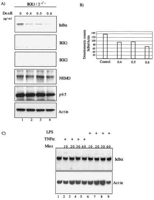
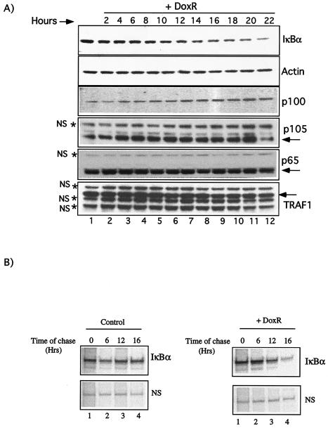
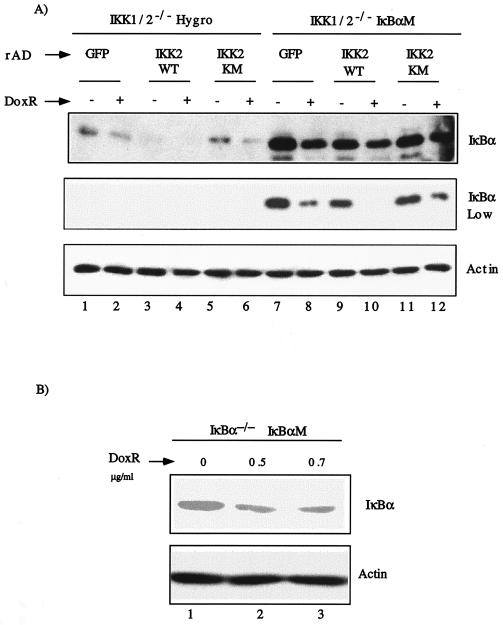
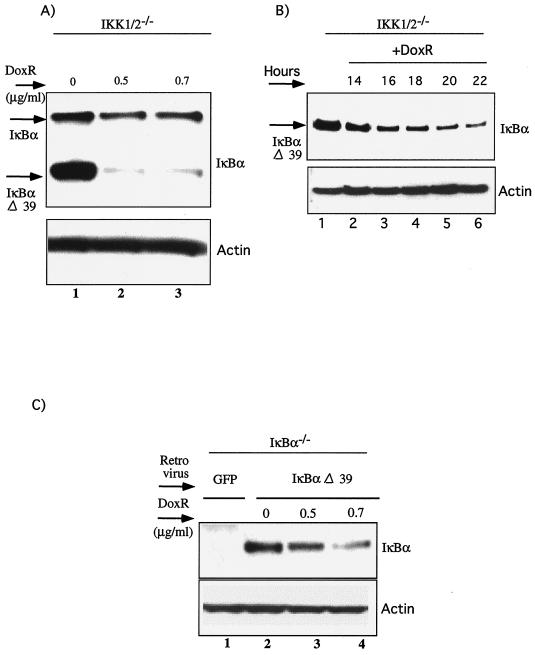
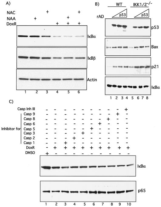
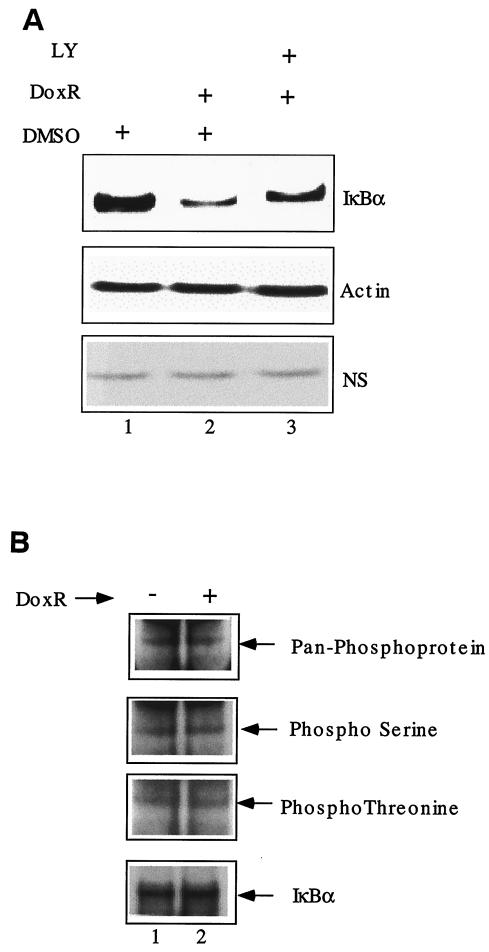
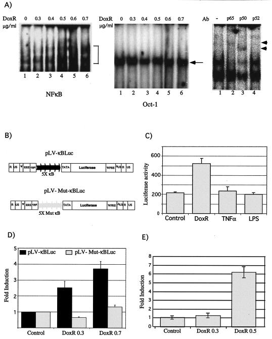
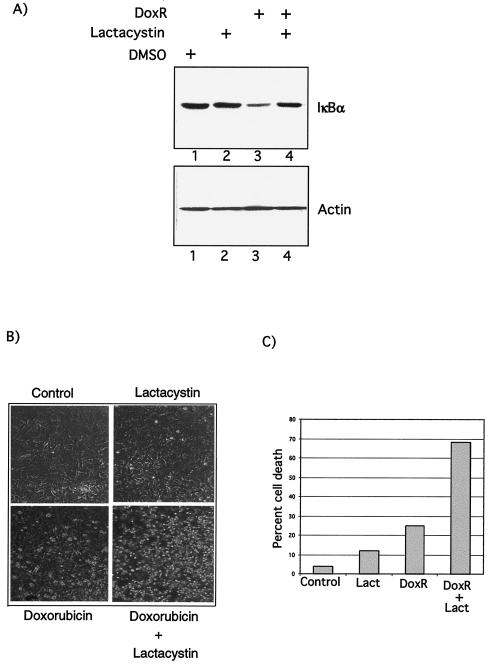
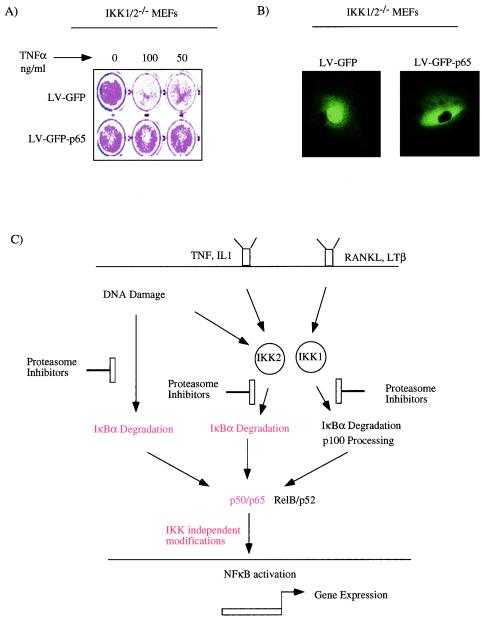
Antiapoptotic activity of NF-kappaB in tumors contributes to acquisition of resistance to chemotherapy. Degradation of IkappaB is a seminal step in activation of NF-kappaB. The IkappaB kinases, IKK1 and IKK2, have been implicated in both IkappaB degradation and subsequent modifications of NFkappaB. Using mouse embryo fibroblasts (MEFs) devoid of both IKK1 and IKK2 genes (IKK1/2(-/-)), we document a novel IkappaB degradation mechanism. We show that this degradation induced by a chemotherapeutic agent, doxorubicin (DoxR), does not require the classical serine 32 and 36 phosphorylation or the PEST domain of IkappaBalpha. Degradation of IkappaBalpha is partially blocked by phosphatidylinositol 3-kinase inhibitor LY294002 and is mediated by the proteasome. Free NF-kappaB generated by DoxR-induced IkappaB degradation in IKK1/2(-/-) cells is able to activate chromatin based NF-kappaB reporter gene and expression of the endogenous target gene, IkappaBalpha. These results also imply that modification of NF-kappaB by IKK1 or IKK2 either prior or subsequent to its release from IkappaB is not essential for NF-kappaB-mediated gene expression at least in response to DNA damage. In addition, DoxR-induced cell death in IKK1/2(-/-) MEFs is enhanced by simultaneous inhibition of NF-kappaB activation by blocking the proteasome activity. These results reveal an additional pathway of activating NF-kappaB during the course of anticancer therapy and provide a mechanistic basis for the observation that proteasome inhibitors could be used as adjuvants in chemotherapy.


Figures

FIG. 1.
FIG. 1.
DoxR-induced IκBα degradation in IKK1/2−/− MEFs. (A) IKK1/2−/− MEFs were plated in six-well plates and allowed to reach confluence. Cells were treated with indicated doses of DoxR and Western blot was performed 24 h later. (B) Densitometric quantitation of IκBα degradation was carried out by using NIH image software. The ratio of IκBα to actin has been plotted on the y axis. (C) Confluent cells grown in six-well dishes were treated with TNF-α or LPS for the indicated amounts of time. Whole-cell lysates were prepared and subjected to Western blot analysis with the indicated antibodies.
FIG. 2.
FIG. 2.
Time course of DoxR-induced IκBα degradation in IKK1/2−/− MEFs. (A) Confluent IKK1/2−/− MEFs were treated with 0.7 μg of DoxR/ml for the indicated time in hours, and a Western blot was performed with the indicated antibodies. The levels of nonspecific proteins (NS) are indicated by asterisks. (B) Pulse-chase analysis of IκBα levels in the absence or presence of DoxR. IKK1/2−/− MEFs were starved and subsequently labeled with 35S protein labeling mix (NEN). Chase with or without 0.7 μg of DoxR/ml was performed for the indicated durations. The levels of a nonspecific (NS) protein are shown as a loading control.
FIG. 3.
FIG. 3.
IκBα degradation in response to DoxR does not require phosphorylations on serine 32 and 36 and in the PEST domain. (A) IKK1/2−/− MEFs were infected with either pLXSH or pLXSH-IκBαM retroviruses and stably selected using hygromycin. Mixed populations of IKK1/2−/− Hygro (lanes 1 to 6) and IKK1/2−/− IκBαM cells (lanes 7 to 12) were infected with rAD, expressing GFP (rAD-GFP), IKK2WT (rAD-IKK2WT), or IKK2KM (rAD-IKK2KM). At 48 h postinfection cells were either left untreated or treated with 0.4 μg of DoxR/ml. Expression of IKK2 and IκBα proteins was measured by using the Flag M2 and IκBα (C21) antibodies, respectively. Actin levels were used to normalize for protein loading. (B) IκBα−/− MEFs were infected with retroviruses expressing the dominant-negative form of IκBα, IκBαM. Stable pools of cells were generated as described previously and plated in six-well dishes. Confluent cells were treated with 0.5 and 0.7 μg of DoxR/ml for 24 h. Lysates were probed with C21 antibody to detect IκBαM.
FIG. 4.
FIG. 4.
IκBα degradation in response to DoxR does not require the PEST domain. (A) IKK1/2−/− MEFs were infected with retroviruses expressing IκBαΔ39, which is lacking the last 39 amino acids of IκBα. Cells were selected on hygromycin, mixed pools of cells were treated with the indicated doses of DoxR for 24 h, and IκBα degradation was evaluated by probing with IκBα C15 antibody. (B) Time course of IκBαΔ39 degradation in IKK1/2−/− MEFs. Confluent cells were treated with 0.5 μg of DoxR/ml for the indicated durations in hours and Western blot analysis for IκBαΔ39 was performed with IκBα C15 antibody. (C) IκBα−/− MEFs were infected with retroviruses expressing either GFP (as a control) or a virus expressing the IκBαΔ39. Stable pools of cells were generated as described previously and plated in six-well dishes. Confluent cells were treated with 0.5 and 0.7 μg of DoxR/ml for 24 h. Lysates were probed with C15 antibody to detect IκBαΔ39.
FIG. 5.
FIG. 5.
IκBα degradation by DoxR is not mediated by ROS, p53, or caspases. (A) Confluent IKK1/2−/− MEFs were treated with 0.7 μg of DoxR/ml and 10 mM NAA or NAC as indicated. After 24 h the cells were lysed, and Western blots were performed. (B) IKK1/2−/− MEFs were cultured in six-well dishes and infected with increasing MOIs of rAD-p53 or rAD-GFP in 300 μl of DMEM with 5% fetal calf serum and antibiotics. The plates were rocked every 10 min, and the infection was allowed to proceed for 2 h, after which the cells were replenished with medium without removing the virus. This medium was changed after overnight incubation at 37°C with fresh medium. Cells were lysed and analyzed by Western blot analysis between 48 and 60 h postinfection. (C) IKK1/2−/− MEFs were treated either with DoxR alone or in combination with various caspase inhibitors, as indicated, for 24 h and then analyzed by Western blotting. Rows: Casp 1, caspase-1 inhibitor VI, Z-YVAD-FMK; Casp 2, caspase-2 inhibitor I, Z-VDVAD-FMK; Casp 3, caspase-3 inhibitor II, Z-DEVD-FMK; Casp 5, caspase-5 inhibitor I, Z-WEHD-FMK; Casp 6, caspase-6 inhibitor I, Z-VEID-FMK; Casp 8, caspase-8 inhibitor II, Z-IETD-FMK; Casp 9, caspase-9 inhibitor I, Z-LEHD-FMK. All are cell-permeable, irreversible, caspase inhibitors of various caspases as indicated by their number. Caspase inhibitor III (Casp Ihn III), Boc-d-FMK, is a broad-spectrum caspase inhibitor. More information regarding these inhibitors can be obtained on the Calbiochem information sheet.
FIG. 6.
FIG. 6.
Role of other kinases in DoxR-mediated IκBα degradation. (A) IKK1/2−/− MEFs were treated with 0.7 μg of DoxR/ml, alone or in combination with LY294002 (80 μM). Western blot analysis was performed 24 h later. The levels of actin and a nonspecific (NS) protein were used as loading controls. (B) IKK1/2−/− MEFs were treated with 0.7 μg of DoxR/ml, along with the proteasome inhibitor lactacystin (20 μM), for 10 h. Treated and untreated cells were lysed, and IκBα was immunoprecipitated. The immunoprecipitated material was analyzed by Western blot with the indicated phospho-specific antibodies. The levels of total IκBα were analyzed by using the C21 antibody.
FIG. 7.
FIG. 7.
NF-κB activity in response to DoxR. (A) IKK1/2−/− MEFs were treated with indicated doses of DoxR for 12 h and analyzed for gel shift activity by using a NF-κB consensus oligonucleotide (left panel) or Oct-1 oligonucleotide (middle panel). Supershift analysis with antibodies to p50, p65, and p52 was performed on extracts derived from cells treated with 0.7 μg of DoxR/ml (right panel). (B) Construction of LV-κBluc and LV-Mut-κBluc vectors. The positions of the κB binding sites, along with the TATA box, have been indicated. (C) IKK1/2−/− MEFs were transduced with LV-κBLuc, and pools of these cells were treated for 18 h with DoxR (0.3 μg/ml), TNF-α (10 ng/ml), and LPS (30 ng/ml). Luciferase activity was evaluated in a 96-well plate reader. (D) IKK1/2−/− MEFs were either transduced with LV-κBLuc or LV-Mut-κBluc vectors, and pools of these cells were treated for 18 h with DoxR before luciferase activity was measured. (E) Real-time PCR analysis for IκBα. IKK1/2−/− MEFs were treated with the indicated doses of DoxR, and the levels of IκBα and actin messages were determined by real-time PCR analysis. The ratios of IκBα levels to actin levels were quantitated and are indicated as a fold change on the y axis.
FIG. 8.
FIG. 8.
Functional relevance of NF-κB activity induced by DoxR. (A) DoxR-mediated IκBα degradation requires the proteasome. IKK1/2−/− MEFs were treated either with dimethyl sulfoxide, lactacystin (20 μM), or 0.7 μg of DoxR/ml alone or in combination as indicated. Lysates were prepared 24 h after treatment, and Western blot analysis was performed as described previously. (B) IKK1/2−/− MEFs were treated either with DoxR (0.7 μg/ml), lactacystin (20 μM), or a combination of the two drugs. Cells were visualized 24 h later. Detached and apoptotic cells are more refractile than the attached cells. (C) Quantitation of cell death induced by DoxR and lactacystin. Cell survival of attached and detached cells was measured after 24 to 30 h of treatment by using a trypan blue viability assay. This figure is a representative of three independent assays.
FIG. 9.
FIG. 9.
(A) Free p65 can protect against TNF-α-induced apoptosis in the absence of IKK activity. IKK1/2−/− MEFs transduced with LV-GFP or LV-GFP-p65 were treated with TNF-α, 48 h after transduction. Cell survival was estimated by staining the cells with crystal violet 48 h after TNF-α treatment. (B) Subcellular localization of GFP-p65. Live IKK1/2−/− cells transduced with LV-GFP or LV-GFP-p65 were imaged at ×60 magnification by using a deconvolution microscope. (C) Model based on our studies. Cytokines such as TNF-α and interleukin-1 primarily employ IKK2, and lymphotoxin β (LTβ) and RANK ligand (RANKL) use IKK1 to activate NF-κB. Unlike these stimuli, DoxR can activate NF-κB in an IKK1- and IKK2-independent manner. Despite their different origins, all of the pathways of NF-κB activation are blocked by proteasome inhibitors. Based on data presented in this report, functional activation by p65/p50 complexes (shown in red) generated after TNF-α signaling (A) or DoxR induction (Fig. 7C and E) do not require further modification by IKK1 or IKK2.

Similar articles

Cited by

References

    1. Baichwal, V. R., and P. A. Baeuerle. 1997. Activate NF-κB or die? Curr. Biol. 7:R94-R96. - PubMed
    1. Bender, K., M. Gottlicher, S. Whiteside, H. J. Rahmsdorf, and P. Herrlich. 1998. Sequential DNA damage-independent and -dependent activation of NF-κB by UV. EMBO J. 17:5170-5181. - PMC - PubMed
    1. Bonnard, M., C. Mirtsos, S. Suzuki, K. Graham, J. Huang, M. Ng, A. Itie, A. Wakeham, A. Shahinian, W. J. Henzel, A. J. Elia, W. Shillinglaw, T. W. Mak, Z. Cao, and W. C. Yeh. 2000. Deficiency of T2K leads to apoptotic liver degeneration and impaired NF-κB-dependent gene transcription. EMBO J. 19:4976-4985. - PMC - PubMed
    1. Brown, K., S. Gerstberger, L. Carlson, G. Franzoso, and U. Siebenlist. 1995. Control of IκB-alpha proteolysis by site-specific, signal-induced phosphorylation. Science 267:1485-1488. - PubMed
    1. Chen, G., P. Cao, and D. V. Goeddel. 2002. TNF-induced recruitment and activation of the IKK complex require Cdc37 and Hsp90. Mol. Cell 9:401-410. - PubMed

Publication types

MeSH terms

Substances

LinkOut - more resources