. 2003 Oct 22;23(29):9547-56.
doi: 10.1523/JNEUROSCI.23-29-09547.2003.

Cross-repressive interaction of the Olig2 and Nkx2.2 transcription factors in developing neural tube associated with formation of a specific physical complex

Affiliations

Cross-repressive interaction of the Olig2 and Nkx2.2 transcription factors in developing neural tube associated with formation of a specific physical complex

Tao Sun et al. J Neurosci. .

Abstract

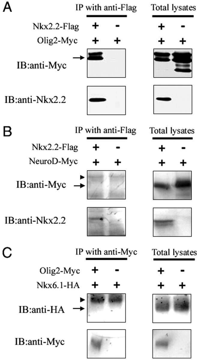
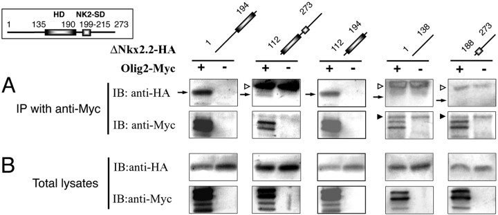
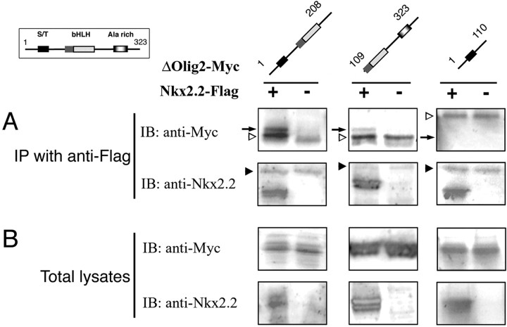
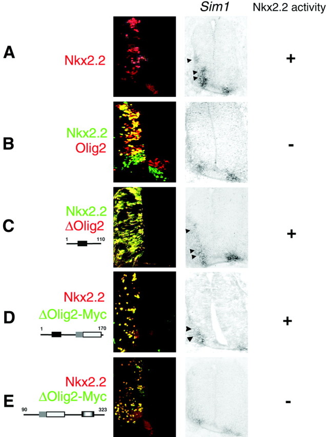
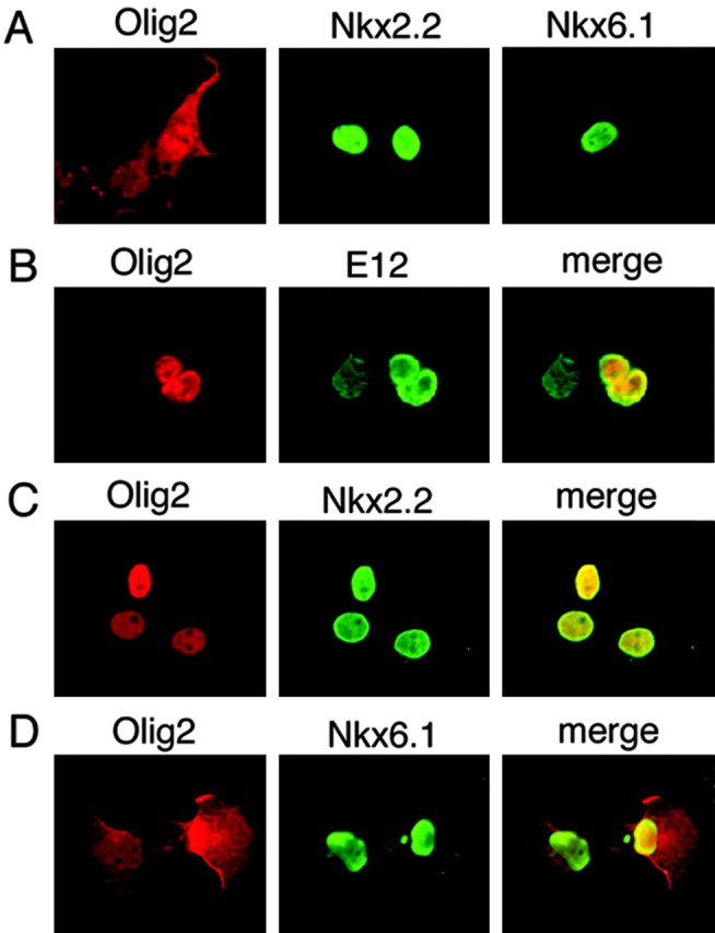
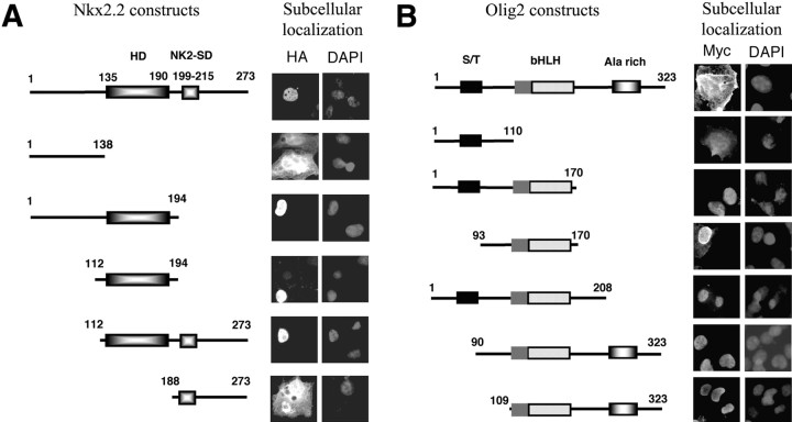
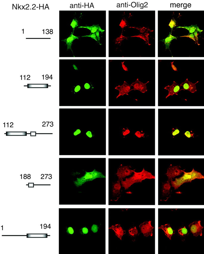
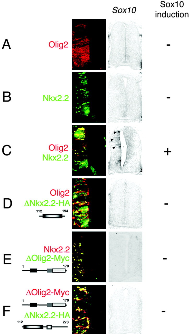
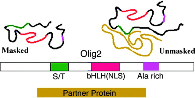
In developing neural tube, the basic helix-loop-helix (bHLH) transcription factor Olig2 interacts with the homeodomain transcription factor Nkx2.2 at two distinct stages. During neuronogenesis, a cross-repressive interaction appears to establish the precise boundary between the p3 and pMN domains. At later times, a cooperative interaction is noted because Nkx2.2 promotes maturation of oligodendrocyte progenitor cells specified by expression of Olig2. We show here that the Olig2 protein can form a physical complex with Nkx2.2 protein in mammalian cells and yeast two-hybrid trap assay. This interaction is specific because Olig2 does not bind to a biologically irrelevant homeodomain transcription factor (Nkx6.1), and Nkx2.2 does not interact with a biologically irrelevant bHLH protein (NeuroD). Deletion mapping analysis suggests that formation of an Olig2-Nkx2.2 physical complex is insufficient for the induction of oligodendrocyte progenitors in developing spine; however, the protein-protein interaction observed might be important for the cross-repressive interaction between Olig2 and Nkx2.2 that helps to establish the pMN-p3 boundary in the developing spinal cord.


Figures

Figure 1.
Figure 1.
Specific physical interaction between Nkx2.2 and Olig2. A, Cos7 cells were cotransfected with expression constructs encoding Flag-tagged Nkx2.2 and Myc-Tagged Olig2. Western blots from anti-Flag immunoprecipitates (IP) and also total cell lysates were immunoblotted (IB) with anti-Myc and anti-Nkx2.2 antibodies. Olig2 was immunoprecipitated by full-length Nkx2.2 (arrow). Note that Olig2 protein is distributed within four separate bands in total cell lysates. Two of these bands may reflect alternate translation initiation sites in the Olig2 cDNA sequence (Lu et al., 2000), and the other two may reflect degradation-post-translational modification products. B, Cos7 cells were cotransfected with expression constructs encoding Flag-tagged Nkx2.2 and Myc-Tagged NeuroD. Western blots from anti-Flag immunoprecipitates and also from total cell lysates were immunoblotted with anti-Myc and anti-Nkx2.2 antibodies. NeuroD was not immunoprecipitated by the full-length Nkx2.2 (arrow indicates the expected NeuroD band, and arrowhead indicates nonspecific IgG band.). C, Cos7 cells were cotransfected with expression constructs encoding Myc-tagged Olig2 and HA-Tagged Nkx6.1. Western blots from anti-Myc immunoprecipitates and also from total cell lysates were immunoblotted with anti-HA and anti-Myc antibodies. Olig2 fails to coimmunoprecipitate with Nkx6.1 (arrow indicates the expected Nkx6.1 band, and arrowhead indicates nonspecific IgG band).
Figure 2.
Figure 2.
The homeodomain of Nkx2.2 is both necessary and sufficient for physical interaction with Olig2. A, Cos7 cells were cotransfected with expression constructs encoding Myc-tagged full-length Olig2 and HA-tagged deletions of Nkx2.2. Western blots of the anti-Myc coimmunoprecipitates were immunoblotted with anti-HA and anti-Myc antibodies. The scheme of full-length Nkx2.2 is shown on the top left, and schemes of Nkx2.2 deletions are shown above each pair of lanes. Arrows indicate the predicted positions of coimmunoprecipitated Nkx2.2 deletion fragments. Nonspecific bands of IgG heavy chain and light chain are indicated by black arrowheads and white arrowheads, respectively. As shown, deletion mutations of Nkx2.2 containing HD domains can be coimmunoprecipitated by the full-length Olig2, whereas deletion mutants that lack HD cannot be coimmunoprecipitated. B, Western blots of total cell lysates were immunoblotted with anti-HA and anti-Myc antibodies.
Figure 3.
Figure 3.
The bHLH domain of Olig2 is both necessary and sufficient for physical interaction with Nkx2.2. A, Full-length and deletion mutations of Olig2 (all Myc tagged) were cotransfected with the Flag-tagged full-length Nkx2.2 into Cos7 cells. Anti-Flag immunoprecipitates were immunoblotted with anti-Flag and anti-Myc antibodies as in Figure 2. The scheme of full-length Olig2 is shown on the top left, and schemes of Olig2 deletions are shown on the top. The predicted positions of coimmunoprecipitated Olig2 deletion fragments are indicated by arrow. Nonspecific bands of IgG heavy chain and light chain are indicated by black arrowheads and white arrowheads, respectively. As shown, Olig2 mutations containing bHLH domains are coimmunoprecipitated by the full-length Nkx2.2. B, Western blots of total cell lysates were immunoblotted with anti-Myc and anti-Nkx2.2 antibodies. The doublet of Nkx2.2 bands shown here may reflect partial degradation of the Flag-tagged Nkx2.2 during handling or a distortion of thick gel during transferring on the membrane.
Figure 8.
Figure 8.
Interaction domains of Olig2 and Nkx2.2 are sufficient for cross-repressive function. Expression constructs encoding full-length Nkx2.2 (with Flag tag) and full-length Olig2 and its mutations with Myc tag were co-electroporated into the neural tube of stage 10-12 chick embryos. After 48 hr, embryos (stage 21-23) were harvested for analysis. Adjacent sections were immunolabeled with polyclonal (red) or monoclonal (green) antibodies targeted to the Nkx2.2 or Olig constructs as indicated and hybridized in situ with a probe targeted to mRNA for chick cSim1. Ectopic expression was directed to the left half of the neural tube, and the contralateral (non-electroporated) side served as a control. Schemes of deletions of Olig2 and Nkx2.2 with amino acid count are shown on the left. The level of induction of Sim1 as visualized by in situ hybridization is summarized by + (induction) or - (no induction) on the right. The embryos shown and results summarized are representative of at least five separate embryos in each case. A, The ectopic expression of full-length Nkx2.2 induces Sim1 expression. B, Cross-repressive interaction: co-electroporation of Nkx2.2 and Olig2 inhibits induction of Sim1. C, Olig2 lacking the bHLH domain is not sufficient for cross-repressive interaction with Nkx2.2. As shown, co-electroporation of the ΔOlig2 (1-110 aa) lacking the bHLH domain with full-length Nkx2.2 does not inhibit Sim1 induction by the Nkx2.2. D, Olig2 fragment containing the amino terminal and bHLH domains is sufficient for cross-repressive interaction. Note however that loss of the C-terminal domain attenuates the repression somewhat. E, Olig2 fragment containing the C terminal and bHLH domains is sufficient for cross-repressive interaction.
Figure 4.
Figure 4.
Olig2 and Nkx2.2 interact within mammalian cells. A, Full-length Olig2, Nkx2.2, and HA-tagged Nkx6.1 were transfected into Cos7 cells individually. Transfected cells were labeled with anti-Olig2 antibodies (red), anti-Nkx2.2 antibodies (green), and anti-HA antibodies (green). Olig2 is localized in the cytoplasm and nucleus, whereas Nkx2.2 and Nkx6.1 are within the nuclei. B, Full-length Olig2 and HA-tagged E12 were cotransfected into Cos7 cells. Transfected cells were labeled with anti-Olig2 antibodies (red) and anti-HA antibodies to visualize E12 (green). Olig2 is recruited into nuclei by cotransfection with E12 (yellow in overlay). C, Full-length Olig2 and Nkx2.2 were cotransfected into Cos7 cells. Transfected cells were labeled with anti-Olig2 antibodies (red) and anti-Nkx2.2 antibodies (green). Olig2 is localized into nuclei by cotransfection with Nkx2.2 (yellow in overlay). D, Full-length Olig2 and Nkx6.1 were cotransfected into Cos7 cells. Transfected cells were labeled with anti-Olig2 antibodies (red) and anti-HA antibodies to visualize Nkx6.1 (green). Nkx6.1 fails to bring Olig2 into nuclei in cotransfected cells.
Figure 5.
Figure 5.
Mapping Nkx2.2 and Olig2 domains that specify subcellular localization of the wild-type proteins. A, Schemes of a series of Nkx2.2 deletion mutations. Full-length Nkx2.2 with Flag tag and deletion mutations with HA tag are illustrated schematically according to amino acid content. The HD (135-190 aa) and the NK2-SD (199-215 aa) are labeled separately. Subcellular localizations of each deletion fragment in transfected Cos7 cells were established by using anti-HA antibodies and DAPI to visualize nuclei. As shown, the HD domain of Nkx2.2 is essential for nuclear localization. B, Schemes of a series of Olig2 deletion mutations. The full-length Olig2 and each deletion mutation with Myc tag are illustrated according to amino acid content. The bHLH domain, S/T, and Ala-rich domain are labeled separately. Subcellular localization of each deletion fragment in transfected Cos7 cells was visualized by using anti-Myc antibodies and DAPI to visualize nuclei. Full-length Olig2 is localized in the cytoplasm and the nucleus, whereas all deletions of Olig2 with bHLH domains are localized in nuclei.
Figure 6.
Figure 6.
The HD and NK2-specific domains of Nkx2.2 are required for relocalization of full-length Olig2 into the nucleus. A series of Nkx2.2 deletion mutations with HA tag and full-length Olig2 with Myc tag were cotransfected into Cos7 cells. Transfected cells were labeled with anti-Olig2 antibodies (red) and anti-HA antibodies (green). Schemes of Nkx2.2 deletion mutations are shown on the left. As shown, only Nkx2.2 mutations with the HD and the NK2-specific domains can bring Olig2 into the nuclei of Cos7 cells.
Figure 7.
Figure 7.
Interaction domains of Olig2 and Nkx2.2 are not sufficient for cooperative biological function. Expression constructs encoding full-length Olig2 (with Myc tag), Nkx2.2 (with HA tag) and their deletion mutations were electroporated into the neural tube of stage 10-12 chick embryos. At 48 hr after electroporation, the embryos (stage 21-23) were harvested for analysis. Adjacent sections were immunolabeled with polyclonal or monoclonal antibodies against the Olig2 or Nkx2.2 expression products in red or green as indicated. Ectopic expression was directed to the left half of the neural tube, and the contralateral (non-electroporated) side served as a control. The embryos shown and the data summarized represent results from at least five analyzed embryos in all cases. Schemes of the Olig2 and Nkx2.2 expression constructs are shown to the left. The level of induction of cSox10 as visualized by in situ hybridization is summarized by the + (induction) and the - (no induction) on the right. A, The ectopic expression of full-length Olig2 does not induce Sox10 expression. B, The ectopic expression of full-length Nkx2.2 does not induce Sox10 expression. C, Cooperative interaction: co-electroporation of Nkx2.2 and Olig2 induces Sox10 expression. D, The homeodomain of Nkx2.2 is not sufficient for cooperative interaction with Olig2. As shown, co-electroporation of the ΔNkx2.2 (112-194 aa) with full-length Olig2 does not induce Sox10. E, Olig2 lacking the C terminus is not sufficient for cooperative interaction with Nkx2.2. As shown, co-electroporation of the ΔOlig2 (1-170 aa) with full-length Nkx2.2 does not induce Sox10. F, Interaction of the homeodomain of Nkx2.2 and the bHLH domain of Olig2 is not sufficient for the induction of Sox10. Co-electroporation of ΔOlig2 (1-170 aa) containing the bHLH domain and ΔNkx2.2 (112-273 aa) lacking the N terminus does not induce Sox10 expression.
Figure 9.
Figure 9.
An occluded NLS motif in Olig2. Our current model of Olig2 proteins is shown. The bHLH domain, S/T domain, and Ala-rich domain are labeled separately. The model suggests that Olig2 has a functional NLS that may possibly be the bHLH motif itself. Olig2 monomers fold in such a way that the bHLH-NLS motif is occluded. The motif is unmasked by (1) interaction with bHLH partner proteins such as E12 (Fig. 4), (2) interaction with co-regulator proteins such as Nkx2.2 (Fig. 4), or deletion of flanking amino acids (Fig. 5).

Similar articles

Cited by

References

    1. Bertrand N, Castro DS, Guillemot F ( 2002) Proneural genes and the specification of neural cell types. Nat Rev Neurosci 3: 517-530. - PubMed
    1. Briscoe J, Sussel L, Serup P, Hartigan-O'Connor D, Jessell TM, Rubenstein JL, Ericson J ( 1999) Homeobox gene Nkx2.2 and specification of neuronal identity by graded Sonic hedgehog signaling. Nature 398: 622-627. - PubMed
    1. Briscoe J, Pierani A, Jessell TM, Ericson J ( 2000) A homeodomain protein code specifies progenitor cell identity and neuronal fate in the ventral neural tube. Cell 101: 435-445. - PubMed
    1. Chien CT, Hsiao CD, Jan LY, Jan YN ( 1996) Neuronal type information encoded in the basic-helix-loop-helix domain of proneural genes. Proc Natl Acad Sci USA 93: 13239-13244. - PMC - PubMed
    1. Ericson J, Rashbass P, Schedl A, Brenner-Morton S, Kawakami A, van Heyningen V, Jessell TM, Briscoe J ( 1997) Pax6 controls progenitor cell identity and neuronal fate in response to graded Shh signaling. Cell 90: 169-180. - PubMed

Publication types

MeSH terms