

eIF2B-related disorders: antenatal onset and involvement of multiple organs
- PMID: 14566705
- PMCID: PMC1180499
- DOI: 10.1086/379524
eIF2B-related disorders: antenatal onset and involvement of multiple organs
Abstract
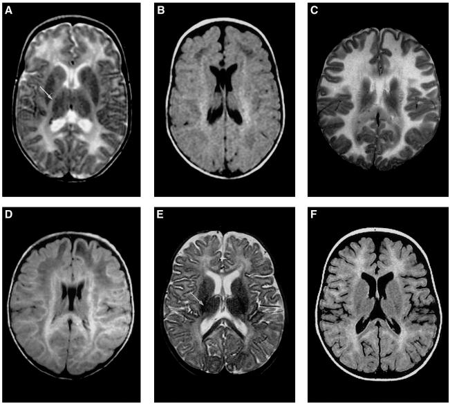
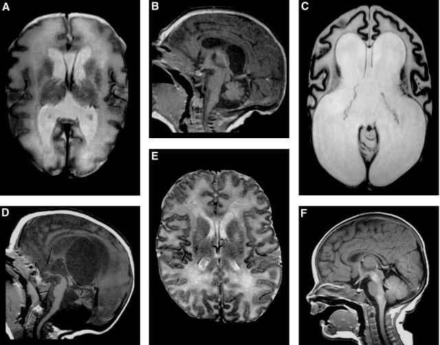
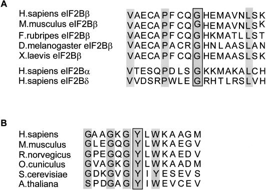
Leukoencephalopathy with vanishing white matter, also called "childhood ataxia with central nervous system hypomyelination," is the first human disease related to mutations in any of the five genes encoding subunits of eukaryotic initiation factor eIF2B or any translation factor at all. eIF2B is essential in all cells of the body for protein synthesis and the regulation of this protein synthesis under different stress conditions. It is surprising that mutations in the eIF2B genes have been reported to lead to abnormalities of the white matter of the brain only, although it has been shown recently that ovarian failure may accompany the leukoencephalopathy. Another surprising observation is that the onset of the disease varies from early childhood to adulthood, with the exception of Cree leukoencephalopathy, a disease related to a particular mutation in one of the eIF2B genes, which invariably has its onset within the first year of life. We analyzed the eIF2B genes of nine patients with an antenatal- or early-infantile-onset encephalopathy and an early demise and found mutations in eight of the patients. In addition to signs of a serious encephalopathy, we found oligohydramnios, intrauterine growth retardation, cataracts, pancreatitis, hepatosplenomegaly, hypoplasia of the kidneys, and ovarian dysgenesis. Until now, no evidence had been found for a genotype-phenotype correlation, but the consistently severe phenotype in affected siblings among our patients and in Cree encephalopathy patients suggests an influence of the genotype on the phenotype.
Figures
Similar articles
-
Decreased guanine nucleotide exchange factor activity in eIF2B-mutated patients.Eur J Hum Genet. 2004 Jul;12(7):561-6. doi: 10.1038/sj.ejhg.5201189. Eur J Hum Genet. 2004. PMID: 15054402
-
Mutations in each of the five subunits of translation initiation factor eIF2B can cause leukoencephalopathy with vanishing white matter.Ann Neurol. 2002 Feb;51(2):264-70. doi: 10.1002/ana.10112. Ann Neurol. 2002. PMID: 11835386
-
A case of ovarioleukodystrophy without eIF2B mutations.J Neurol Sci. 2008 May 15;268(1-2):183-6. doi: 10.1016/j.jns.2007.10.027. Epub 2007 Dec 3. J Neurol Sci. 2008. PMID: 18061208
-
The large spectrum of eIF2B-related diseases.Biochem Soc Trans. 2006 Feb;34(Pt 1):22-9. doi: 10.1042/BST20060022. Biochem Soc Trans. 2006. PMID: 16246171 Review.
-
Vanishing white matter disease.Lancet Neurol. 2006 May;5(5):413-23. doi: 10.1016/S1474-4422(06)70440-9. Lancet Neurol. 2006. PMID: 16632312 Review.
Cited by
-
Foetal onset of EIF2B related disorder in two siblings: cerebellar hypoplasia with absent Bergmann glia and severe hypomyelination.Acta Neuropathol Commun. 2020 Apr 15;8(1):48. doi: 10.1186/s40478-020-00929-2. Acta Neuropathol Commun. 2020. PMID: 32293553 Free PMC article.
-
Astrocytes are central in the pathomechanisms of vanishing white matter.J Clin Invest. 2016 Apr 1;126(4):1512-24. doi: 10.1172/JCI83908. Epub 2016 Mar 14. J Clin Invest. 2016. PMID: 26974157 Free PMC article.
-
Role of Saccharomyces cerevisiae TAN1 (tRNA acetyltransferase) in eukaryotic initiation factor 2B (eIF2B)-mediated translation control and stress response.3 Biotech. 2017 Jul;7(3):223. doi: 10.1007/s13205-017-0857-8. Epub 2017 Jul 4. 3 Biotech. 2017. PMID: 28677085 Free PMC article.
-
Vanishing white matter: Eukaryotic initiation factor 2B model and the impact of missense mutations.Mol Genet Genomic Med. 2021 Mar;9(3):e1593. doi: 10.1002/mgg3.1593. Epub 2021 Jan 12. Mol Genet Genomic Med. 2021. PMID: 33432707 Free PMC article.
-
An autopsy case of infantile-onset vanishing white matter disease related to an EIF2B2 mutation (V85E) in a hemizygous region.Int J Clin Exp Pathol. 2014 May 15;7(6):3355-62. eCollection 2014. Int J Clin Exp Pathol. 2014. PMID: 25031760 Free PMC article.
References
Electronic-Database Information
-
- Online Mendelian Inheritance in Man (OMIM), http://www.ncbi.nlm.nih.gov/Omim/ (for VWM and Wolcott-Rallison syndrome)
-
- VU University Medical Center site, http://www.vumc.nl/whitematter
References
-
- Barkovich AJ, Kjos BO, Jackson DE, Norman D (1988) Normal maturation of the neonatal and infant brain: MR imaging at 1.5 T. Radiology 166:173–180 - PubMed
-
- Bin-Abbas B, Al-Mulhim A, Al-Ashwal A (2002) Wolcott-Rallison syndrome in two siblings with isolated central hypothyroidism. Am J Med Genet 111:187–190 - PubMed
-
- Black DN, Booth F, Watters GV, Andermann E, Dumont C, Halliday WC, Hoogstraten J, Kabay ME, Kaplan P, Meagher-Villemure K, Michaud J, O’Gorman G (1988) Leukoencephalopathy among native Indian infants in northern Quebec and Manitoba. Ann Neurol 24:490–496 - PubMed
-
- Boltshauser E, Barth PG, Troost D, Martin E, Stallmach T (2002) “Vanishing white matter” and ovarian dysgenesis in an infant with cerebro-oculo-facio-skeletal phenotype. Neuropediatrics 33:57–62 - PubMed
-
- Brück W, Herms J, Brockmann K, Schulz-Schaeffer W, Hanefeld F (2001) Myelinopathia centralis diffusa (vanishing white matter disease): evidence of apoptotic oligodendrocyte degeneration in early lesion development. Ann Neurol 50:532–536 - PubMed
Publication types
MeSH terms
Substances
LinkOut - more resources
Full Text Sources
Medical
Molecular Biology Databases
