Differential regulation of midbrain dopaminergic neuron development by Wnt-1, Wnt-3a, and Wnt-5a
- PMID: 14557550
- PMCID: PMC240689
- DOI: 10.1073/pnas.1534900100
Differential regulation of midbrain dopaminergic neuron development by Wnt-1, Wnt-3a, and Wnt-5a
Erratum in
- Proc Natl Acad Sci U S A. 2004 Nov 16;101(46):16390
Abstract
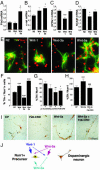
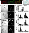
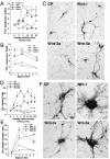
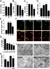
The Wnts are a family of glycoproteins that regulate cell proliferation, fate decisions, and differentiation. In our study, we examined the contribution of Wnts to the development of ventral midbrain (VM) dopaminergic (DA) neurons. Our results show that beta-catenin is expressed in DA precursor cells and that beta-catenin signaling takes place in these cells, as assessed in TOPGAL [Tcf optimal-promoter beta-galactosidase] reporter mice. We also found that Wnt-1, -3a, and -5a expression is differentially regulated during development and that partially purified Wnts distinctively regulate VM development. Wnt-3a promoted the proliferation of precursor cells expressing the orphan nuclear receptor-related factor 1 (Nurr1) but did not increase the number of tyrosine hydroxylase-positive neurons. Instead, Wnt-1 and -5a increased the number of rat midbrain DA neurons in rat embryonic day 14.5 precursor cultures by two distinct mechanisms. Wnt-1 predominantly increased the proliferation of Nurr1+ precursors, up-regulated cyclins D1 and D3, and down-regulated p27 and p57 mRNAs. In contrast, Wnt-5a primarily increased the proportion of Nurr1+ precursors that acquired a neuronal DA phenotype and up-regulated the expression of Ptx3 and c-ret mRNA. Moreover, the soluble cysteine-rich domain of Frizzled-8 (a Wnt inhibitor) blocked endogenous Wnts and the effects of Wnt-1 and -5a on proliferation and the acquisition of a DA phenotype in precursor cultures. These findings indicate that Wnts are key regulators of proliferation and differentiation of DA precursors during VM neurogenesis and that different Wnts have specific and unique activity profiles.
Figures


Similar articles
-
Dynamic temporal and cell type-specific expression of Wnt signaling components in the developing midbrain.Exp Cell Res. 2006 May 15;312(9):1626-36. doi: 10.1016/j.yexcr.2006.01.032. Epub 2006 Feb 28. Exp Cell Res. 2006. PMID: 16510140
-
GSK-3beta inhibition/beta-catenin stabilization in ventral midbrain precursors increases differentiation into dopamine neurons.J Cell Sci. 2004 Nov 15;117(Pt 24):5731-7. doi: 10.1242/jcs.01505. Epub 2004 Nov 2. J Cell Sci. 2004. PMID: 15522889
-
Transformation by Wnt family proteins correlates with regulation of beta-catenin.Cell Growth Differ. 1997 Dec;8(12):1349-58. Cell Growth Differ. 1997. PMID: 9419423
-
Function of Wnts in dopaminergic neuron development.Neurodegener Dis. 2006;3(1-2):5-11. doi: 10.1159/000092086. Neurodegener Dis. 2006. PMID: 16909030 Review.
-
Caught up in a Wnt storm: Wnt signaling in cancer.Biochim Biophys Acta. 2003 Jun 5;1653(1):1-24. doi: 10.1016/s0304-419x(03)00005-2. Biochim Biophys Acta. 2003. PMID: 12781368 Review.
Cited by
-
Activation of Wnt/β-catenin pathway by exogenous Wnt1 protects SH-SY5Y cells against 6-hydroxydopamine toxicity.J Mol Neurosci. 2013 Jan;49(1):105-15. doi: 10.1007/s12031-012-9900-8. Epub 2012 Oct 11. J Mol Neurosci. 2013. PMID: 23065334
-
Expression of early developmental markers predicts the efficiency of embryonic stem cell differentiation into midbrain dopaminergic neurons.Stem Cells Dev. 2013 Feb 1;22(3):397-411. doi: 10.1089/scd.2012.0238. Epub 2012 Sep 20. Stem Cells Dev. 2013. PMID: 22889265 Free PMC article.
-
Dopaminergic neuronal cluster size is determined during early forebrain patterning.Development. 2008 Oct;135(20):3401-13. doi: 10.1242/dev.024232. Epub 2008 Sep 17. Development. 2008. PMID: 18799544 Free PMC article.
-
Targeting of neurotrophic factors, their receptors, and signaling pathways in the developmental neurotoxicity of organophosphates in vivo and in vitro.Brain Res Bull. 2008 Jul 1;76(4):424-38. doi: 10.1016/j.brainresbull.2008.01.001. Epub 2008 Feb 1. Brain Res Bull. 2008. PMID: 18502319 Free PMC article.
-
Wnt5a-dopamine D2 receptor interactions regulate dopamine neuron development via extracellular signal-regulated kinase (ERK) activation.J Biol Chem. 2011 May 6;286(18):15641-51. doi: 10.1074/jbc.M110.188078. Epub 2011 Mar 15. J Biol Chem. 2011. PMID: 21454669 Free PMC article.
References
-
- Zetterstrom, R. H., Solomin, L., Jansson, L., Hoffer, B. J., Olson, L. & Perlmann, T. (1997) Science 276 248–250. - PubMed
-
- Castillo, S. O., Baffi, J. S., Palkovits, M., Goldstein, D. S., Kopin, I. J., Witta, J., Magnuson, M. A. & Nikodem, V. M. (1998) Mol. Cell. Neurosci. 11 36–46. - PubMed
-
- Le, W., Conneely, O. M., Zou, L., He, Y., Saucedo-Cardenas, O., Jankovic, J., Mosier, D. R. & Appel, S. H. (1999) Exp. Neurol. 159 451–458. - PubMed
-
- Smidt, M. P., Asbreuk, C. H., Cox, J. J., Chen, H., Johnson, R. L. & Burbach, J. P. (2000) Nat. Neurosci. 3 337–341. - PubMed
Publication types
MeSH terms
Substances
Grants and funding
LinkOut - more resources
Full Text Sources
Other Literature Sources
Molecular Biology Databases
Miscellaneous

