. 2003 Oct 15;22(20):5370-81.
doi: 10.1093/emboj/cdg532.

Mitochondrial translocation contact sites: separation of dynamic and stabilizing elements in formation of a TOM-TIM-preprotein supercomplex

Affiliations

Mitochondrial translocation contact sites: separation of dynamic and stabilizing elements in formation of a TOM-TIM-preprotein supercomplex

Agnieszka Chacinska et al. EMBO J. .

Abstract

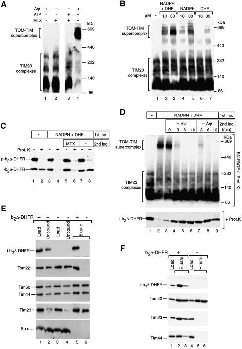
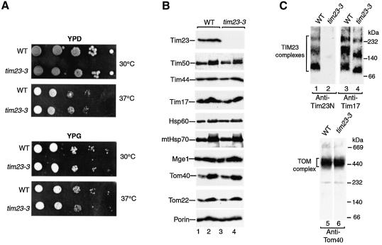
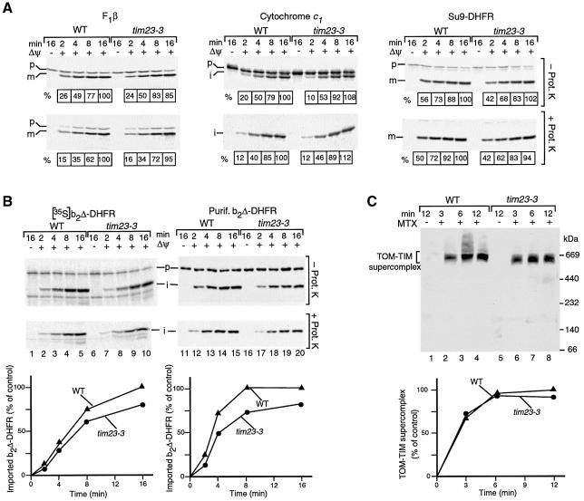
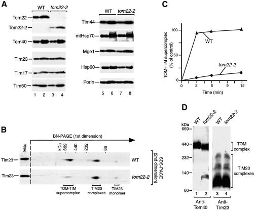
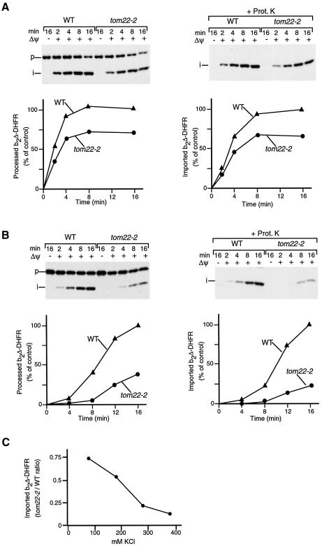
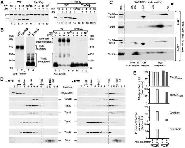
Preproteins with N-terminal presequences are imported into mitochondria at translocation contact sites that include the translocase of the outer membrane (TOM complex) and the presequence translocase of the inner membrane (TIM23 complex). Little is known about the functional cooperation of these translocases. We have characterized translocation contact sites by a productive TOM-TIM-preprotein supercomplex to address the role of three translocase subunits that expose domains to the intermembrane space (IMS). The IMS domain of the receptor Tom22 is required for stabilization of the translocation contact site supercomplex. Surprisingly, the N-terminal segment of the channel Tim23, which tethers the TIM23 complex to the outer membrane, is dispensable for both protein import and generation of the TOM-TIM supercomplex. Tim50, with its large IMS domain, is crucial for generation but not for stabilization of the supercomplex. Thus, Tim50 functions as a dynamic factor and the IMS domain of Tom22 represents a stabilizing element in formation of a productive translocation contact site supercomplex.


Figures

None
Fig. 1. The TOM–TIM supercomplex represents a productive translocation intermediate. (A) Formation of the supercomplex. Purified b2Δ-DHFR was incubated with wild-type yeast mitochondria for 15 min at 25°C in the presence or absence of MTX. For sample 3, Δψ was dissipated before the import reaction. For samples 1 and 2, the import assay was depleted of ATP. After the import reaction, the mitochondria were reisolated, lyzed with digitonin and subjected to BN–PAGE. Analysis was performed by immunodecoration with Tim23-specific antibodies. (B) Generation of the supercomplex in the presence of NADPH and DHF. b2Δ-DHFR was imported into isolated mitochondria for 12 min at 25°C, followed by BN–PAGE and immunodecoration for Tim23. (C) Two-step import assay. Purified b2Δ-DHFR was incubated with mitochondria for 15 min at 25°C in the presence or absence of 30 µM NADPH and 30 µM DHF (first incubation). Upon reisolation, the mitochondria of samples 5–8 were resuspended in fresh import buffer and incubated for a further 10 min at 25°C (second incubation). Where indicated, the mitochondria were treated with proteinase K. The samples were separated by SDS–PAGE and immunodecorated with DHFR-specific antibodies. p, precursor; i, processed (intermediate-sized) form of b2Δ-DHFR. (D) Purified b2Δ-DHFR was arrested in mitochondria in the presence or absence of 30 µM NADPH and 30 µM DHF. Upon reisolation, the mitochondria of samples 3–9 were resuspended in fresh import buffer and incubated for the indicated times at 25°C in the presence or absence of a Δψ as indicated. The samples were divided in half. One portion was subjected to BN–PAGE and decorated with Tim23-specific antibodies (upper panel). The other portion was treated with proteinase K, separated by SDS–PAGE and decorated with DHFR-specific antibodies (lower panel). (E) Affinity-purification of the supercomplex via Tim23ProtA. Where indicated, b2Δ-DHFR was accumulated in yeast mitochondria carrying Tim23ProtA in the presence of MTX. The mitochondria were reisolated, lyzed with digitonin, and subjected to IgG affinity chromatography, SDS–PAGE and immunodecoration. Thirty percent of the load and unbound material and 100% of the eluate are shown. (F) Affinity-purification of the supercomplex via Tom22His10. Where indicated, b2Δ-DHFR was accumulated in mitochondria carrying Tom22His10 in the presence of MTX. The reisolated mitochondria were lyzed with digitonin, and subjected to Ni-NTA affinity chromatography, SDS–PAGE and immunodecoration. Ten percent of load and 100% of the eluate fractions are shown.
None
Fig. 2. A yeast mutant lacking the N-terminal 50 residues of Tim23. (A) Growth of tim23-3 mutant cells is indistinguishable from wild-type cells. Wild-type (WT) and tim23-3 cells were subjected to consecutive 10-fold dilutions, spotted on fermentable medium (YPD) or non-fermentable medium (YPG) and grown for 3 days at the indicated temperatures. (B) Protein composition of tim23-3 mitochondria. Wild-type mitochondria and tim23-3 mitochondria were subjected to SDS–PAGE and immunodecoration. Odd-numbered and even-numbered samples, 10 and 20 µg mitochondrial protein, respectively. (C) BN–PAGE of digitonin-lyzed wild-type and tim23-3 mitochondria and immunodecoration.
None
Fig. 3. A connection of mitochondrial outer and inner membranes via Tim23 is not critical for formation of the TOM–TIM supercomplex. (A) Import of preproteins into tim23-3 mitochondria. The 35S-labeled precursors of F1-ATPase subunit β (F1β), cytochrome c1 and Su9-DHFR were incubated with wild-type (WT) or tim23-3 mitochondria at 25°C. Where indicated, the mitochondria were treated with proteinase K after the import reaction. The mitochondria were reisolated and analyzed by SDS–PAGE and digital autoradiography. The amount of processed protein in energized wild-type mitochondria after an import time of 16 min was set to 100%, respectively. p, precursor; i, m, processed forms (intermediate-sized, mature) of the protein. (B35S-labeled precursor of b2Δ-DHFR (left panel) or purified b2Δ-DHFR (right panel) were imported into wild-type or tim23-3 mitochondria. The amount of protease-protected, processed i-b2Δ-DHFR in energized wild-type mitochondria after an import time of 16 min was set to 100% (control). (C) Formation of the TOM–TIM supercomplex. Purified b2Δ-DHFR was incubated with wild-type or tim23-3 mitochondria in the presence of MTX unless indicated otherwise. The mitochondria were reisolated, lyzed with digitonin and subjected to BN–PAGE and immunodecoration with antibodies directed against DHFR. The amount of supercomplex formed after an incubation of 12 min in wild-type mitochondria was set to 100% (control).
None
Fig. 4. The IMS domain of Tom22 is required for stabilization of the TOM–TIM supercomplex. (A) Protein composition of tom22-2 mitochondria. Wild-type (WT) and tom22-2 mitochondria (odd-numbered and even-numbered samples, 10 and 20 µg protein, respectively) were subjected to SDS–PAGE and immunodecoration. (B) Strong reduction of the supercomplex in tom22-2 mitochondria. Purified b2Δ-DHFR was accumulated in wild-type or tom22-2 mitochondria in the presence of MTX for 15 min at 25°C. The mitochondria were reisolated, lyzed with digitonin and subjected to 2D PAGE (BN–PAGE followed by SDS–PAGE) and immunodecoration with antibodies directed against Tim23. (C) Quantification of supercomplex formation. The import experiment was performed as described for (B) with different import times. The amount of supercomplex formed in wild-type mitochondria after 12 min was set to 100% (control). (D) Stability of TOM and TIM23 complexes in tom22-2 mitochondria. Digitonin-lyzed mitochondria (70 µg protein) were subjected to BN–PAGE and immunodecoration.
None
Fig. 5. Protein import into tom22-2 mitochondria. (A) Import of purified b2Δ-DHFR into wild-type (WT) or tom22-2 mitochondria (80 mM KCl in import buffer). Where indicated, the mitochondria were treated with proteinase K after the import reaction. The mitochondria were reisolated and analyzed by SDS–PAGE. The amount of processed (i-form) of b2Δ-DHFR in energized wild-type mitochondria after a 16 min import was set to 100% (control). (B) Import of purified b2Δ-DHFR in the presence of 250 mM KCl. The experiment was performed as described for (A). (C) Dependence of b2Δ-DHFR import on the ionic strength. The experiment was performed as described above at the indicated concentrations of KCl. The ratio of imported protein in tom22-2 mitochondria to wild-type mitochondria is shown.
None
Fig. 6. The TOM–TIM supercomplex is not dissociated by a release of Tim50. (A) Import of b2Δ-DHFR is inhibited by depletion of Tim50. Purified b2Δ-DHFR was imported into Tim50-depleted or wild-type (WT) mitochondria. The reisolated mitochondria were separated by SDS–PAGE, followed by immunodecoration with antibodies against DHFR. (B) Generation of the supercomplex is blocked in Tim50-depleted mitochondria. Samples 1–4, isolated wild-type mitochondria and Tim50-depleted mitochondria (70 µg protein each) were lyzed with digitonin and subjected to BN–PAGE and immunodecoration as indicated (absence of preprotein). Samples 5–12, purified b2Δ-DHFR was arrested in wild-type or Tim50-depleted mitochondria (70 µg protein) in the presence of MTX, unless indicated otherwise. The mitochondria were reisolated and subjected to BN–PAGE and immunodecoration with Tim23-specific antibodies. (C) The supercomplex separated by BN–PAGE does not contain Tim50. Purified b2Δ-DHFR was imported into wild-type mitochondria in the presence or absence of MTX for 15 min at 25°C. Protein complexes were separated by 2D PAGE (BN–PAGE followed by SDS–PAGE). Tim50, Tom40 and Tim23 were detected by immunodecoration. (D) Sucrose gradient analysis of the supercomplex. Purified b2Δ-DHFR was imported into wild-type mitochondria in the presence or absence of MTX. The mitochondria were reisolated and lyzed with digitonin. Protein complexes were separated by sucrose gradient centrifugation. Fractions were collected, separated by SDS–PAGE and individual proteins were detected by immunodecoration (15 out of 20 fractions are shown). (E) Quantitative assessment of the presence of Tim50 in the supercomplex. Where indicated, b2Δ-DHFR was accumulated (acc.) in energized mitochondria in the presence of MTX. Top panel, Tim23ProtA mitochondria; second panel, Tom22His10 mitochondria; third and fourth panels, wild-type mitochondria. The mitochondria were reisolated and lyzed with digitonin and subjected to either IgG-affinity chromatography (top panel), Ni-NTA affinity chromatography (second panel), sucrose gradient centrifugation (third panel) or BN–PAGE (fourth panel). The amounts of Tim23 and Tim50 were determined by immunodecoration. First and second panels, the recovery of affinity- purified Tim23 from mitochondria with accumulated b2Δ-DHFR was set to 100% (control). Third and fourth panels, the total amount of solubilized Tim23 and Tim50 was set to 100% (control), respectively.

Similar articles

Cited by

References

    1. Abe Y., Shodai,T., Muto,T., Mihara,K., Torii,H., Nishikawa,S., Endo,T. and Kohda,D. (2000) Structural basis of presequence recognition by the mitochondrial protein import receptor Tom20. Cell, 100, 551–560. - PubMed
    1. Arnold I., Pfeiffer,K., Neupert,W., Stuart,R.A. and Schägger,H. (1998) Yeast mitochondrial F1F0-ATP synthase exists as a dimer: identification of three dimer-specific subunits. EMBO J., 17, 7170–7178. - PMC - PubMed
    1. Bauer M.F., Sirrenberg,C., Neupert,W. and Brunner,M. (1996) Role of Tim23 as voltage sensor and presequence receptor in protein import into mitochondria. Cell, 87, 33–41. - PubMed
    1. Bauer M.F., Hofmann,S., Neupert,W. and Brunner,M. (2000) Protein translocation into mitochondria: the role of TIM complexes. Trends Cell Biol., 10, 25–31. - PubMed
    1. Bolliger L., Junne,T., Schatz,G. and Lithgow,T. (1995) Acidic receptor domains on both sides of the outer membrane mediate translocation of precursor proteins into yeast mitochondria. EMBO J., 14, 6318–6326. - PMC - PubMed

Publication types

MeSH terms

Substances