. 2003 Oct 1;22(19):5102-14.
doi: 10.1093/emboj/cdg490.

MO25alpha/beta interact with STRADalpha/beta enhancing their ability to bind, activate and localize LKB1 in the cytoplasm

Affiliations

MO25alpha/beta interact with STRADalpha/beta enhancing their ability to bind, activate and localize LKB1 in the cytoplasm

Jérôme Boudeau et al. EMBO J. .

Abstract

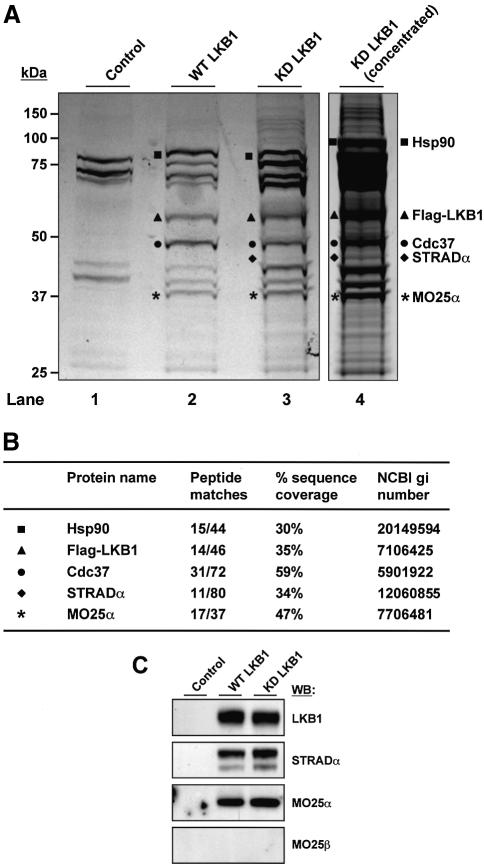
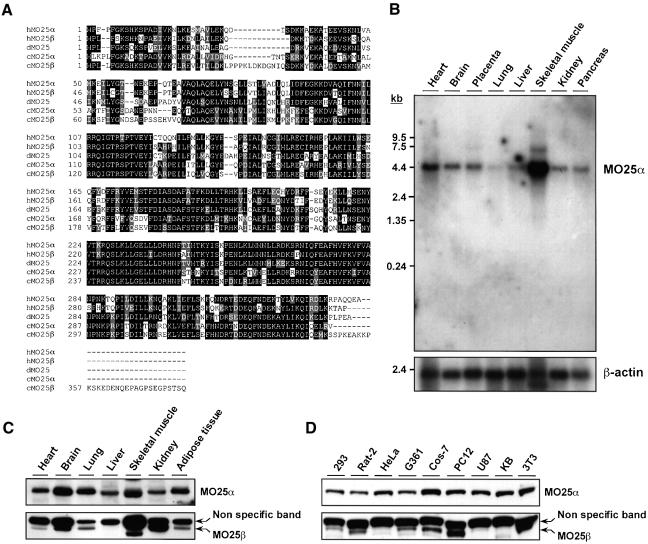
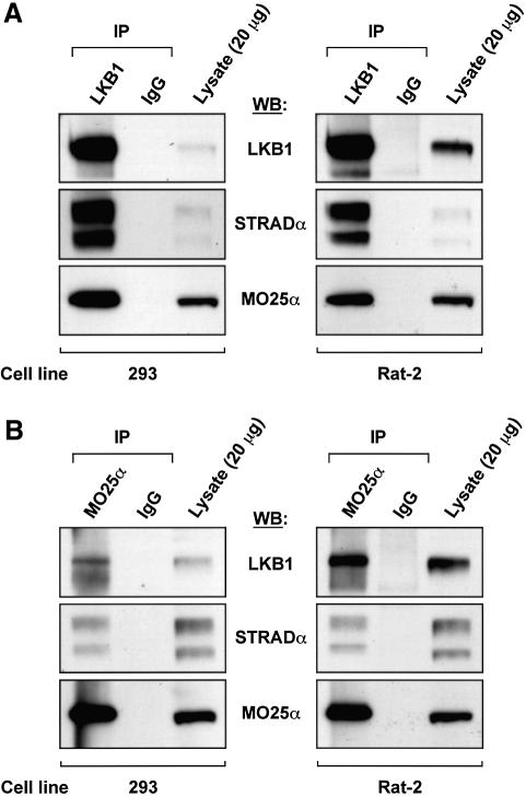
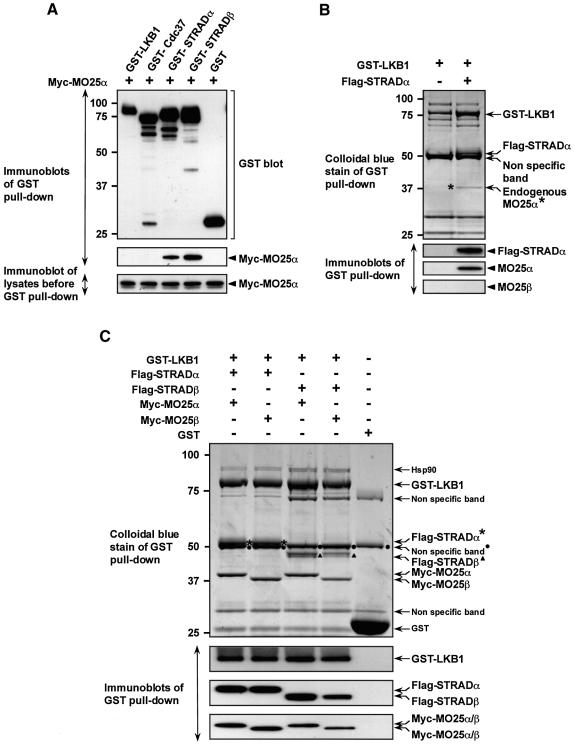
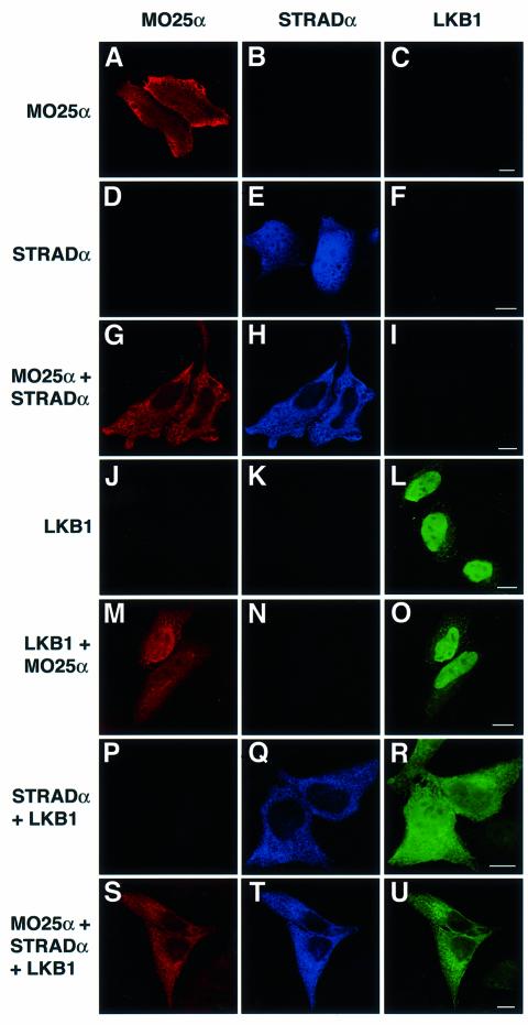
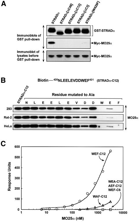
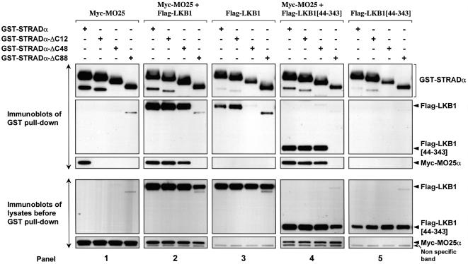
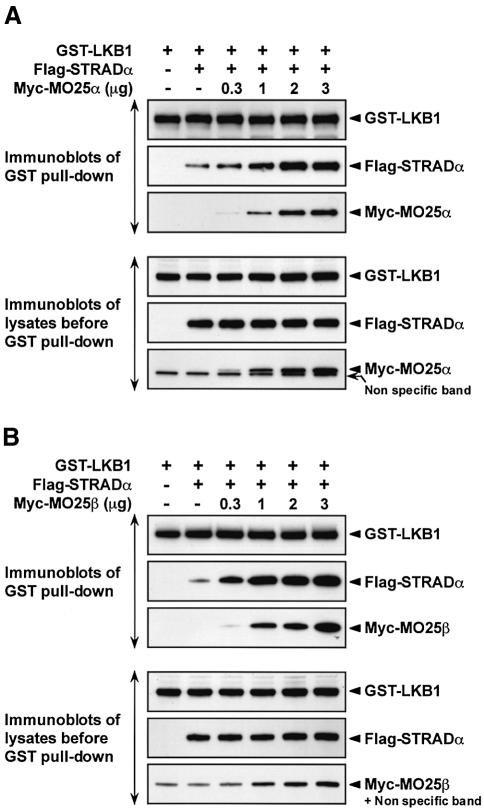
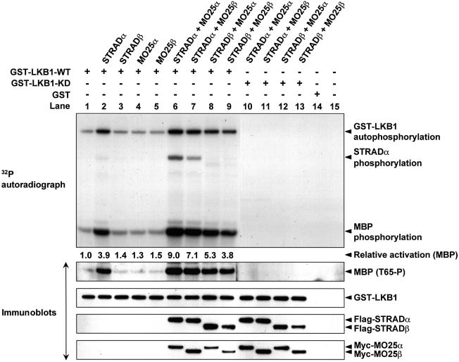
Mutations in the LKB1 protein kinase result in the inherited Peutz Jeghers cancer syndrome. LKB1 has been implicated in regulating cell proliferation and polarity although little is known about how this enzyme is regulated. We recently showed that LKB1 is activated through its interaction with STRADalpha, a catalytically deficient pseudokinase. Here we show that endogenous LKB1-STRADalpha complex is associated with a protein of unknown function, termed MO25alpha, through the interaction of MO25alpha with the last three residues of STRADalpha. MO25alpha and STRADalpha anchor LKB1 in the cytoplasm, excluding it from the nucleus. Moreover, MO25alpha enhances the formation of the LKB1-STRADalpha complex in vivo, stimulating the catalytic activity of LKB1 approximately 10-fold. We demonstrate that the related STRADbeta and MO25beta isoforms are also able to stabilize LKB1 in an active complex and that it is possible to isolate complexes of LKB1 bound to STRAD and MO25 isoforms, in which the subunits are present in equimolar amounts. Our results indicate that MO25 may function as a scaffolding component of the LKB1-STRAD complex and plays a crucial role in regulating LKB1 activity and cellular localization.


Figures

None
Fig. 1. Association of MO25α with LKB1. (A) Cell lysates derived from control parental HeLa cells or HeLa cells stably expressing N-terminal Flag epitope-tagged wild-type (WT) or kinase-dead (KD) LKB1 were passed through an anti-Flag M2–affinity agarose column, LKB1 eluted with the Flag peptide and the samples concentrated as described in Materials and methods. The samples were electrophoresed on a polyacrylamide gel and the protein bands visualized following colloidal Coomassie Blue staining. Protein bands unique to the wild-type and kinase-dead LKB1 preparations are indicated. (B) The colloidal Coomassie Blue-stained bands labelled as indicated in (A) were excised from the gel, the proteins digested in gel with trypsin, and their identities were determined by tryptic peptide mass-spectral fingerprint. The identity of STRADα and MO25α were confirmed by LC–MS/MS sequence analysis on a Q-TOF2 mass spectrometer. The number of tryptic peptides, percentage of sequence coverage and NCBI gi accession numbers for each protein identified are indicated. (C) The samples purified in (A) were immunoblotted with the indicated antibodies. Identical results were obtained following two independent purifications of LKB1 from HeLa cells.
None
Fig. 2. Amino acid sequence and tissue distribution patterns of MO25α and MO25β isoforms. (A) Amino acid sequence alignment of the human MO25α (NCBI accession No. NP_057373) and MO25β (NCBI accession No. CAC37735) isoforms as well as C.elegans MO25α (NCBI accession No. CAB16486) and MO25β (NCBI accession No. NP_508691) and Drosophila MO25 (NCBI accession No. P91891) putative homologues. Conserved residues are boxed in black, and homologous residues are shaded in grey. Sequence alignments were performed using the CLUSTALW and BOXSHADE programmes at http://www.ch.embnet.org/ using standard parameters. (B) A 32P-labelled fragment of the MO25α cDNA was used to probe a northern blot containing polyadenylated RNA isolated from the indicated human tissues. The membrane was autoradiographed, and the MO25α probe was observed to hybridize to a 4.2-kb message, identical to the size predicted for the MO25α message from database analysis. As a loading control, the northern blot was hybridized with a β-actin probe. (C and D) The indicated mouse tissue (C) or cell (D) extracts containing 20 µg of total cell protein were immunoblotted with anti-MO25α and anti-MO25β antibodies.
None
Fig. 3. Endogenous LKB1 is associated with MO25α. (A) LKB1 was immunoprecipitated from 2 mg of the indicated lysates using 10 µg of anti-LKB1 antibody covalently coupled to protein G–Sepharose, and the immunoprecipitates were immunoblotted with the indicated antibodies. As a control, immunoprecipitations were also performed in parallel experiments with pre-immune IgG antibodies covalently coupled to protein G–Sepharose. In each gel, 20 µg of total cell lysate was also immunoblotted in parallel. (B) MO25α was immunoprecipitated as above except that 15 µg of the MO25α antibody was employed. The results shown are from a single experiment that was repeated three times with similar results.
None
Fig. 4. MO25α is associated with LKB1 through STRAD. (A) 293 cells were transfected with plasmids encoding for the expression of the indicated GST fusion proteins together with Myc-MO25α. Thirty-six hours post-transfection, the GST-tagged proteins were affinity purified from the cell lysates using glutathione–Sepharose as described in Materials and methods. Similar amounts of the purified GST fusion proteins were subjected to SDS–PAGE and immunoblotted with anti-Myc antibody to detect co-purified Myc-MO25α, or with anti-GST antibody to ensure that comparable amounts of the GST-tagged proteins were present in each lane (upper and middle panels). Total cell lysates (5 µg) prior to affinity purification were also subjected to immunoblotting with anti-Myc antibody to ensure that Myc-MO25α was expressed at similar levels in each co-transfection (lower panel). (B) N-terminal GST-tagged LKB1 was expressed in 293 cells in the presence or absence of Flag-STRADα and purified as above. The purified proteins were subjected to SDS–PAGE and visualized by colloidal Coomassie Blue staining (upper panel). The faint protein band indicated, corresponding to an endogenous protein of 40-kDa (marked *), was identified by tryptic peptide mass-spectral fingerprint as endogenously expressed MO25α (with 38% sequence coverage). The purified proteins were also immunoblotted with the indicated antibodies (lower panels). (C) As above except that GST–LKB1 was expressed in 293 cells together with the indicated isoforms of Flag-STRAD and Myc-MO25 and the proteins visualized by colloidal Coomassie Blue staining were also immunoblotted with the indicated antibodies. For all panels, similar results were obtained in at least three separate experiments.
None
Fig. 5. MO25α and STRADα anchor LKB1 in the cytoplasm. HeLa cells were transfected with the indicated constructs encoding for the expression of Myc-MO25α, Flag-STRADα and GFP–LKB1. Twenty-four hours post-transfection the cells were fixed in 4% (by vol) paraformaldehyde and immunostained with anti-MO25α antibody to detect MO25α (TR anti-sheep secondary antibody, red channel) and anti-Flag to detect STRADα (Cy5 anti-mouse secondary antibody, blue channel). GFP–LKB1 localization was visualized directly through the GFP fluorescence (green channel). The cells were imaged using a Zeiss LSM 510 META confocal microscope. The cells shown are representative images obtained in three separate experiments. The scale bars correspond to 10 µm.
None
Fig. 6. MO25α recognizes the C-terminal three residues of STRADα. (A) N-terminal GST-tagged wild-type STRADα or the indicated mutants of STRADα were expressed in 293 cells together with Myc-MO25α, and 36 h post-transfection the STRADα proteins were affinity purified from the cell lysates using glutathione–Sepharose. Similar amounts of the purified proteins were subjected to SDS–PAGE and immunoblotting with anti-Myc antibody to detect co-purified Myc-MO25α, or with anti-GST antibody to ensure that comparable amounts of the GST-tagged proteins were present in each lane (upper and middle panels). Five micrograms of the total cell lysates prior to affinity purification were also subjected to immunoblotting with anti-Myc antibody to ensure that Myc-MO25α was expressed at similar levels in each condition (lower panel). (B) The indicated cell lysates (0.5 mg) were incubated with 5 µg of an N-terminal biotinylated peptide encompassing either the C-terminal 12 residues of STRADα conjugated to streptavidin–Sepharose (NLEELEVDDWEF, termed STRADα-C12) or mutants of this peptide in which the indicated residues were individually mutated to Ala. Following isolation and washing of the beads, the samples were subjected to SDS–PAGE and immunoblotted with an anti-MO25α antibody. (C) Binding of bacterially expressed MO25α to the indicated peptides was analysed by surface plasmon resonance BiaCore analysis as described in Materials and methods. Binding was analysed over a range of MO25α concentrations (6.25–3200 nM) and the response level at the steady-state binding was plotted versus the log of the MO25α concentration. The estimated Kd for the STRADα-C12 peptide was obtained by fitting the data to the formula [m1 X m0/(m0 + m2)] using Kaleidagraph software and the Kd was calculated to be 850 nM. WEF-C12 corresponds to NLEELEVDDWEF, WEA-C12 corresponds to NLEELEVDDWEA, AEF-C12 corresponds to NLEELEVDDAEF, WAF-C12 corresponds to NLEELEVDDWAF, WEF-C6 corresponds to VDDWEF.
None
Fig. 7. Binding of LKB1 to STRADα creates novel binding site(s) for MO25α. The indicated forms of GST–STRADα were co-expressed in 293 cells together with Myc-MO25α and/or wild-type Flag-LKB1 or the isolated LKB1 catalytic domain (LKB1[44–343]). Thirty-six hours post-transfection, the GST–STRADα proteins were affinity purified, subjected to SDS–PAGE and immunoblotting with anti-Myc antibody to detect Myc-MO25α, anti-Flag antibody to detect co-purified Flag-LKB1 and Flag-LKB1[44–343], or with anti-GST antibody to detect STRADα forms (upper panels). Five micrograms of total cell lysates prior to affinity purification were also subjected to immunoblotting with anti-Myc and anti-Flag antibodies to ensure that MO25α and LKB1 were expressed at similar levels in each co-transfection (lower panels). Similar results were obtained in three separate experiments.
None
Fig. 8. MO25 isoforms stabilize the LKB1–STRADα complex. (A) 293 cells were transfected with 3 µg of the DNA construct encoding for expression of GST–LKB1, in the presence or absence of 3 µg of the Flag-STRADα construct and in the absence or presence of the indicated amounts of Myc-MO25α construct. Thirty-six hours post- transfection, GST–LKB1 was affinity purified from the cell lysates and immunoblotted with appropriate epitope antibodies to detect LKB1, STRADα and MO25α. Five micrograms of total cell lysates prior to affinity purification were also immunoblotted with the indicated antibodies (lower panels). (B) As above except that the MO25β construct was employed instead of the MO25α construct. Similar results were obtained in three separate experiments.
None
Fig. 9. Activation of LKB1 by association with STRAD and MO25 isoforms. 293 cells were transfected with constructs encoding GST–LKB1 in the presence or absence of constructs encoding the indicated isoforms of STRAD and MO25. Thirty-six hours post-transfection, GST–LKB1 was affinity purified and assayed for autophosphorylation and transphosphorylation of MBP as described in the Materials and methods. The reactions were electrophoresed and the gel autoradiographed (top panel). Phosphorylation of MBP by LKB1 at Thr65 was also monitored by immunoblotting with a phosphospecific antibody (T65-P), which recognizes MBP phosphorylated by LKB1 at this residue. Each reaction was also immunoblotted with the appropriate epitope tag antibodies to monitor levels of LKB1, STRAD and MO25 isoforms. Similar results were obtained in three separate experiments.
None
Fig. 10. siRNA knockdown of endogenous MO25α destabilizes the LKB1–STRADα complex. (A) 293 cells were either not transfected or transfected with 8 µg of the empty pSUPER vector or the pSUPER vector encoding the pS1 or pS2 siRNA sequences. At the indicated times post-transfection, cells were lysed and 20 µg of lysate immunoblotted with the indicated antibodies. (B and C) As above except that 72 h post-transfection cells were lysed and the lysates were either immunoblotted with the indicated antibodies (B) or LKB1 was immunoprecipitated from 0.5 mg of the indicated lysates using 5 µg of anti-LKB1 antibody covalently coupled to protein G–Sepharose (C). The immunoprecipitates were immunoblotted with the indicated antibodies. (D) As above except that 72 h post-transfection cells were lysed and LKB1 immunoprecipitated from 0.5 mg of the indicated lysates using 5 µg of anti-LKB1 antibody or 5 µg of pre-immune antibody covalently coupled to protein G–Sepharose. The immunoprecipitates were assayed for transphosphorylation of MBP as described in the Materials and methods. The reactions were electrophoresed and phosphorylation of MBP by LKB1 at Thr65 was monitored by immunoblotting with a phosphospecific antibody (T65-P), which recognizes MBP phosphorylated by LKB1 at this residue. Similar results were obtained in three separate experiments.

Similar articles

Cited by

References

    1. Baas A.F., Boudeau,J., Sapkota,G.P., Smit,L., Medema,R., Morrice,N.A., Alessi,D.R. and Clevers,H.C. (2003) Activation of the tumour suppressor kinase LKB1 by the STE20-like pseudokinase STRAD. EMBO J., 22, 3062–3072. - PMC - PubMed
    1. Bardeesy N., Sinha,M., Hezel,A.F., Signoretti,S., Hathaway,N.A., Sharpless,N.E., Loda,M., Carrasco,D.R. and DePinho,R.A. (2002) Loss of the Lkb1 tumour suppressor provokes intestinal polyposis but resistance to transformation. Nature, 419, 162–167. - PubMed
    1. Bidlingmaier S., Weiss,E.L., Seidel,C., Drubin,D.G. and Snyder,M. (2001) The Cbk1p pathway is important for polarized cell growth and cell separation in Saccharomyces cerevisiae. Mol. Cell. Biol., 21, 2449–2462. - PMC - PubMed
    1. Boudeau J., Deak,M., Lawlor,M.A., Morrice,N.A. and Alessi,D.R. (2003a) Heat-shock protein 90 and Cdc37 interact with LKB1 and regulate its stability. Biochem. J., 370, 849–857. - PMC - PubMed
    1. Boudeau J., Kieloch,A., Alessi,D.R., Stella,A., Guanti,G. and Resta,N. (2003b) Functional analysis of LKB1/STK11 mutants and two aberrant isoforms found in Peutz-Jeghers syndrome patients. Hum. Mutat., 21, 172. - PubMed

Publication types

MeSH terms

Substances