. 2003 Oct;77(20):10957-74.
doi: 10.1128/jvi.77.20.10957-10974.2003.

In vitro proteolytic processing of the MD145 norovirus ORF1 nonstructural polyprotein yields stable precursors and products similar to those detected in calicivirus-infected cells

Affiliations

In vitro proteolytic processing of the MD145 norovirus ORF1 nonstructural polyprotein yields stable precursors and products similar to those detected in calicivirus-infected cells

Gaël Belliot et al. J Virol. 2003 Oct.

Abstract

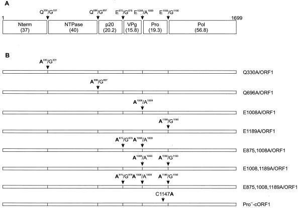
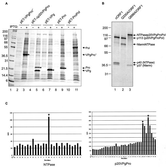
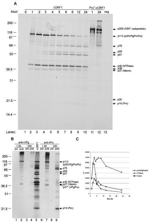
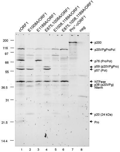
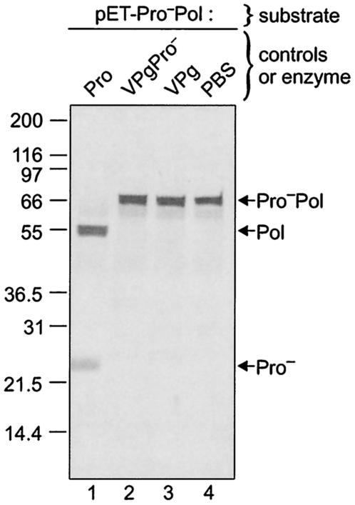
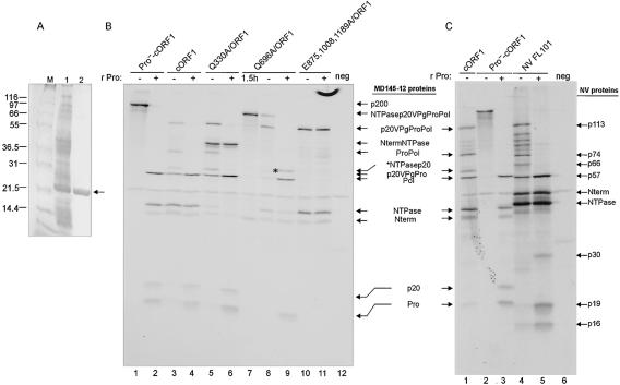
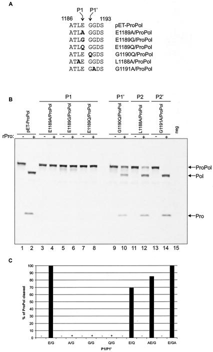
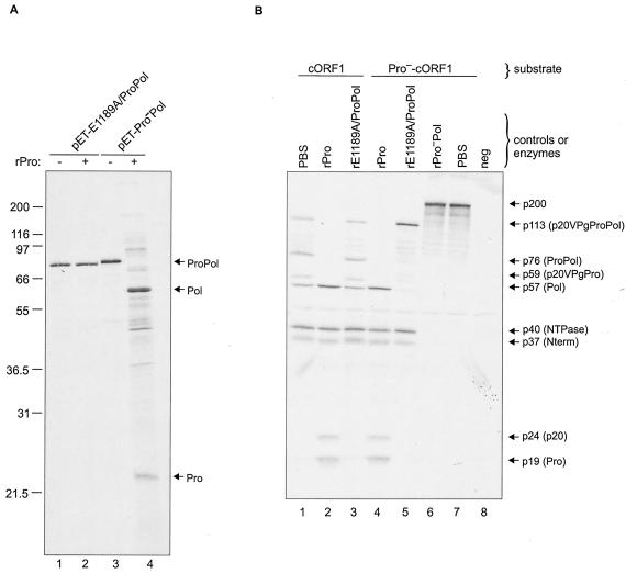
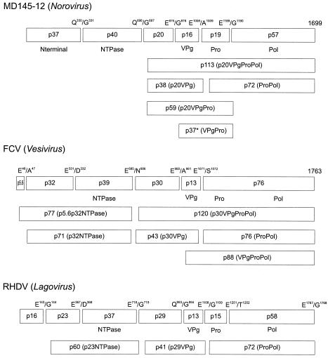
The MD145-12 strain (GII/4) is a member of the genus Norovirus in the Caliciviridae and was detected in a patient with acute gastroenteritis in a Maryland nursing home. The open reading frame 1 (ORF1) (encoding the nonstructural polyprotein) was cloned as a consensus sequence into various expression vectors, and a proteolytic cleavage map was determined. The virus-encoded cysteine proteinase mediated at least five cleavages (Q(330)/G(331), Q(696)/G(697), E(875)/G(876), E(1008)/A(1009), and E(1189)/G(1190)) in the ORF1 polyprotein in the following order: N-terminal protein; nucleoside triphosphatase; 20-kDa protein (p20); virus protein, genome linked (VPg); proteinase (Pro); polymerase (Pol). A time course analysis of proteolytic processing of the MD145-12 ORF1 polyprotein in an in vitro coupled transcription and translation assay allowed the identification of stable precursors and final mapped cleavage products. Stable precursors included p20VPg (analogous to the 3AB of the picornaviruses) and ProPol (analogous to the 3CD of the picornaviruses). Less stable processing intermediates were identified as p20VPgProPol, p20VPgPro, and VPgPro. The MD145-12 Pro and ProPol proteins were expressed in bacteria as active forms of the proteinase and used to further characterize their substrate specificities in trans cleavage assays. The MD145-12 Pro was able to cleave its five mapped cleavage sites in trans and, in addition, could mediate trans cleavage of the Norwalk virus (GI/I) ORF1 polyprotein into a similar proteolytic processing profile. Taken together, our data establish a model for proteolytic processing in the noroviruses that is consistent with nonstructural precursors and products identified in studies of caliciviruses that replicate in cell culture systems.


Figures

FIG. 1.
FIG. 1.
Predicted cleavage sites of the MD145-12 ORF1 polyprotein and constructs designed to study the proteolytic processing of the entire polyprotein. (A) Map of the cleavage sites for ORF1 of the MD145-12 norovirus based on the previously identified cleavage sites of the Southampton, Camberwell, and Chiba viruses (16, 18, 27, 28). Sequences of the dipeptide cleavage sites and their positions are indicated by arrows. The calculated molecular mass (shown in parentheses) of each protein is given in kilodaltons. The designation of the potential cleavage products is indicated inside boxes and is adapted from Liu et al. (18) and Green et al. (9). (B) Diagram of ORF1 constructs. The cORF1 construct corresponds to a consensus sequence of the entire ORF1. For other constructs, the mutated cleavage sites are indicated, and amino acid changes are in bold. The Pro-cORF1 construct has a Cys to Ala substitution at position 1147, which inactivated the proteinase (16, 29).
FIG. 2.
FIG. 2.
Mapping study of the MD145-12 norovirus ORF1 polyprotein. (A) Coomassie blue-stained bacterially expressed proteins used for N-terminal sequence analysis and trans cleavage assays. The constructs indicated above the gel were used to transform the strain BL21(DE3) of E. coli. Protein expression was induced as described in Materials and Methods, and the soluble proteins were separated in a 10 to 20% Tris-Gly polyacrylamide gel. The even- and odd-numbered lanes are soluble cellular fractions before (minus sign) and after (plus sign) IPTG induction, respectively. The proteins having a His6 tag are indicated by an asterisk. The proteins of interest are indicated by arrows on the right. For this figure and the following, Pro indicates an inactivated proteinase, in which the Cys of the active site has been replaced by an Ala residue at position 1147. In a parallel experiment, proteins in the soluble fractions in lanes 5 and 11 were used to determine the N termini of the proteinase, VPg, and polymerase. The migration of VPg in lane 7 is slightly slower than in lane 5 because of an engineered His6 tag. Lane 1 contains Mark XII protein molecular weight marker (Invitrogen). Mark XII was used in all subsequent experiments. (B) Site-directed mutagenesis of the Q330/G331 and Q696/G697 MD145-12 ORF1 cleavage sites. In vitro translation assays with the plasmid constructs cORF1, Q330A/ORF1, and Q696A/ORF1 in lanes 1, 2, and 3, respectively, were performed for 1.5 h at 30°C in the presence of [35S]Met. For each construct, an aliquot of 5 μl was separated in a 10 to 20% Tris-Gly polyacrylamide gel by SDS-PAGE, and 35S-labeled products were detected by autoradiography. The deduced identity of each protein is indicated on the right. (C) Radioactive microsequencing mapping of the Q330/G331 and Q696/G697 cleavage sites. An in vitro translation reaction from the construct pET-ΔNtermNTPasep20VPgPro was performed in the presence of [35S]Met. The cleavage products NTPase and p20VPgPro were resolved by SDS-PAGE and electroblotted to a ProBlott membrane. For both proteins, an N-terminal sequencing analysis by Edman degradation was performed, and the released radioactivity (indicated in counts per minute) was determined for each cycle. Peaks corresponding to labeled Met residues are indicated by arrows. Amino acid sequences of the ORF1 polyprotein matching the radioactive profile are indicated below each graph.
FIG. 3.
FIG. 3.
Kinetic analysis of the MD145-12 ORF1 polyprotein processing in TNT. (A) A TNT master mix using the cORF1 construct was aliquoted into 25-μl fractions that were incubated at 30°C for 1 to 24 h (lanes 1 to 10). A negative control for the processing was provided by the Pro-cORF1 construct incubated in TNT reactions for 1 and 24 h at 30°C (lanes 11 and 12, respectively). Ten microliters of each fraction was resolved in a 12% Tris-Gly polyacrylamide gel. 35S-labeled products were detected by autoradiography. An additional control (neg) for these experiments included a TNT reaction with the pSPORT1 vector (lane 13). Protein designations are adjacent to the arrows. (B) Immunoprecipitation of the cORF1 in vitro translation products with antisera raised against the recombinant VPg or Pro proteins expressed in bacteria. The in vitro translation products were incubated either for 1.5 h or 24 h prior to immunoprecipitation analysis with preimmunization (lanes 1, 3, 6, and 8) or postimmunization sera (lanes 2, 4, 7, and 9). The precipitated products were separated in a 12% polyacrylamide gel prior to autoradiography. The positive control in lane 5 was 5 μl of a cORF1 in vitro translation mixture that was incubated 24 h at 30°C. The proteins of interest are indicated on the right with arrows. The asterisks mark the putative VPgPro complex. (C) The relative quantities of the Nterm, NTPase, and p20VPgProPol protein were calculated for each time point using a phosphorimager. Intensity values are given in arbitrary unit per Met residue (AU). This ratio is the relative light intensity divided by the number of Met residues present in the proteins Nterm (6 residues), NTPase (10 residues), or p20VPgProPol (26 residues).
FIG. 4.
FIG. 4.
Site-directed mutagenesis of the E875/G876, E1008/A1009, and E1189/G1190 cleavage sites. In vitro translation assays containing the plasmid constructs indicated (lanes 1 to 7) were performed overnight at 30°C in the presence of [35S]Met. For each construct, 10 μl of a 25-μl reaction mixture was separated in a 12% Tris-Gly polyacrylamide gel. The in vitro translation product of pSPORT1 (lane 8) was included as the negative control (neg). 35S-labeled products were detected by autoradiography. Relevant proteins are indicated by arrows. It should be noted that the p20 protein showed an observed mass of 24 kDa. The constructs used in this experiment are diagrammed in Fig. 1.
FIG. 5.
FIG. 5.
Development of a trans cleavage assay for detection of active forms of the viral proteinase. The radiolabeled ProPol precursor protein was generated from the pET-ProPol construct by TNT. Five microliters was used as a substrate in the trans cleavage assay. The enzymatic activity present in the Pro, VPgPro, and VPg bacterial total cell lysates was assayed. Following overnight incubation at 30°C, the reaction assays were resolved in a 10 to 20% Tris-Gly polyacrylamide gel, and the 35S-labeled translation products were detected by autoradiography. The negative control (lane 4) consisted of 5 μl of the in vitro translation reaction mixture incubated with 5 μl of PBS (pH 7.4) overnight at 30°C. The cleavage products Pol and Pro as well as the uncleaved ProPol complex are indicated by arrows on the right.
FIG. 6.
FIG. 6.
trans cleavage activity of purified recombinant proteinase on native or variant forms of the ORF1 polyprotein. (A) SDS-PAGE analysis of the MD145-12 rPro before (lane 1) and after (lane 2) Ni-NTA purification. Proteins were stained with Coomassie blue, and the proteinase is indicated by an arrow. Lane M contains the protein molecular weight marker. (B) TNT reactions using the constructs (lanes 1 to 11) indicated above each lane were performed for 1.5 h at 30°C in the presence of [35S]Met. Five microliters of the TNT reaction mixture was incubated with either 5 μl of PBS (minus sign) or 5 μl of PBS containing 2 μg of purified rPro (plus sign). In lane 7, the TNT reaction mixture was incubated for 1.5 h at 30°C and immediately stored at −20°C without proteinase treatment. The negative control (neg) was a TNT reaction mixture containing pSPORT1. trans cleavage assays were analyzed by electrophoresis in a 12% Tris-Gly polyacrylamide gel prior to autoradiography. The proteins of interest are marked by arrows on the right. The asterisk in lane 9 indicates the putative NTPasep20 protein. (C) TNT reactions using the constructs (lanes 1 to 5) indicated above each lane were performed overnight at 30°C in the presence of [35S]Met. The NV FL101 plasmid (lanes 4 and 5) is a full-length cDNA clone of Norwalk virus (NV) (GI/1). Five microliters of the TNT reaction mixture was incubated with either 5 μl of PBS at pH 7.4 (minus sign) or 5 μl of PBS containing 2 μg of MD145-12 rPro (plus sign). The negative control (neg, lane 6) was a TNT reaction mixture containing pSPORT1. trans cleavage assays were analyzed by electrophoresis in a 12% Tris-Gly polyacrylamide gel prior to autoradiography. The proteins of interest for MD145-12 and NV are indicated on the left and right of the gel, respectively.
FIG. 7.
FIG. 7.
Mutational analysis of the ProPol dipeptide cleavage site in the trans cleavage assay. (A) Partial alignment of the ProPol cleavage site E1189/G1190 mutations. Changed amino acids are shown in bold type. Amino acids at the position P1 and P1′ of the cleavage site are indicated by arrows. (B) TNT reactions were conducted in a final volume of 25 μl for 1.5 h at 30°C, using the constructs indicated above the gel. For each construct, 5 μl of TNT reaction mixture was incubated with 5 μl of PBS (odd-numbered lanes, minus sign) or with 2 μg of MD145-12 rPro (even-numbered lanes, plus sign). The TNT product derived from pET-ProPol is shown in lanes 1 and 2. The negative control (neg) in lane 15 is an in vitro translation reaction mixture containing the pET-28b plasmid. Cleavage assays were resolved in 10 to 20% Tris-Gly polyacrylamide gels prior to autoradiography. ProPol, Pol, and Pro are indicated. (C) The cleavage efficiency of the ProPol precursor was determined by phosphorimaging. The intensity for the ProPol bands was determined in arbitrary units before (AUPBS) and after (AUPro) proteinase treatment. For each construct, the calculation of the cleavage efficiency was possible since the amount of [35S]Met was the same in the presence or absence of proteinase. The percentage of cleaved ProPol for the trans cleavage assay was calculated by the following formula: (1-AUPro/AUPBS) × 100.
FIG. 8.
FIG. 8.
Proteinase activity of the ProPol complex. (A) Five microliters of the TNT products derived from constructs pET-E1189A/ProPol (lane 1) and pET-ProPol (lane 3) was incubated 24 h with 5 μl of PBS to monitor for cis cleavage activity. The TNT products were also incubated with 2 μg of rPro to detect trans cleavage (lanes 2 and 4, plus signs). ProPol, Pol, and Pro proteins are indicated by arrows. (B) The cORF1 (lanes 1, 2, and 3) and Pro-cORF1 constructs (lanes 4 to 7) were translated in vitro for 1.5 h at 30°C. trans cleavage assays were performed 24 h at 30°C. For each construct, an aliquot of 5 μl was incubated with either 5 μl of PBS (lanes 1 and 7) or 2 μg of rPro (lanes 2 and 4). The cORF1 and Pro-cORF1 translation products were also incubated with 2 μg of bacterially expressed and partially purified rE1189A/ProPol (lanes 3 and 5) or 2 μg of rProPol (lane 6). A TNT reaction mixture containing pSPORT1 was included as a control (lane 8). The cleavage assays were analyzed in a 12% Tris-Gly polyacrylamide gel prior to autoradiography. The proteins of interest are indicated with arrows.
FIG. 9.
FIG. 9.
Comparison of the nonstructural polyprotein cleavage maps of MD145-12 (Norovirus), feline calicivirus (FCV) (Vesivirus), and rabbit hemorrhagic disease virus (RHDV) (Lagovirus) (14, 19) and their known processing intermediates. Each cleavage product is designated by the letter “p” followed by its molecular mass. Functional domains and cleavage sites are indicated.
FIG. 10.
FIG. 10.
Model for temporal proteolytic processing of the MD145-12 ORF1 polyprotein by the viral proteinase. Active dipeptide cleavage sites and their locations are indicated. Primary cleavages at Q330/G331 and Q696/G697 release the N-terminal and NTPase proteins. Two possible secondary cleavage pathways involve processing of the p20VPgProPol precursor. In one secondary pathway, the proteinase may cleave at E1008/A1009 to release p20VPg and ProPol. In the other pathway (shown with the dotted arrows), the proteinase may cleave at E1189/G1190 to release p20VPgPro and Pol. Additional processing of the released precursors occurs over time. Likely cis cleavages executed by the proteinase are shown immediately below the indicated precursor. White boxes indicate proteins in which the observed levels decrease over time. Gray boxes indicate proteins that remain present or accumulate over time. The designation for each protein based on molecular mass is indicated in parentheses.

Similar articles

Cited by

References

    1. Ando, T., J. S. Noel, and R. L. Fankhauser. 2000. Genetic classification of “Norwalk-like viruses.” J. Infect. Dis. 181(Suppl. 2):S336-S348. - PubMed
    1. Blair, W. S., X. Li, and B. L. Semler. 1993. A cellular cofactor facilitates efficient 3CD cleavage of the poliovirus P1 precursor. J. Virol. 67:2336-2343. - PMC - PubMed
    1. Blair, W. S., and B. L. Semler. 1991. Role for the P4 amino acid residue in substrate utilization by the poliovirus 3CD proteinase. J. Virol. 65:6111-6123. - PMC - PubMed
    1. Boniotti, B., C. Wirblich, M. Sibilia, G. Meyers, H. J. Thiel, and C. Rossi. 1994. Identification and characterization of a 3C-like protease from rabbit hemorrhagic disease virus, a calicivirus. J. Virol. 68:6487-6495. - PMC - PubMed
    1. Bradford, M. M. 1976. A rapid and sensitive method for the quantitation of microgram quantities of protein utilizing the principle of protein-dye binding. Anal. Biochem. 72:248-254. - PubMed

Publication types

LinkOut - more resources