. 2003 Oct;163(4):1417-28.
doi: 10.1016/S0002-9440(10)63499-2.

Vascular endothelial growth factor modulates skeletal myoblast function

Affiliations

Vascular endothelial growth factor modulates skeletal myoblast function

Antonia Germani et al. Am J Pathol. 2003 Oct.

Abstract

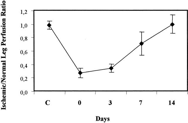
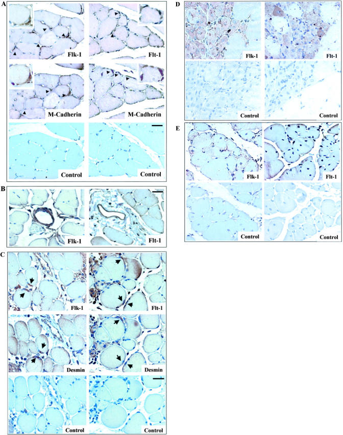
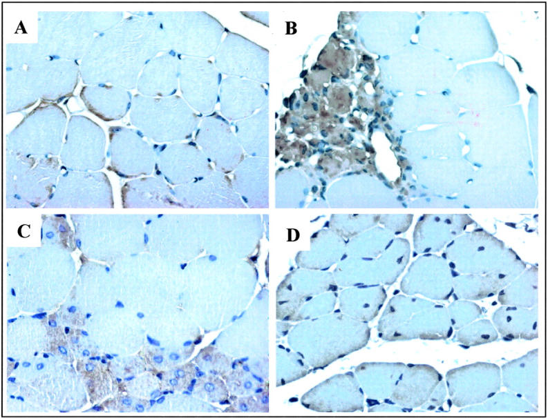
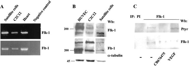
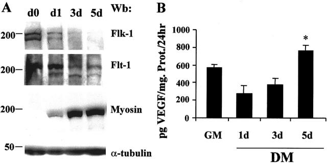
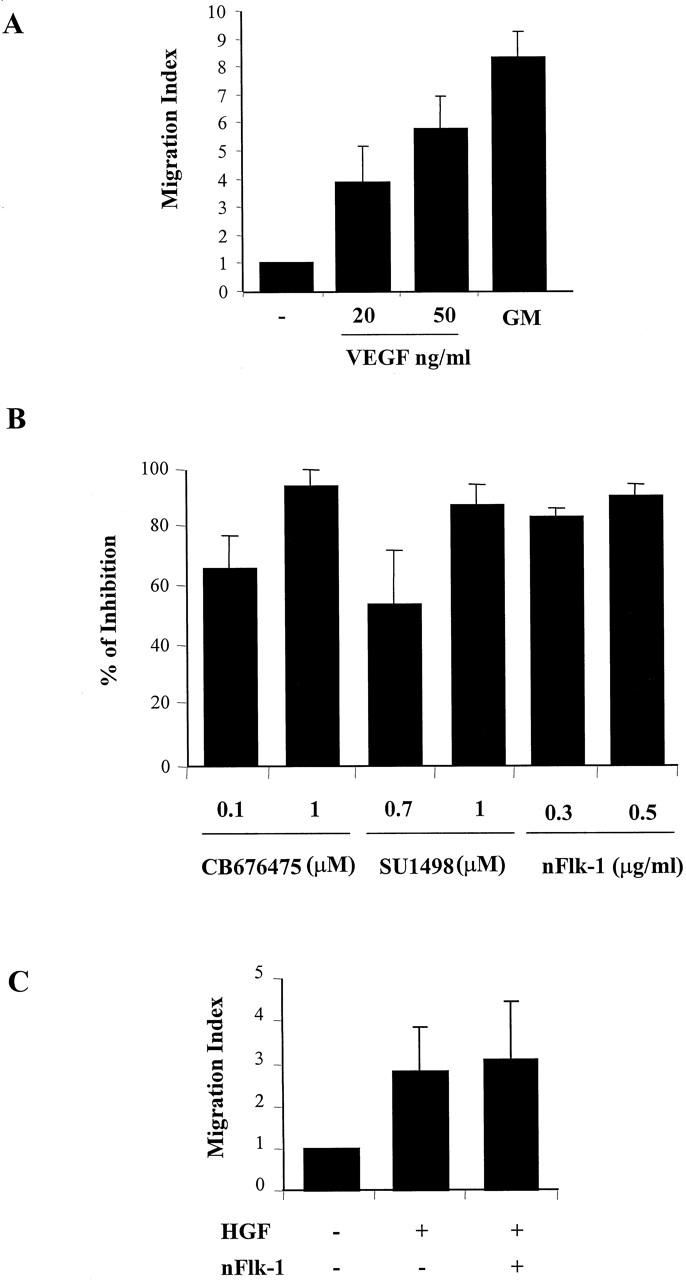
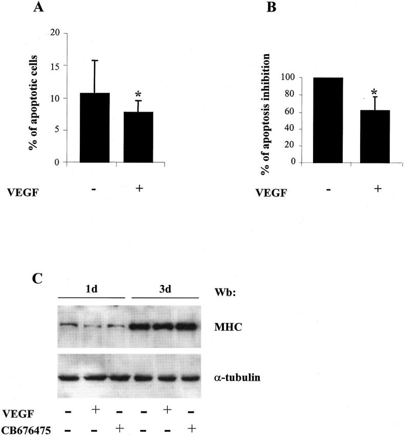
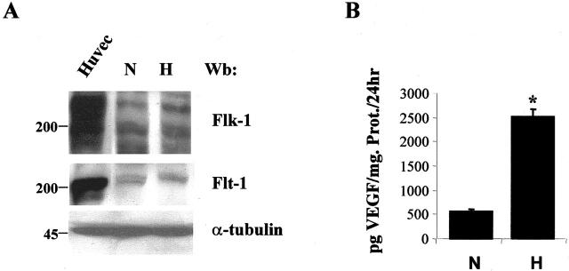
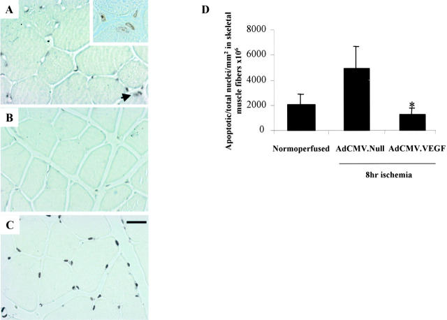
Vascular endothelial growth factor (VEGF) expression is enhanced in ischemic skeletal muscle and is thought to play a key role in the angiogenic response to ischemia. However, it is still unknown whether, in addition to new blood vessel growth, VEGF modulates skeletal muscle cell function. In the present study immunohistochemical analysis showed that, in normoperfused mouse hindlimb, VEGF and its receptors Flk-1 and Flt-1 were expressed mostly in quiescent satellite cells. Unilateral hindlimb ischemia was induced by left femoral artery ligation. At day 3 and day 7 after the induction of ischemia, Flk-1 and Flt-1 were expressed in regenerating muscle fibers and VEGF expression by these fibers was markedly enhanced. Additional in vitro experiments showed that in growing medium both cultured satellite cells and myoblast cell line C2C12 expressed VEGF and its receptors. Under these conditions, Flk-1 receptor exhibited constitutive tyrosine phosphorylation that was increased by VEGF treatment. During myogenic differentiation Flk-1 and Flt-1 were down-regulated. In a modified Boyden Chamber assay, VEGF enhanced C2C12 myoblasts migration approximately fivefold. Moreover, VEGF administration to differentiating C2C12 myoblasts prevented apoptosis, while inhibition of VEGF signaling either with selective VEGF receptor inhibitors (SU1498 and CB676475) or a neutralizing Flk-1 antibody, enhanced cell death approximately 3.5-fold. Finally, adenovirus-mediated VEGF(165) gene transfer inhibited ischemia-induced apoptosis in skeletal muscle. These results support a role for VEGF in myoblast migration and survival, and suggest a novel autocrine role of VEGF in skeletal muscle repair during ischemia.


Figures

Figure 1.
Figure 1.
Quantitative evaluation of blood flow recovery after hindlimb ischemia. LDPI was used to quantify both right and left hindlimb perfusion, preoperatively (C), immediately after femoral artery ligation (0), and at the indicated time points, postoperatively. Analysis was performed by calculating the average perfusion of each ischemic and non-ischemic foot and expressing it as a ratio of left (ischemic) to right (normoperfused) foot.
Figure 2.
Figure 2.
Expression of VEGF and its receptors in skeletal muscle cells in vivo. Flk-1 and Flt-1 expression in normoperfused mouse skeletal muscle (A) and in vascular structures (B). Serial muscle sections were immunostained for Flk-1 and Flt-1. Positive cells, indicated by arrowheads, were identified as satellite cells by their immunoreactivity with M-cadherin antibody. Insets show higher-power photomicrographs of satellite cell. Control immunostaining was performed by omitting the primary antibody. Magnification, ×40 (inset ×100); bar, 25 μm. Time-course of Flk-1 and Flt-1 expression (C to E). Serial sections from hindlimbs were obtained at 3 days (C), 7 days (D), and 14 days (E) following the induction of ischemia. Flk-1 and Flt-1 were expressed in activated satellite cells as identified by desmin labeling (C); 7 days after ischemia Flk-1 and Flt-1 were expressed in regenerating myotubes (D) and the expression of both receptors decreased at day 14 (E), when the regenerative process was nearly complete. Magnification, ×40; bar, 25 μm.
Figure 3.
Figure 3.
VEGF expression in skeletal muscle cells in vivo. Time-course of VEGF expression in mouse ischemic hindlimb. A: VEGF immunostaining was observed in satellite cells of normal skeletal muscle (A). VEGF protein was detected in satellite cells at day 3 (B) and in regenerating fibers at day 7 (C) after femoral artery ligation. The immunostaining decreased in regenerating fibers at 14 days after ischemic injury (D). Magnification, ×40; bar, 25 μm.
Figure 4.
Figure 4.
Flk-1 and Flt-1 expression in myogenic cells in vitro. A: RT-PCR analysis of Flk-1 and Flt-1 expression in skeletal muscle cell culture. Total RNAs (1 μg) extracted from C2C12 cells, satellite cells, and newborn mice heart (positive control) were used for reverse transcription. PCR analysis was carried out using specific primers for Flk-1 and Flt-1. Negative control represents RT-PCR of C2C12 cells RNA without oligonucleotides. B: Western blot analysis showed the presence of Flk-1 and Flt-1 proteins from satellite cells and C2C12 cells in GM. Total extract from HUVEC was used as a positive control for the expression of both receptors. C: Flk-1 phosphorylation in C2C12 cells. Lysates from C2C12 untreated or treated either with VEGF165 (50 ng/ml) for 5 minutes or CB676475 (1 μmol/L) for 1 hour, were immunoprecipitated with anti-Flk-1 Mab or a preimmune serum (PI). Subsequently, immunoprecipitated proteins were subjected to Western blot analysis with anti-phosphotyrosine (top) and reprobed with antibody to Flk-1 (bottom).
Figure 5.
Figure 5.
Expression of VEGF and its receptors during myogenic differentiation. A: Western blot analysis of total C2C12 cell lysates shows that Flk-1 and Flt-1 proteins decreased progressively over a 5-day time period when cells in GM at day 0 (d0) were changed to DM. In agreement with the myogenic differentiation of these cells, MyHC expression increased progressively over the same time period. Western blot analysis with anti α-tubulin antibody was performed on the same membrane to confirm equal loading of the lanes. In these experiments myoblasts cultured in GM were 80% confluent when they were switched to DM. B: ELISA determination of VEGF production from proliferating and differentiating C2C12 cells. At the onset of differentiation VEGF level decreased and over a 5-day time period in DM was significantly higher to that found in GM. Culture medium was changed every 24 hours and VEGF levels in conditioned media were determined after 1 day of culture in GM and at day 1, 3, and 5 of culture in DM. Results represent mean ± SD of six experiments. The asterisk indicates a P ≤ 0.05 vs. GM.
Figure 6.
Figure 6.
Chemotaxis of C2C12 myoblasts in response to VEGF. A: C2C12 (2 × 10⁴) were placed in upper compartment of the modified Boyden chambers. VEGF165 at the indicated concentration was added to the lower compartment and incubated for 6 hours at 37°C. GM was used as a positive control. After staining with Giemsa solution, migrated cells were quantified by counting nuclei in five random microscope fields (×40). The data are expressed as the fold increase in the number of migrated cells relative to the number of migrated cells in the absence of factor (migration index) and are the means ± SD of at least four independent experiments performed in triplicate. B: Effect of Flk-1 and Flt-1 inhibitors on VEGF-mediated C2C12 migration. C2C12 cells were incubated with the indicated concentration of CB676475, SU1498, and nFlk-1 Mab and placed in the upper chamber. VEGF (20 ng/ml) was added to the lower chamber and quantification of migrated cells was performed as described in (A). The data are expressed as % inhibition of migration index. Results represent mean ± SD of three independent experiments performed in triplicate. C: Effect of Flk-1 inhibitors on HGF-induced C2C12 migration. C2C12 cells were incubated with 0.5 μg/ml of nFlk-1 in the upper chamber and HGF (15 ng/ml) was added to the lower chamber. Results represent the mean ± SD of three experiments.
Figure 7.
Figure 7.
Effect of VEGF on differentiation-induced apoptosis of C2C12 myoblasts. C2C12 myoblasts were plated at 10 cells/60-mm diameter dish and cultured for 72 hours in DM without or with 20 ng/ml of VEGF165. A: TUNEL labeling was used to detect apoptotic myoblasts in the cultures. Apoptotic nuclei were counted in 20 random fields at ×40 magnification and expressed as a percentage of total nuclei. Results represent mean ± SD of five independent experiments. The asterisk indicates a P ≤ 0.01. B: ELISA quantification of histone-associated fragments in C2C12 cultures. Inhibition of apoptosis was reported as a % of optical density reduction between untreated and VEGF-treated C2C12 cells. Results represent mean ± SD of six independent experiments. The asterisk indicates a P ≤ 0.001. C: Effect of VEGF and CD676475 treatment on C2C12 myogenic differentiation. Western blot analysis of total extract from C2C12 cells treated for the indicated time-points either with VEGF or CB676475. Myogenic differentiation was assessed as MyHC expression with MF20 Mab. The same filter was probed with anti α-tubulin Mab to show equal protein concentration (lower panel).
Figure 8.
Figure 8.
Effect of hypoxia on the expression of VEGF and its receptors by C2C12 myoblasts. A: Cell lysates were prepared from C2C12 cultured in DM cells and kept either in normoxia or hypoxia for 48 hours and subjected to Western blot analysis using anti-Flk-1 and anti-Flt-1 Mabs. The same membrane was probed with anti α-tubulin antibody to confirm equal loading of the lanes. B: ELISA determination of VEGF production from normoxic and hypoxic C2C12 cells. CM from 1 day culture of C2C12 in normoxia and hypoxia conditions were collected. VEGF production was detected by ELISA as described in Materials and Methods. Results represent mean ± SD of four experiments. The asterisk indicates a P ≤ 0.05.
Figure 9.
Figure 9.
Effect of Flk-1 and Flt-1 inactivation on hypoxia-mediated inhibition of C2C12 apoptosis. C2C12 myoblasts were plated in GM at 2 × 10 cells/60-mm diameter dish for 24 hours. Thereafter cells were switched to DM and cultured either in normoxic or hypoxic conditions for 48 hours. nFlk-1 (0.5 μg/ml) was added to the culture medium for the entire period of treatment. TUNEL labeling was used to detect apoptotic myoblasts. A: Micrographs: left panels illustrate the fluorescent TUNEL images from a representative experiment while right panels illustrate Hoechst staining of the same cells. B: Quantification of apoptotic cells obtained in the experimental conditions described for A. TUNEL-positive cells and total Hoechst-stained nuclei were counted on 20 fields for each experiment. Results represent mean ± SD of six independent experiments. The asterisk indicates a P ≤ 0.001.
Figure 10.
Figure 10.
In vivo effect of Ad.VEGF on ischemia-induced skeletal muscle apoptosis. Apoptosis was measured by TUNEL assay 8 hours after femoral artery ligation. Representative sections of ischemic adductor muscles treated with AdCMV.Null (A), AdCMV.VEGF165 (B), or DNAsi as a positive control (C). Arrowhead indicates apoptotic nuclei. Inset shows a higher-power photomicrograph of TUNEL-positive skeletal muscle nuclei indicated by the arrowhead. Magnification ×40; bar 25 μm. D: Bar graph of the mean TUNEL-positive skeletal muscle nuclei number/mm ×10 cells from normoperfused and ischemic skeletal muscle injected either with Ad.CMV.Null or Ad.CMV.VEGF. The asterisk indicates a P < 0.05 vs. AdCMV.Null.

Similar articles

Cited by

References

    1. Matsumoto T, Claesson-Welsh L: VEGF receptor signal transduction. Sci STKE 2001, 112:1-17 - PubMed
    1. Asahara T, Takahashi T, Masuda H, Kalka C, Chen D, Iwaguro H, Inai Y, Silver M, Isner JM: VEGF contributes to postnatal neovascularization by mobilizing bone marrow-derived endothelial progenitor cells. EMBO J 1999, 18:3964-3972 - PMC - PubMed
    1. Kalka C, Masuda H, Takahashi T, Gordon R, Tepper O, Gravereaux E, Pieczek A, Iwaguro H, Hayashi SI, Isner JM, Asahara T: Vascular endothelial growth factor(165) gene transfer augments circulating endothelial progenitor cells in human subjects. Circ Res 2000, 86:1198-1202 - PubMed
    1. Kalka C, Tehrani H, Laudenberg B, Vale PR, Isner JM, Asahara T, Symes JF: VEGF gene transfer mobilizes endothelial progenitor cells in patients with inoperable coronary disease. Ann Thorac Surg 2000, 70:829-834 - PubMed
    1. Tischer E, Mitchell R, Hartman T, Silva M, Gospodarowicz D, Fiddes JC, Abraham JA: The human gene for vascular endothelial growth factor: multiple protein forms are encoded through alternative exon splicing. J Biol Chem 1991, 266:11947-11954 - PubMed

MeSH terms

Substances

LinkOut - more resources