

Transforming growth factor beta in cardiovascular development and function
- PMID: 12948523
- PMCID: PMC3855389
- DOI: 10.1016/s1359-6101(03)00044-3
Transforming growth factor beta in cardiovascular development and function
Abstract
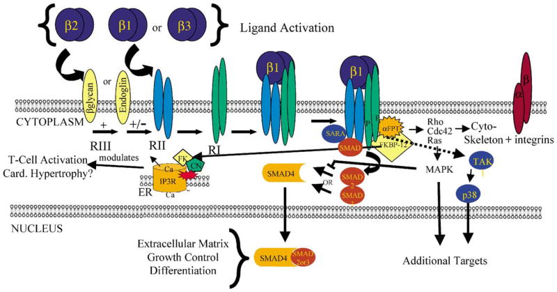
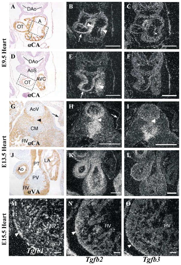
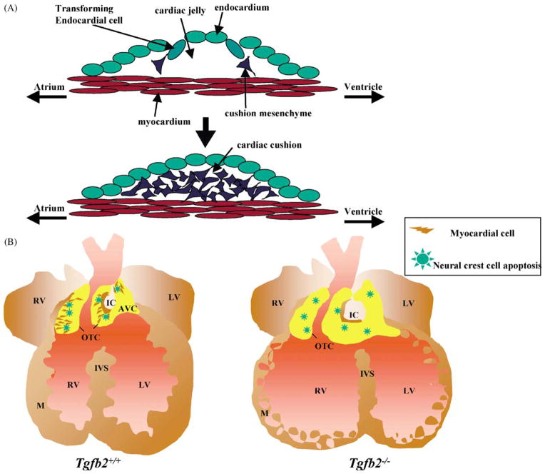
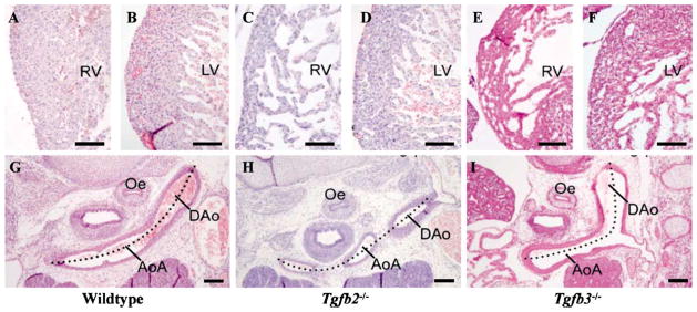
Transforming growth factor betas (TGFbetas) are pleiotropic cytokines involved in many biological processes. Genetic engineering and tissue explanation studies have revealed specific non-overlapping roles for TGFbeta ligands and their signaling molecules in development and in normal function of the cardiovascular system in the adult. In the embryo, TGFbetas appear to be involved in epithelial-mesenchymal transformations (EMT) during endocardial cushion formation, and in epicardial epithelial-mesenchymal transformations essential for coronary vasculature, ventricular myocardial development and compaction. In the adult, TGFbetas are involved in cardiac hypertrophy, vascular remodeling and regulation of the renal renin-angiotensin system. The evidence for TGFbeta activities during cardiovascular development and physiologic function will be given and areas which need further investigation will be discussed.
Figures
Similar articles
-
Induction of endocardial cushion tissue in the avian heart is regulated, in part, by TGFbeta-3-mediated autocrine signaling.Dev Biol. 1997 Aug 1;188(1):64-74. doi: 10.1006/dbio.1997.8637. Dev Biol. 1997. PMID: 9245512
-
Activin receptor-like kinase 2 and Smad6 regulate epithelial-mesenchymal transformation during cardiac valve formation.Dev Biol. 2005 Apr 1;280(1):201-10. doi: 10.1016/j.ydbio.2004.12.037. Dev Biol. 2005. PMID: 15766759
-
Roles of TGFbeta and BMP during valvulo-septal endocardial cushion formation.Anat Sci Int. 2009 Sep;84(3):77-87. doi: 10.1007/s12565-009-0027-0. Epub 2009 Mar 14. Anat Sci Int. 2009. PMID: 19288174 Review.
-
Bmp2 is essential for cardiac cushion epithelial-mesenchymal transition and myocardial patterning.Development. 2005 Dec;132(24):5601-11. doi: 10.1242/dev.02156. Development. 2005. PMID: 16314491
-
Mechanisms involved in valvuloseptal endocardial cushion formation in early cardiogenesis: roles of transforming growth factor (TGF)-beta and bone morphogenetic protein (BMP).Anat Rec. 2000 Feb 1;258(2):119-27. doi: 10.1002/(SICI)1097-0185(20000201)258:2<119::AID-AR1>3.0.CO;2-U. Anat Rec. 2000. PMID: 10645959 Review.
Cited by
-
Inhibition of TGF-β/Smad3 Signaling Disrupts Cardiomyocyte Cell Cycle Progression and Epithelial-Mesenchymal Transition-Like Response During Ventricle Regeneration.Front Cell Dev Biol. 2021 Mar 16;9:632372. doi: 10.3389/fcell.2021.632372. eCollection 2021. Front Cell Dev Biol. 2021. PMID: 33816481 Free PMC article.
-
Generation of mice with a conditional allele for the transforming growth factor beta3 gene.Genesis. 2012 Jan;50(1):59-66. doi: 10.1002/dvg.20789. Epub 2012 Jan 6. Genesis. 2012. PMID: 22223248 Free PMC article.
-
First evidence of maternally inherited mosaicism in TGFBR1 and subtle primary myocardial changes in Loeys-Dietz syndrome: a case report.BMC Med Genet. 2018 Sep 15;19(1):170. doi: 10.1186/s12881-018-0661-2. BMC Med Genet. 2018. PMID: 30219046 Free PMC article.
-
Modeling Human Heart Development and Congenital Defects Using Organoids: How Close Are We?J Cardiovasc Dev Dis. 2022 Apr 21;9(5):125. doi: 10.3390/jcdd9050125. J Cardiovasc Dev Dis. 2022. PMID: 35621836 Free PMC article. Review.
-
Generation of mice with a conditional allele for transforming growth factor beta 1 gene.Genesis. 2009 Jun;47(6):423-31. doi: 10.1002/dvg.20516. Genesis. 2009. PMID: 19415629 Free PMC article.
References
-
- Massague J, Chen YG. Controlling TGF-beta signaling. Genes Dev. 2000;14:627–44. - PubMed
Publication types
MeSH terms
Substances
Grants and funding
LinkOut - more resources
Full Text Sources
Other Literature Sources
